Friday, March 31, 2023

Should Exclamation Points Be Used in Scientific Writing? Yes!

Exclamation points are rare in scientific writing, but I like them. Occasionally Russ Hobbie and I use them in Intermediate Physics for Medicine and Biology. They’re fine as long as you don’t overdo it.

Let’s see what my favorite books about writing say.

The Elements of Style, by Strunk and White, superimposed on Intermediate Physics for Medicine and Biology.
The Elements of Style,
by Strunk and White.

Strunk and White (The Elements of Style): “Do not attempt to emphasize simple statements by using a mark of exclamation…The exclamation mark is to be reserved for use after true exclamations or commands.”

On Writing Well, by William Zinsser, superimposed on Intermediate Physics for Medicine and Biology.
On Writing Well,
by William Zinsser.

Zinsser (On Writing Well): “Don’t use it unless you must to achieve a certain effect. It has a gushy aura…Resist using an exclamation point to notify the reader that you are making a joke or being ironic…Humor is best achieved by understatement, and there’s nothing subtle about an exclamation point.”

Dreyer's English, by Benjamin Dreyer, superimposed on Intermediate Physics for Medicine and Biology.
  Dreyer's English,
by Benjamin Dreyer.

Dreyer (Dreyer’s English): “Go light on the exclamation points. When overused, they’re bossy, hectoring, and, ultimately, wearying. Some writers recommend that you should use no more than a dozen exclamation points per book; others insist that you should use no more than a dozen exclamation points in a lifetime.”

Plain Words, by Sir Ernest Gowers, superimposed on Intermediate Physics for Medicine and Biology.
Plain Words,
by Sir Ernest Gowers.

Gowers (Plain Words): His disdain for the exclamation point is so complete he doesn’t even acknowledge that it exists.

Below I list eleven places where exclamation points appear in IPMB. In each case, I indicate if we should keep the exclamation point or toss it out. Note: I don’t include any exclamation point that indicates a factorial, such as 4! = 1 × 2 × 3 × 4 = 24; that’s a mathematical symbol, not punctuation.

Page 29: Homework Problem 43 in Chapter 1 says “Suppose a student asked you, ‘How can blood be moving more slowly in a capillary than in the aorta? … The capillary has a much smaller cross-sectional area than the aorta. Therefore, the blood should move faster in the capillary than in the aorta!” The exclamation point belongs to the hypothetical student, not to Russ and me. If you don’t like it, take it up with the student (and beware, those students tend to be gushy). Keep.

Page 44: In Chapter 2, Russ and I write “Moreover, commercial graphing software does not impose this constraint on log–log plots, so it is becoming less and less likely that you can determine the exponent by glancing at the plot. Be careful!” The exclamation point is a warning. Keep.

Page 51: In the references at the end of Chapter 2, we cite Albert Bartlett’s delightful book “The Essential Exponential!” The exponential point is Bartlett’s, and is part of the title (like Oklahoma!). Keep.

Page 53: In the introduction to Chapter 3, Russ and I explain why statistical methods are so useful in thermodynamics. We estimate how long is required to simulate all the particles in a cubic millimeter of blood. We conclude “If a computer can do 1012 operations/s, then the complete calculation for a single time interval will require 108 s or 3 years!” In other words, a really long time. My feelings on this one are mixed. Toss.

Page 270: In a footnote in Chapter 10, we write “Strictly speaking, (dV/dt)alveoli is not the derivative of a function V. (It always has a positive value, and the lungs are not expanding without limit!) We use the notation to remind ourselves that it is the rate of air exchange in the alveoli.” If I could delete only one exclamation point from IPMB, this would it. The thought of those lungs getting bigger and bigger is just disturbing. Toss.

Page 308: In Chapter 11, Russ and I compare two methods for doing a least squares fit of an exponentially decaying function. Method 1: take the logarithm of the data and then do a linear fit. Method 2: do a nonlinear fit to the original data. We end the analysis with a moral: “Use nonlinear least squares, Method 2!” Keep.

Page 388: A section in Chapter 14 has tricky units. We remind the reader to “Be careful with units!” Exclamation points are legitimate when issuing a command or warning. Perhaps, however, we shouldn’t have used both bold and an exclamation point. Keep.

Page 484: When explaining the linear-quadratic model for radiation damage, Russ and I concoct an illustrative but unrealistic example. We then warn the reader “(This is not realistic!).” I wonder if we should’ve made a more realistic example and avoided the exclamation point. Toss.

Page 497: In Homework Problem 8 of Chapter 16, Russ and I discuss x-ray imaging devices used to fit kids’ shoes, common in the 1940s. “These marvelous units were operated by people who had no concept of radiation safety and aimed a beam of x-rays upward through the feet and right at the reproductive organs of the children!” My vote is to keep this exclamation point at all costs! Let’s even keep the sarcasm. Keep.

Page 587: In Appendix H, we use an exclamation point when trying to make a joke. We had already developed the binomial probability distribution using p for the probability of success and q for the probability of failure. Then we write “Suppose that we do N independent tests, and suppose that in healthy people, the probability that each test is abnormal is p. (In our vocabulary, having an abnormal test is ‘success’!).” Lame. Toss.

Page 593: Appendix I has a footnote containing my favorite exclamation point in Intermediate Physics for Medicine and Biology. It appears when we introduce Stirling’s formula: ln n! ≈ n ln n - n. “For more about Stirling’s formula, see N. D. Mermin (1994) Stirling’s formula! Am J Phys 52: 362–365.” The exclamation point is a pun. Keep!

Friday, March 24, 2023

Three New Reviews

Over the last couple years, I’ve been writing lots of review articles. In the last few weeks three have been published. All of them are open access, so you can read them without a subscription.

Can MRI be Used as a Sensor to Record Neural Activity?

Can MRI be Used as a Sensor
to Record Neural Activity?
This review asks the question “Can MRI be Used as a Sensor to Record Neural Activity?” The article is published in the journal Sensors (Volume 23, Article Number 1337). The abstract is reproduced below.
Magnetic resonance provides exquisite anatomical images and functional MRI monitors physiological activity by recording blood oxygenation. This review attempts to answer the following question: Can MRI be used as a sensor to directly record neural behavior? It considers MRI sensing of electrical activity in the heart and in peripheral nerves before turning to the central topic: recording of brain activity. The primary hypothesis is that bioelectric current produced by a nerve or muscle creates a magnetic field that influences the magnetic resonance signal, although other mechanisms for detection are also considered. Recent studies have provided evidence that using MRI to sense neural activity is possible under ideal conditions. Whether it can be used routinely to provide functional information about brain processes in people remains an open question. The review concludes with a survey of artificial intelligence techniques that have been applied to functional MRI and may be appropriate for MRI sensing of neural activity.

Parts of the review may be familiar to readers of this blog. For instance, in June of 2016 I wrote about Yoshio Okada’s experiment to measure neural activation in a brain cerebellum of a turtle, in August 2019 I described Allen Song’s use of spin-lock methods to record brain activity, and in April 2020 I discussed J. H. Nagel’s 1984 abstract that may have been the first to report using MRI to image action currents. All these topics are featured in my review article. In addition, I analyzed my calculation, performed with graduate student Dan Xu, of the magnetic field produced inside the heart, and I reviewed my work with friend and colleague Ranjith Wijesinghe, from Ball State University, on MRI detection of bioelectrical activity in the brain and peripheral nerves. At the end of the review, I examined the use of artificial intelligence to interpret this type of MRI data. I don’t really know much about artificial intelligence, but the journal wanted me to address this topic so I did. With AI making so much news these days (ChatGPT was recently on the cover of TIME magazine!), I’m glad I included it.

Readers of Intermediate Physics for Medicine and Biology will find this review to be a useful extension of Section 18.12 (“Functional MRI”), especially the last paragraph of that section beginning with “Much recent research has focused on using MRI to image neural activity directly, rather than through changes in blood flow...”

Magneto-Acoustic Imaging in Biology

Magneto-Acoustic Imaging in Biology
Next is “Magneto-Acoustic Imaging in Biology,” published in the journal Applied Sciences (Volume 13, Article Number 3877). The abstract states

This review examines the use of magneto-acoustic methods to measure electrical conductivity. It focuses on two techniques developed in the last two decades: Magneto-Acoustic Tomography with Magnetic Induction (MAT-MI) and Magneto-Acousto-Electrical Tomography (MAET). These developments have the potential to change the way medical doctors image biological tissue.
The only place in IPMB where Russ Hobbie and I talked about these topics is in Homework Problem 31 in Chapter 8, which analyzes a simple example of MAT-MI.

A Mathematical Model of Mechanotransduction

A Mathematical Model of Mechanotransduction
Finally comes “A Mathematical Model of Mechanotransduction” in the new journal Academia Biology (Volume 1; I can’t figure out what the article number is?!).

This article reviews the mechanical bidomain model, a mathematical description of how the extracellular matrix and intracellular cytoskeleton of cardiac tissue are coupled by integrin membrane proteins. The fundamental hypothesis is that the difference between the intracellular and extracellular displacements drives mechanotransduction. A one-dimensional example illustrates the model, which is then extended to two or three dimensions. In a few cases, the bidomain equations can be solved analytically, demonstrating how tissue motion can be divided into two parts: monodomain displacements that are the same in both spaces and therefore do not contribute to mechanotransduction, and bidomain displacements that cause mechanotransduction. The model contains a length constant that depends on the intracellular and extracellular shear moduli and the integrin spring constant. Bidomain effects often occur within a few length constants of the tissue edge. Unequal anisotropy ratios in the intra- and extracellular spaces can modulate mechanotransduction. Insight into model predictions is supplied by simple analytical examples, such as the shearing of a slab of cardiac tissue or the contraction of a tissue sheet. Computational methods for solving the model equations are described, and precursors to the model are reviewed. Potential applications are discussed, such as predicting growth and remodeling in the diseased heart, analyzing stretch-induced arrhythmias, modeling shear forces in a vessel caused by blood flow, examining the role of mechanical forces in engineered sheets of tissue, studying differentiation in colonies of stem cells, and characterizing the response to localized forces applied to nanoparticles.

This review is similar to my article that I discussed in a blog post about a year ago, but better. I originally published it as a manuscript on the bioRxiv, the preprint server for biology, but it received little attention. I hope this version does better. If you want to read this article, download the pdf instead of reading it online. The equations are all messed up on the journal website, but they look fine in the file.

If you put these three reviews together with my previous ones about magnetic stimulation and the bidomain model of cardiac electrophysiology, you have a pretty good summary of the topics I’ve worked on throughout my career. Are there more reviews coming? I’m working feverishly to finish one more. For now, I’ll let you guess the topic. I hope it’ll come out later this year.

Friday, March 17, 2023

Daylight Saving Time

Why Should We Abolish Daylight Saving Time?
 J. Biol. Rhythms, 34:227–230, 2019.
 
Last Sunday, we all switched from standard time to daylight saving time, losing an hour of sleep in the process. Should we stop this changing of clocks every six months? We have three options: 1) we can continue to switch between standard time in the winter and daylight saving time in the summer (our current practice), 2) we can change to permanent daylight saving time (a change that the Senate has passed, but has not yet been approved by the House of Representatives), or 3) we can change to permanent standard time. A position paper from the Society of Research on Biological Rhythms addresses this issue. The citation and abstract are given below.
Roenneberg T, Wirz-Justice A, Skene DJ, et al. (2019) “Why Should We Abolish Daylight Saving Time?Journal of Biological Rhythms, Volume 34, Pages 227–230.

Local and national governments around the world are currently considering the elimination of the annual switch to and from Daylight Saving Time (DST). As an international organization of scientists dedicated to studying circadian and other biological rhythms, the Society for Research on Biological Rhythms (SRBR) engaged experts in the field to write a Position Paper on the consequences of choosing to live on DST or Standard Time (ST). The authors take the position that, based on comparisons of large populations living in DST or ST or on western versus eastern edges of time zones, the advantages of permanent ST outweigh switching to DST annually or permanently. Four peer reviewers provided expert critiques of the initial submission, and the SRBR Executive Board approved the revised manuscript as a Position Paper to help educate the public in their evaluation of current legislative actions to end DST.

Biological oscillations are complicated. Readers of Chapter 10 in Intermediate Physics for Medicine and Biology know that nonlinear dynamics makes resetting an oscillator’s phase difficult, and that driving a nonlinear oscillator can lead to complex, and sometimes even chaotic, behavior. I’m glad that the Society for Research on Biological Rhythms sought advice from experts about this issue.

Roenneberg et al.’s paper focuses on health issues related to our three clocks: the sun clock, our body clock, and our social clock (the clock set by society). The authors summarize the problem of synchronizing these three clocks in this way: 

We live according to the same social clock time within a time zone, but as long as we still can see the natural day (through windows or on our way to or from work or school), our body clocks still follow more or less the time of the sun clock.

Is this a problem? Apparently so. 

We know that DST increases the time difference between the social clock and the body clock. More and more studies show that time differences between the social clock and the body clock challenge our health, are associated with decreased life expectancy, shorten sleep, cause mental and cognitive problems, and contribute to the many sleep disturbances.

I removed the references from this quote, but the authors support these claims by citing many research studies. Note that these problems do not arise only from the change back-and-forth between standard and daylight saving time in the spring and fall. Even permanent daylight saving time would cause chronic health problems.

My preference is permanent standard time. I live in Rochester Hills, Michigan (a suburb of Detroit), which is in the western part of the Eastern Time Zone, so I was surprised to read that 

the further west people live within a time zone, the more health problems they may experience and the shorter they live on average.

Yikes! 

Michigan is also in the northern part of the United States, where differences in the length of day and night throughout the year are exaggerated compared to our southern neighbors. This year in early January the sun rose in Detroit just after 8 am. Change to year-round daylight saving time and we would have sunrise at 9 am. For a morning person like me, that’s a lot of darkness before the sun comes up. 

My personal preference, however, isn’t important. I’m sure there are others who feel just as strongly that year-round daylight saving time is better than year-round standard time. What impresses me is that Roenneberg et al. make a strong case favoring permanent standard time as being better for society overall. They conclude that

if we want to improve human health, we should not fight against our body clock, and therefore, we should abandon DST and return to Standard Time (which is when the sun clock time most closely matches the social clock time) throughout the year.

I agree!

And if Congress refuses to move to year-round standard time, I would rather keep things as they are now (changing with the seasons) rather than have year-round daylight saving time.

Friday, March 10, 2023

Physics Girl has Long Covid

I’m a fan of Dianna Cowern, better known as Physics Girl, who makes Youtube videos about physics that would be helpful for readers of Intermediate Physics for Medicine and Biology. Three years ago I featured several of her videos in a blog post.

Cowern is suffering from a severe case of long Covid. I’m going to turn this week’s post over to Simone Giertz for an update on Cowern’s health.

An update on Dianna's health, by Simone Giertz.


Friday, March 3, 2023

Cobalt Blues: The Story of Leonard Grimmett, the Man Behind the First Cobalt-60 Unit in the United States

Cobalt Blues,
by Peter Almond.
I recently read Cobalt Blues: The Story of Leonard Grimmett, the Man Behind the First Cobalt-60 Unit in the United States (Springer, 2013), written by Peter Almond. The treatment of cancer using the isotope cobalt-60 is now obsolete, but in the era just after World War II it was cutting-edge technology. In his prologue, Almond writes
[The British medical physicist Leonard George] Grimmett was an expert in the use of radium to treat cancer and in the safe handling and measurement of radiation and radioactive materials in clinical situations. He had spent the best part of his career devising better, safer, and more efficient ways to treat cancer with radiation and he remained in England during [World War II]... Then in 1948 while working for UNESCO in Paris he received an offer he could not refuse the, “…post as physicist to a new ‘Cancer Research Institute and Atomic Center’ in The University of Texas”, one of the original universities in the ORINS [Oak Ridge Institute of Nuclear Studies] consortium. Thus was set in motion the events that would lead Grimmett to Houston, Texas and to be the first person to publish, in 1950, the design of a cobalt-60 radiation therapy unit for the treatment of cancer. For the next 25 years cobalt-60 units would be the mainstay of cancer radiation therapy, treating millions of patients worldwide. Grimmett, however, would not live to see the completion of his work. This is his story.
Grimmett is a fascinating guy. As a young boy he learned to play the piano and was quite good. “He had worked his way through college playing for the silent movies, but with the advent of the ‘talkies,’ he had lost his income. He went to work at Westminster Hospital.” At Westminster and other hospitals he helped develop cancer treatment machines using radium, and later he established the medical physics program at the renowned M. D. Anderson Cancer Center. But he had other talents. He was a pilot, a scriptwriter, a gemologist, and jeweler. He’s remembered today primarily for developing a cobalt-60 therapy machine. Almond writes
It is not known for sure who first had the idea of replacing the radium in teletherapy units with a more suitable and less-expensive artificial radioactive substance. Grimmett, however, had been thinking about it for some years before he went to Houston, and a case can be made that he was the first.

What motivated him to use cobalt? “What Grimmett was looking for was an artificial radioactive isotope with gamma ray energies of 1–5 MeV with as long a half-life as possible that could be made in large quantities at a reasonable price.” He considered using sodium-24 for therapy. After 24Na beta decays it emits two gamma rays with energies of 4.1 and 1.4 MeV (see Fig. 17.9 of Intermediate Physics for Medicine and Biology). However, the half-life of 24Na is only 15 hours. 

The idea that cobalt-60 might be a suitable replacement for radium first occurred to Grimmett while he was reading Physical Review in an air-raid shelter during World War II… Later, after the war, he would have read the paper by J. S. Mitchell in the December 1946 issue of the British Journal of Radiology [82]. This is often cited as the paper that initiated the cobalt-60 era. Mitchell specifically mentions cobalt-60 as a replacement for radium beam therapy, and he gave the half-life as 5.3 years and the gamma ray energies as 1.3 and 1.1 MeV. He also reported that it could be produced in “the pile” (nuclear reactor).
Why did Almond title his book Cobalt Blues? Grimmett had trouble obtaining the needed cobalt-60. It is a by-product of nuclear reactors. He first tried the reactor at Oak Ridge, but ended up getting it from a reactor on Chalk River in Canada. Incidentally, the book cover of Cobalt Blues is a lovely cobalt blue.

Grimmett was not the only person trying to use cobalt-60 to treat cancer. Almond briefly describes the other groups, including one in Canada by Harold Johns, and tries to sort out the various priority claims.

Unfortunately, Grimmett died unexpectedly and never saw his unit in use. His obituary in the Houston Chronicle begins

Doctor Grimmett, Cancer Expert, Dies Suddenly 
Dr. Leonard G. Grimmett, 49, eminent physicist whose work in cancer research at M.D. Anderson Hospital, opened a whole new field of treatment of cancer, died of a heart attack at 1:10 a.m. Sunday at his home, 3238 Ewing.
I enjoyed Almond’s book. I learned much about the early years of the M. D. Anderson Cancer Center and about the issues that must be considered when building radiation therapy units. Readers of IPMB will find Cobalt Blues fascinating.

Friday, February 24, 2023

A Simple Mathematical Function Representing the Intracellular Action Potential

In Problem 14 of Chapter 7 in Intermediate Physics for Medicine and Biology, Russ Hobbie and I chose a strange-looking function to represent the intracellular potential along a nerve axon, vi(x). It’s zero everywhere except in the range −a < x < a, where it’s


Why this function? Well, it has several nice properties, which I’ll leave for you to explore in this new homework problem.
Section 7.4

Problem 14 ¼. For the intracellular potential, vi(x), given in Problem 14
(a) show that vi(x) is an even function,
(b) evaluate vi(x) at x = 0,
(c) show that vi(x) and dvi(x)/dx are continuous at x = 0, a/2 and a, and
(d) plot vi(x), dvi(x)/dx, and d2vi(x)/dx2 as functions of x, over the range −2a < x < 2a.
This representation of vi(x) has a shape like that of an action potential. Other functions also have a similar shape, such as a Gaussian. But our function is nice because it’s non-zero over only a finite region (−a < x < a) and it’s represented by a simple, low-order polynomial rather than a special function. An even simpler function for vi(x) would be triangular waveform, like that shown in Figure 7.4 of IPMB. However, that function has a discontinuous derivative and therefore its second derivative is infinite at discrete points (delta functions), making it tricky (but not too tricky) to deal with when calculating the extracellular potential (Eq. 7.21). Our function in Problem 14 ¼ has a discontinuous but finite second derivative.

The main disadvantage of the function in Problem 14 ¼ is that the depolarization phase of the “action potential” has the same shape as the repolarization phase. In a real nerve, the upstroke is usually briefer than the downstroke. The next new homework problem asks you to design a new function vi(x) that does not suffer from this limitation.
Section 7.4

Problem 14 ½. Design a piecewise continuous mathematical function for the intracellular potential along a nerve axon, vi(x), having the following properties. 
(a) vi(x) is zero outside the region −a < x < 2a
(b) vi(x) and its derivative dvi(x)/dx are continuous. 
(c) vi(x) is maximum and equal to one at x = 0. 
(d) vi(x) can be represented by a polynomial bi + ci x + di x2, where i refers to four regions: 
        i = 1,    −a < x < −a/2 
        i = 2, −a/2 < x < 0 
        i = 3,      0 < x < a
        i = 4,      a < x < 2a.
Finally, here’s another function that I’m particularly fond of.
Section 7.4

Problem 14 ¾. Consider a function that is zero everywhere except in the region −a < x < 2a, where it is

(a) Plot vi(x) versus x over the region −a < x < 2a,
(b) Show that vi(x) and its derivative are each continuous. 
(c) Calculate the maximum value of vi(x).
Simple functions like those described in this post rarely capture the full behavior of biological phenomena. Instead, they are “toy models” that build insight. They are valuable tools when describing biological phenomena mathematically.

Friday, February 17, 2023

Abraham Liboff (1927–2023)

Abe Liboff, in his office at Oakland University.
Abe Liboff, in his
office at Oakland University
Oakland University
physicist Abe Liboff died recently. A notice from President Ora Hirsch Pescovitz, published on the OU website, stated:
It is with deep sadness that I inform you of the death of Professor Emeritus Abraham Liboff who passed away on January 9, 2023. Dr. Liboff joined the Oakland University community in the Department of Physics on August 15, 1972, where he served until his retirement in August 2000.

During his tenure here at OU, Dr. Liboff was Chair of the Department of Physics. He is credited with 111 research publications, more than two dozen patents and nearly 3,400 scholarly citations during his career.

I arrived at OU in 1998, so his time at OU and mine overlapped by a couple years. I remember having a delightful breakfast with him during my job interview. He was one of the founders of OU’s medical physics PhD program that I directed for 15 years. His office was just a few doors down the hall from mine and he helped me get started at Oakland. I’ll miss him.

Although I loved the man, I didn’t love Abe’s cyclotron resonance theory of how magnetic fields interact with biological tissue. It’s difficult to reconcile admiration for a scientist with rejection of his scientific contributions. Rather than trying to explain Abe’s theory, I’ll quote the abstract from his article “Geomagnetic Cyclotron Resonance in Living Cells,” published in the Journal of Biological Physics (Volume 13, Pages 99–102, 1985).

Although considerable experimental evidence now exists to indicate that low-frequency magnetic fields influence living cells, the mode of coupling remains a mystery. We propose a radical new model for electromagnetic interactions with cells, one resulting from a cyclotron resonance mechanism attached to ions moving through transmembrane channels. It is shown that the cyclotron resonance condition on such ions readily leads to a predicted ELF-coupling at geomagnetic levels. This model quantitatively explains the results reported by Blackman et al. (1984), identifying the focus of magnetic interaction in these experiments as K+ charge carriers. The cyclotron resonance concept is consistent with recent indications showing that many membrane channels have helical configurations. This model is quite testable, can probably be applied to other circulating charge components within the cell and, most important, leads to the feasibility of direct resonant electromagnetic energy transfer to selected compartments of the cell.
In my book Are Electromagnetic Fields Making Me Ill? I didn’t have the heart to attack Abe in print. When discussing cyclotron resonance effects, I cited the work of Carl Blackman instead, who proposed a similar theory. What’s the problem with this idea? If you calculate the cyclotron frequency of a calcium ion in the earth’s magnetic field, you get about 23 Hz (see Eq. 8.5 in Intermediate Physics for Medicine and Biology). However, the thermal speed of a calcium ion at body temperature is about 440 m/s (Eq. 4.12 in IPMB). At that speed, the radius of the cyclotron orbit would be 3 meters (roughly ten feet)! The mean free path of a ion in water, however, is about an angstrom, which means the ion will suffer more than a billion collisions in one orbit; these interactions should swamp any cyclotron motion. Moreover, ion channels have a size of about 100 angstroms. In order to have a orbital radius similar to the size of a ion channel, the calcium ion would need to be moving extremely fast, which means it would have a kinetic energy vastly larger than the thermal energy. The theory just doesn’t work.

Since Liboff isn’t around to defend himself, I’ll let Louis Slesin—the editor and publisher of Microwave News—tell Abe’s side of the story. Read Slesin’s Reminiscence on the Occasion of Abe Liboff’s 90th Birthday. Although I don’t agree with Slesin on much, we both concur that Abe was a “wonderful and generous man.” If you want to hear about cyclotron resonance straight from the horse’s mouth, you can hear Abe talk about his career and work in a series of videos posted on the Seqex YouTube channel. (Seqex is a company that sells products based on Abe’s theories.) Below I link to the most interesting of these videos, in which Abe tells how he conceived of his cyclotron resonance idea.

Abe Liboff discussing the cyclotron resonance theory.
https://www.youtube.com/watch?v=YL-wqJ-PMAQ&list=PLCO-VktC6wofkMeEeZknT9Y4WhMnP76Ee&index=6

Friday, February 10, 2023

Water, Cavendish, and Lavoisier

To understand biological physics, you must know the properties of water. In Intermediate Physics for Medicine and Biology, Russ Hobbie and I discuss water’s density, compressibility, viscosity, heat capacity, surface tension, thermal conductivity, dielectric constant, and index of refraction. It’s behavior is critical for osmosis, diffusion, absorption of x-rays, propagation of ultrasonic waves, and magnetic resonance imaging.

In the very first section of IPMB, about distances and sizes, we say
At the 1-nm scale and below, we reach the world of small molecules and individual atoms. Water is the most common molecule in our body. It consists of two atoms of hydrogen and one of oxygen. The distance between adjacent atoms in water is about 0.1 nm.
Every schoolchild learns that water is H2O. But how do we know that water is made from hydrogen and oxygen? In other words, how did we first learn that water is not an element itself, but is a compound of two elements?

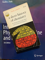
A Short History of Chemistry, by Isaac Asimov, superimposed on Intermediate Physics for Medicine and Biology.
A Short History of Chemistry,
by Isaac Asimov.
I’ll let Isaac Asimov tell the story. In A Short Histor of Chemistry, he writes
In 1783 [English scientist Henry] Cavendish was … working with his inflammable gas… He burned some of it and studied the consequences. He found that the vapors produced by the burning condensed to form a liquid that, on investigation, proved to be nothing more or less than water.

This was a crucially important experiment. In the first place, it was another hard blow at the Greek theory of the elements [air, water, earth, fire], for it showed that water was not a simple substance but was the sole product of the combination of two gases.

[French chemist Antoine] Lavoisier, hearing of the experiment, named Cavendish’s gas, hydrogen (“water-producer”) and pointed out that hydrogen burned by combining with oxygen and that therefore water was a hydrogen-oxygen combination.
Asimov's Biographical Encyclopedia
of Science and Technology
,
by Isaac Asimov.
So now you know. But these two brilliant scientists had tragic fates. You can find the stories in Asimov’s Biographical Encyclopedia of Science and Technology.

Cavendish (1731-1810):
[Cavendish] was excessively shy and absent-minded. He almost never spoke and when he did it was with a sort of stammer… He build a separate entrance to his house so he could come and leave alone… he even literally insisted on dying alone.

The eccentric had one and only one love, and that was scientific research. He spent almost sixty years in exclusive preoccupation with it. It was a pure love, too, for he did not care whether his findings were published, whether he got credit, or anything beyond the fact that he was sating his own curiosity. He wrote no books and published only twenty articles altogether. As a result, much of what he did remained unknown until years after his death…
Lavoisier (1743-1794):
In the same year that [Lavoisier’s] textbook [Elementary Treatise on Chemistry] appeared the French Revolution broke out. By 1792 the radical antimonarchists were in control…. Lavoisier… was guillotined on May 8, 1794, and buried in an unmarked grave. Two months later the radicals were overthrown. His was the most deplorable single casualty of the revolution.

50 Prominent Medical Physicists

Fifty Outstanding Medical Physicists superimposed on Intermediate Physics for Medicine and Biology.
Fifty Outstanding Medical Physicists.
In 2014, to mark its 50th anniversary, the International Organization for Medical Physics published a list of 50 medical physicists who have made an outstanding contribution to the advancement of medical physics over the last 50 years. I thought it would be interesting to see how many Russ Hobbie and I mention in Intermediate Physics for Medicine and Biology.

Hardly any. Only 11 of these 50 prominent medical physicists appear in IPMB.
  1. Peter R. Almond, of the M. D. Anderson Cancer Center, helped develop cancer treatments using photons, electrons, and neutrons. IPMB cites his paper “Review of Electron Beam Therapy Physics” (Physics in Medicine & Biology, Volume 51, Pages R455–R489, 2006) coauthored with Kenneth Hogstrom
  2. F. Herbert Attix worked at the Naval Research Laboratory and then the University of Wisconsin. IPMB cites Attix a dozen times, primarily for his landmark textbook Introduction to Radiological Physics and Radiation Dosimetry
  3. John Cameron was the father of the medical physics PhD program at the University of Wisconsin. IPMB cites his book Physics of the Body
  4. Aaron Fenster, of the University of Western Ontario, co-founded a microCT company eventually sold to GE Healthcare. Russ and I cite his paper with Paul Carson about the “Evolution of Ultrasound Physics and the Role of Medical Physicists in the AAPM and its Journal in that Evolution” (Medical Physics, Volume 36, Pages 411–428, 2008). 
  5. William Hendee, of the Medical College of Wisconsin, coauthored over thirty books. IPMB cites one of them, written with E. Russell Ritenour: Medical Imaging Physics
  6. Godfrey Hounsfield won a Nobel Prize for developing X-ray computed tomography, a central topic in Chapters 12 and 16 of IPMB
  7. Willi Kalender, of the University of Erlangen-Nuremberg, introduced volumetric spiral computer tomography. IPMB cites his book Computed Tomography: Fundamentals, System Technology, Image Quality and Applications
  8. Paul Lauterbur won a Nobel Prize for the invention of magnetic resonance imaging, the main topic of Chapter 18 in IPMB
  9. Peter Mansfield shared the Nobel Prize with Lauterbur, primarily for his contributions to MRI, including echo-planar imaging
  10. Colon Orton, of Wayne State University, studied radiotherapy and was active in the American Association of Physicists in Medicine. In IPMB, Russ and I cite three of his “Point/Counterpoint” articles in Medical Physics, for which he served as moderator. 
  11. Peter Wells, of Cardiff University, pioneered techniques using ultrasound imaging, the topic of Chapter 13 in IPMB. Russ and I cite his paper “Ultrasound Imaging” (Physics in Medicine and Biology, Volume 51, Pages R83–R98, 2006).
Another eight don’t appear in IPMB but have been mentioned in this blog: Anders Brahme, John Cunningham, Charles Mistretta, Ervin Podgorsak, Madan Rehani, Jean-Claude Rosenwald, Jacob Van Dyk, and Steve Webb. I’m not familiar with the other 31. I guess I don’t know as much as I thought I did. 😮 You can find the entire list here
 
Enjoy! 

*************************

As I was posting this article on my blog, it occurred to me that the list of 50 medical physicists came out about ten years ago, and that I ought to update it to 60 outstanding medical physicists in the last 60 years. Here are my additional ten. I tried to honor the spirit of the list by restricting myself to those who worked in the era from 1963 to 2023, but I couldn’t resist going back just a little further to select a few who worked in the 1950s.
  1. Paul Callaghan developed microscopic MRI 
  2. Hermann Carr proposed using gradients to image with magnetic resonance
  3. Allan Cormack won the Nobel Prize for his work on computed tomography
  4. Raymond Damadian performed early work on developing MRI
  5. Erwin Hahn discovered the nuclear magnetic resonance spin echo
  6. Eric Hall is the author of Radiobiology for the Radiologist
  7. John Hubbell compiled data on photon cross sections and attenuation coefficients
  8. Harold Johns is a coauthor of The Physics of Radiology
  9. Faiz Khan is the author of The Physics of Radiation Therapy
  10. William Oldendorf was a pioneer in computed tomography

Friday, February 3, 2023

Felix Savart, Biological Physicist

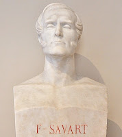
Bust of Félix Savart in the Institut de France.
Bust of Félix Savart
in the Institut de France.
From Wikipedia.
  
I’m fascinated by scientists who make the transition from medicine to physics, which is the opposite of my own transition from physics to medicine. One example is Félix Savart. In this blog post, I provide several excerpts from a 1959 article by Victor McKusick and H. Kenneth Wiskind, titled Félix Savart (1791–1841), Physician-Physicist: Early Studies Pertinent to the Understanding of Murmurs (Journal of the History of Medicine and Allied Sciences, Volume 14, Pages 411–423).

Savart was born in Meziere, France on June 30, 1791. His family had a long history of excelling in engineering, but Savart chose a different path.
Savart decided on a medical career and about 1808 entered the hospital in Metz. From 1810 to 1814 he served as a regimental surgeon in Napoleon’s armies… After discharge from the army, he completed his medical training in Strasbourg, where he received his doctor’s degree in October 1816. The title of his doctorate thesis was "Du cirsocele." The mundane topic of varicocele [enlarged veins in the scrotum] must have had little intrinsic appeal for him, and it is perhaps slight wonder he did not stay in medicine.
I can understand how that topic might drive a person away from the medical profession. For whatever reason, Savart spent little time practicing medicine. Instead, he was interested in physics, and particularly in sound.
In 1817 Savart returned to Metz with the intention of establishing a medical practice… He spent his time “more in fitting out a laboratory and building instruments than in seeing sick people and perusing Hippocrates…” It was during this period that he… began to devote himself specifically to the study of acoustics, a subject which engaged his attention almost exclusively for the remainder of his life.
McKusick and Wiskind compare Savart to three other physicians who made the transition to physics: Hermann von Helmholtz, Thomas Young, and Jean Leonard Marie Poiseuille. When Savart was 28, he made a life-changing trip to Paris.
In 1819 Savart went to Paris… to consult Jean-Baptiste Biot (1774–1862) in connection with his study of the acoustics of musical instruments. This was undoubtedly a turning point in Savart’s career. Biot encouraged and aided Savart in many ways and took him into collaboration in a study of electricity.
Savart’s name appears in Intermediate Physics for Medicine and Biology only when paired with Biot for the Biot-Savart Law. Russ Hobbie and I write
8.2.3 The Biot-Savart Law

In situations where the symmetry of the problem does not allow the [magnetic] field to be calculated from Ampere’s law, it is possible to find the field due to a steady current in a closed circuit using the Biot-Savart law.

Ironically, Savart is remembered among physicists for this one investigation into magnetism rather than a lifetime studying acoustics. 

Savart was an excellent experimentalist and instrument builder. He made careful measurements of the frequencies produced by a trapezoid violin, which a French commission found to be as good as the violins of Stradivarius. McKusick and Wiskind describe one of his more significant inventions: the Savart wheel.

About 1830 Savart invented a toothed wheel for determining the number of vibrations in a given musical tone. He attached tongues of pasteboard to the hoop of the wheel and arranged for these to strike a projecting object as the wheel was turned… [With this invention] Savart [determined] the frequency limits of audibility of sounds for the human ear [see Section 13.4 in IPMB]. He set the low and high values at 8 and 24,000 cycles per second, respectively... The values he determined are of the same order of magnitude as the 16 to 16,000 cycles per second one usually hears quoted now.
Savart also has a unit named for him.
The savart is a unit related to the perceptible change in frequency; 300 savarts are approximately equal to one octave. However, this unit has not enjoyed general acceptance and usage.
Another unit for frequency interval, discussed previously in this blog, is the cent. A savart is about 4 cents.

Savart became of member of the French Academie des Sciences in 1827, a position he held “until his untimely death on 16 March 1841 at the age of fifty years.”

Félix Savart is a biological physicist in the mold of Helmholtz, Young, and Poiseuille. He’s just the sort of interdisciplinary scientist that Russ and I had in mind when writing Intermediate Physics for Medicine and Biology.

Bart Hopkin describes the Savart wheel.
https://www.youtube.com/watch?v=yhen0XGyheY

A Trapezoid violin, designed by Félix Savart.
https://www.youtube.com/watch?v=Q3npNDKkqsc