Friday, November 29, 2013

From Vision to Change: Educational Initiatives and Research at the Intersection of Physics and Biology

A few months ago, the journal CBE—Life Sciences Education published a special issue about education at the intersection of physics and biology. This topic is of great interest to readers of the 4th edition of Intermediate Physics for Medicine and Biology. The special issue was motivated by the American Association for the Advancement of Science report Vision and Change in Undergraduate Biology Education. While there were many interesting articles in this issue, my favorite was the essay “Learning Each Other’s Ropes: Negotiating Interdisciplinary Authenticity” (Volume 12, Pages 175–186, 2013), by Edward Redish and Todd Cooke, both from the University of Maryland. They describe their goals in the paper’s abstract.
A common feature of the recent calls for reform of the undergraduate biology curriculum has been for better coordination between biology and the courses from the allied disciplines of mathematics, chemistry, and physics. Physics has lagged behind math and chemistry in creating new, biologically oriented curricula, although much activity is now taking place, and significant progress is being made. In this essay, we consider a case study: a multiyear conversation between a physicist interested in adapting his physics course for biologists (E.F.R.) and a biologist interested in including more physics in his biology course (T.J.C.). These extended discussions have led us both to a deeper understanding of each other’s discipline and to significant changes in the way we each think about and present our classes. We discuss two examples in detail: the creation of a physics problem on fluid flow for a biology class and the creation of a biologically authentic physics problem on scaling and dimensional analysis. In each case, we see differences in how the two disciplines frame and see value in the tasks. We conclude with some generalizations about how biology and physics look at the world differently that help us navigate the minefield of counterproductive stereotypical responses.
I found this paper to be fascinating, and it will be helpful as Russ Hobbie and I prepare the 5th edition of IPMB. It is interesting that the authors use the word “negotiating” in the title, because I felt that Redish and Cooke were involved in an extended negotiation about how much physics to include in an introductory biology class. This process is not restricted to instruction; I go through an often painful negotiation regarding the emphasis of biology versus physics with the reviewers of almost every research article I’ve ever published. I like the conversational tone of Redish and Cooke’s paper, and how it describes the growth of a close collaboration between a biologist and a physicist, each with a different worldview. Probably the most important contribution of the article is the story of how they uncovered and dealt with their hidden biases (they use the term epistemologies, which is one of those words from the science education literature that I dislike). Readers of this blog may remember Redish; one of my blog entries earlier this year discussed his article in Physics Today about “Reinventing Physics for Life Science Majors.” At first, I was annoyed by Redish and Cooke’s habit of referring to themselves collectively in the first person and individually in the third person (as in, “our physicist” and “our biologist”), but as I read on this technique began to grow on me and in the end I found it endearing. I particularly enjoyed their discussion about the role of problem solving in physics and biology, and what makes a good homework problem.
In our interdisciplinary discussions, we also learned that biologists and physicists had dramatically different views of what makes a good biological example in physics… We came to understand that what would be of value in a physics class is biological authenticity—examples in which solving a physics problem in a biological context gives the student a deeper understanding of why the biological system behaves the way it does.
Russ and I strive to achieve authenticity in our end-of-chapter homework problems. Our book is aimed at an intermediate level--we assume the student is comfortable with calculus--so we may have an easier time constructing nontrivial physics exercises applied to biology than an introductory instructor would, but I sometimes wonder if the biologists and medical doctors find them as useful as we think they are.

Redish and Cooke present a list of “cultural components” of both physics and biology that are illuminating.
Physics: Common Cultural Components
  • Introductory physics classes often stress reasoning from a few fundamental (usually mathematically formulated) principles. 

  • Physicists often stress building a complete understanding of the simplest possible (often highly abstract) examples— “toy models”—and often do not go beyond them at the introductory level.
 
  • Physicists quantify their view of the physical world, model with math, and think with equations, qualitatively as well as quantitatively.
 
  • Physicists concern themselves with constraints that hold no matter what the internal details (conservation laws, center of mass, etc.).
Biology: Common Cultural Components
  • Biology is often incredibly complex. Many biological processes involve the interactions of component parts leading to emergent phenomena, which include the property of life itself. 

  • Most introductory biology does not emphasize quantitative reasoning and problem solving to the extent these are emphasized in introductory physics.
 
  • Biology contains a critical historical constraint in that natural selection can only act on pre-existing molecules, cells, and organisms for generating new solutions.
 
  • Much of introductory biology is descriptive (and introduces a large vocabulary).
 
  • However, biology—even at the introductory level—looks for mechanism and often considers micro–macro connections between the molecules involved and the larger phenomenon.
 
  • Biologists (both professionals and students) focus on and value real examples and structure–function relationships.
As I read these lists, it is clear to me that I am definitely in the physics camp. It would not be an exaggeration to say that a primary goal of IPMB is to force introduce the physicist’s culture, as described above, to students interested in biology and medicine.

I do have one minor criticism. Initially I was impressed by Redish’s analysis of the relationship of flow to pressure in a blood vessel (Hagen-Poiseuille flow), with the appearance of the fourth power of the radius, using simple arguments involving no calculus. Upon further reflection, however, I’m not totally comfortable with the derivation. Here it is in brief:

Fpressure = ΔP A

Fdrag = b L v

Q = A v

where ΔP is the pressure drop, A is the cross-sectional area of the vessel, L is the vessel length, v is the speed (assumed uniform, or plug flow), b is a frictional proportionality constant, and Q is the volume flow. If you set the two forces equal, and eliminate v in favor of Q, you get

ΔP = (b L/A2) Q

The 1/A2 dependence implies that the flow increases as the fourth power of the radius. My concern is this: suppose a student approaches Redish and says “I follow your derivation, but shouldn’t the drag force be proportional to the surface area where the flow contacts the vessel wall? In other words, shouldn’t the drag force be given by Fdrag = c (2πrL) v?” (I use c for the proportionality constant because it now has different units that b.) Of course, if you do the calculation using this expression for the drag force, you get the wrong answer (a 1/r3 dependence)! I wonder if any of his students ever brought this up, and how he responded? The complete derivation is given in Chapter 1 of IPMB, and the central point is that the frictional force depends on dv/dr rather than v, but the analysis uses some rather advanced calculus that would be inappropriate in the introductory biology class that Redish and Cooke consider. The trade-off between simplifying a concept so it is accessible versus being as accurate as possible is always difficult. I don’t know what the best approach would be in this case. (I can always make one recommendation: buy a copy of IPMB!)

Despite this one reservation, I enjoyed Redish and Cooke’s paper very much. Let me give them the last word.
We conclude that the process [of interdisciplinary collaboration aimed at revising the biology introductory course] is significantly more complex than many reformers working largely within their discipline often assume. But the process of learning each other’s ropes—at least to the extent that we can understand each other’s goals and ask each other challenging questions—can be both enlightening and enjoyable. And much to our surprise, we each feel that we have developed a deeper understanding of our own discipline as a result of our discussions.

Friday, November 22, 2013

My Ideal Bookshelf

My Ideal Bookshelf, by Thessaly La Force, with artwork by Jane Mount.
My Ideal Bookshelf,
by Thessaly La Force, with
artwork by Jane Mount.
While browsing at the library recently, I ran across the book My Ideal Bookshelf, edited by Thessaly La Force, with artwork by Jane Mount. Their preface describes the book well.
The assignment sounds straightforward enough. Select a small shelf of books that represent you—the books that have changed your life, that have made you who you are today, you favorite favorites. You begin, perhaps, by walking over to your bookshelf and skimming the spines on the top shelf. You pull down a handful that you remember loving; you grab a couple that you read over and over again. Some you know just by the color of their dust jackets. One is in tatters—it was passed down by your mother—and it’s dog-eared and carefully held together by tape and tenderness. The closer you look, the trickier the task turns out to be….

We asked more than one hundred creative people in a variety of disciplines from around the world to do exactly this. Chefs and architects, writers and fashion designers, filmmakers and ballet dancers all agreed to share their ideal bookshelves.
It’s not the words that make this book is so interesting; it is the pictures. Each contributor had their bookshelf painted. The dust jackets says “Jane Mount’s original paintings of the colorful and delightful book spines and occasional objects d’art from the contributors’ personal bookshelves showcase the selections.” You can probably guess where this is going. I just had to collect the ideal bookshelf for the 4th edition of Intermediate Physics for Medicine and Biology. (Someone had too much time on his hands this week.) Here it is:

My Ideal Bookshelf, for Intermediate Physics for Medicine and Biology.
My Ideal Bookshelf, for
Intermediate Physics for Medicine and Biology.

My illustration does not have the charm of Mount’s quirky paintings (see her create one of these illustrations below). I put mine together in powerpoint, and every book spine is a rectangle. If you like this sort of thing, browse through My Ideal Bookshelf and enjoy her enchanting artwork. If you REALLY like My Ideal Bookshelf, you can pay big bucks and get prints of various book collections (see http://www.idealbookshelf.com).


How did I choose which books to include in my illustration? The book had to be cited in IPMB, it had to deal with either physics or the application of physics to biology, and I had to have access to a copy (either from my own office bookshelf or from the Oakland University library) so I could recall what the spine looked like. The towering blue volume of IPMB dominates the skyline that is my bookshelf. Some great authors are represented, including Edward Purcell, Philip Morrison, Art Winfree, Knut Schmidt-Nielsen, Leon Glass, and Howard Berg. Two volumes of the wonderful Berkeley Physics Course appear. Powers of Ten and The Machinery of Life are both beautifully illustrated, and would be the best choices from my shelf for mathophobes. I wanted to include Strogatz’s Nonlinear Dynamics and Chaos, but I couldn’t reproduce the marbled coloration of the spine using powerpoint.

Do you want to learn more about books related to IPMB? I listed my top ten books in an earlier blog entry, and a catalog of 40 books can be found in an Amazon.com Listmania! list for IPMB that I put together a while back. Click one button on that webpage and you can buy them all (I hope your credit card has a high limit).

Whose collection of books in My Ideal Bookshelf is closest to my taste? I resonated with the bookshelves of Atul Gawande (doctor and writer), John Maeda (graphic designer and computer scientist), and most of all Jonathan Zittrain (legal scholar and professor). For you voyeurs who want to peak further into my personal bookshelf, see another of my Listmania! lists containing my favorite books.

Friday, November 15, 2013

Upcoming Events

I like to use this blog to remind readers of the 4th edition of Intermediate Physics for Medicine and Biology about upcoming events they might enjoy. Here are a few:


1. Each December, the Howard Hughes Medical Institute webcasts its Holiday Lectures on Science. This year, the topic is “Medicine in the Genomic Era.”
Sixty years after James Watson and Francis Crick revealed the structure of the DNA double helix and only a decade after scientists published the first complete read-through of all three billion DNA bases in the human genome, the ability to routinely sequence and analyze individual genomes is revolutionizing the practice of medicine—from how diseases are first diagnosed to how they are treated and managed. In the 2013 Holiday Lectures on Science, Charles L. Sawyers of Memorial Sloan-Kettering Cancer Center and Christopher A. Walsh of Boston Children’s Hospital will reveal the breathtaking pace of discoveries into the genetic causes of various types of cancers and diseases of the nervous system, and discuss the impact of those discoveries on our understanding of normal human development and disease.
Although there may not be a lot of physics in these lectures, the analysis of genomic data is a fine example of how mathematics and medicine are intertwined. The webcast schedule is
December 5th, 2013
Live webcast
10:00 a.m. ET “Sizing up the Brain, Gene by Gene,” by Christopher A. Walsh
11:30 a.m. ET “Cancer as a Genetic Disease,” by Charles L. Sawyers
Re-webcast
10:00 a.m. PT “Sizing up the Brain, Gene by Gene,” by Christopher A. Walsh
11:30 a.m. PT “Cancer as a Genetic Disease,” by Charles L. Sawyers 
December 6th, 2013
Live webcast
10:00 a.m. ET “Decoding the Autism Puzzle,” by Christopher A. Walsh
11:30 a.m. ET “From Cancer Genomics to Cancer Drugs,” by Charles L. Sawyers
Re-webcast
10:00 a.m. PT “Decoding the Autism Puzzle,” by Christopher A. Walsh
11:30 a.m. PT “From Cancer Genomics to Cancer Drugs,” by Charles L. Sawyers
I have seen these HHMI lectures in the past, and they are excellent. Aimed at the layman, the audience consists primarily of young scientists (high school students interested in science, if I recall correctly). They say you should register for the lectures, but I don’t think it costs anything. Enjoy.


2. During the Christmas season, the Royal Institution (home to one of my heroes, Michael Faraday) presents its Christmas Lectures. This year, they are about developmental biology.
The 2013 CHRISTMAS LECTURES® presented by Dr Alison Woollard from the University of Oxford, will explore the frontiers of developmental biology and uncover the remarkable transformation of a single cell into a complex organism. What do these mechanisms tell us about the relationships between all creatures on Earth? And can we harness this knowledge to improve or even extend our own lives? - See more at: http://richannel.org/christmas-lectures/2013/life-fantastic#sthash.eGBmtufx.dpuf
These lectures will take place December 14, 17, and 19, in front of a live audience (again, mainly of students) at the Royal Institution in London. We Yanks who can’t afford to cross the pond will have to wait until January to view the recordings at the Royal Institution website.

If you think there is no physics in developmental biology, then you haven’t read Biological Physics of the Developing Embryo. (Honestly, I haven’t read the whole thing either, but I have skimmed through it and there is a lot of physics there.)


3. This time of year is when students should start thinking about research opportunities for summer 2014. One way to find such opportunities is by looking at the Pathways-to-Science website. I recommend the National Institutes of Health Internship Program in Biomedical Research. Having worked at NIH, I know that it is the best place in the world to do biomedical research. For an undergraduate student (or even high school student), working at NIH is the chance of a lifetime. Apply!

Oakland University, where I work, has a website listing many other summer research programs, such as the many Research Experiences for Undergraduates programs funded by the National Science Foundation. Some of them are for OU students, but we also list many programs that anyone can apply to. Set some time aside over the holiday break to review these programs.


4. I am the Oakland University representative for the Barry M. Goldwater Scholarship, which is the most prestigious honor available for undergraduate science, math, and engineering majors. I suspect many readers of IPMB are top students at their university. If you are currently a sophomore or junior at a United States university, you should ask around and find your Goldwater representative. Hurry, because the deadline is approaching fast! (Sorry, my friends from other countries, but only US citizens and permanent residents can apply.)


5. I just learned about the Science News for Students website. It is interesting, and worth a look. How did we survive without sites like these when I was growing up?


6. This week, Oakland University sent five of our top undergraduate researchers to the Sigma Xi 2013 Student Research Conference. This is a great meeting for young scholars, and undergraduates should explore the possibility of attending with their research mentor. This year’s meeting was November 8 and 9 at the Sheraton Imperial Hotel in Research Triangle Park, North Carolina. I realize I was tardy in announcing this meeting…you just missed this year’s conference. But students should keep the meeting in mind for next year. Plan ahead! I am a big fan of Sigma Xi, the Scientific Research Society.

Friday, November 8, 2013

Tobacco Mosaic Virus

The tobacco mosaic virus.
Table 4.2 in the 4th edition of Intermediate Physics for Medicine and Biology contains list of various particles and their root-mean-square thermal velocities. It is meant to illustrate Equation 4.12, which specifies the rms velocity as a function of a particle’s mass and the temperature. The list contains many common molecules important to life—water, oxygen, glucose, hemoglobin—and even small organisms such as Escherichia coli bacteria. Between the protein hemoglobin and the bacterium E. coli lies the romantically-named tobacco mosaic virus. Why was this particular virus included in the table? One could argue that the table needed at least one virus to fill the large gap between molecules and cells. But the table also includes the bacteriophage, a smaller virus that infects bacteria and that played a leading role in the development of molecular biology. Why then also include the Tobacco mosaic virus? I must admit that the table and its entries predate the 4th edition of the textbook. Russ Hobbie included this table in earlier editions of IPMB, so I can only guess. But a look at the history of biology suggests an answer.

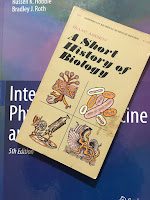
A Short History of Biology, by Isaac Asimov, superimposed on Intermediate Physics for Medicine and BIology.
A Short History of Biology,
by Isaac Asimov.
The tobacco mosaic virus was the first virus discovered. Isaac Asimov describes this discovery in A Short History of Biology
But twentieth-century serology [the study of plasma serum] reserved its most spectacular successes for the battle with microorganisms of a type unknown to [Louis] Pasteur and [Robert] Koch [founders of bacteriology] in their day. Pasteur had failed to find the infective agent of rabies, a clearly infectious disease undoubtedly caused, according to his germ theory, by a microorganism. Pasteur suggested that the microorganism existed but that it was too small to be detected by the techniques of the time. In this, he turned out to be correct.

The fact that an infectious agent might be much smaller than ordinary bacteria was shown to be true in connection with a disease affecting the tobacco plant (“tobacco mosaic disease”). It was known that juice from diseased plants would infect healthy ones and, in 1892, the Russian botanist, Dmitri Iosifovich Ivanovski (1864–1920), showed that the juice remained infective even after it had passed through filters fine enough to keep any known bacterium from passing through. In 1895, this was discovered independently, by the Dutch botanist, Martinus Willem Beijerinck (1851–1931). Beijerinck named the infective agent a “filtrable virus” where virus simply means “poison.” This marked the beginning of the science of virology.
The tobacco mosaic virus has other claims to fame. In 1935 it became the first virus to be crystalized, by American biochemist Wendell Stanley, a feat for which he received the 1946 Nobel Prize in Chemistry. Two years later, English plant pathologist Sir Frederick Bawden showed that the Tobacco mosaic virus contained ribonucleic acid (RNA), suggesting that nucleic acids are crucial for life. Rosalind Franklin, famous for her roll in Watson and Crick’s discovery of the structure of DNA, later studied the structure of the tobacco mosaic virus. In their paper “Tobacco Mosaic Virus: Pioneering Research for a Century” (Plant Cell, Volume 11, Pages 301–308, 1999), Creager et al. write
Tobacco mosaic virus (TMV), as we now know the agent that Beijerinck and others were studying, was the first virus to be identified. Perhaps because of this, research on TMV and other plant viruses has continued to be of profound significance in addressing fundamental questions about the nature of viruses in general. Indeed, TMV as a model system has been at the forefront of virology research to the present time. For example, TMV was the first virus to be chemically purified (Stanley, 1935; Bawden et al., 1936), to be detected in an analytical ultracentrifuge and in an electrophoresis apparatus (Eriksson-Quensel and Svedberg, 1936), and to be visualized in an electron microscope (Kausche et al., 1939). TMV RNA was used in the first decisive experiments showing that nucleic acids carry hereditary information and that nucleic acid alone is sufficient for viral infectivity (Fraenkel-Conrat, 1956; Gierer and Schramm, 1956). The TMV coat protein (CP) was the first virus protein to be sequenced (Anderer et al., 1960; Tsugita et al., 1960), and TMV’s particle structure was among the first to be elucidated in atomic detail (Bloomer et al., 1978; Namba et al., 1989).

TMV’s preeminence has extended into the recombinant era, when the first transgenic plants were constructed using TMV to demonstrate the concept of CP-mediated cross-protection (Abel et al., 1986). TMV was also the first virus shown to encode a cell-to-cell movement protein (MP; Deom et al., 1987). MP binds to RNA (Citovsky et al., 1990), associates with cytoskeletal elements (Heinlein et al., 1995; McLean et al., 1995), and increases the permeability of plasmodesmata to mediate cell-to-cell movement of the virus (Wolf et al., 1989; Waigmann et al., 1994).
What is the “mosaic” part of the virus name mean? A mosaic virus infects plants and causes the leaves to appear speckled. To learn more about this virus, read The Life of a Virus: Tobacco Mosaic Virus as an Experimental Model, 1930–1965 by Angela Creager (I have not read this, but it looks interesting, as does her more recent book Life Atomic: A History of Radioisotopes in Science and Medicine).

Russ must have known exactly what he was doing when he compiled Table 4.2 (and created Fig. 4.12), as he selected perhaps the most important virus historically, and one that has become a model system for microbiology.

Friday, November 1, 2013

Isotopes of Carbon

One of the fundamental ideas of nuclear physics is the isotope. In the 4th edition of Intermediate Physics for Medicine and Biology, Russ Hobbie and I describe isotopes at the start of Chapter 17.
Each atom contains a nucleus about 100,000 times smaller than the atom. The nuclear charge determines the number of electrons in the neutral atom and hence its chemical properties. The nuclear mass determines the mass of the atom. For a given nuclear charge there can be a number of nuclei with different masses or isotopes. If an isotope is unstable, it transforms into another nucleus through radioactive decay.
In order to understand isotopes, consider the isotopes of carbon, one of the most important elements for life.

12C

The most abundant isotope of carbon is 12C. This isotope is stable represents 99% of all carbon. The nucleus of 12C contains six protons (carbon’s atomic number is six) and six neutrons. The nucleus is basically three alpha particles stuck together.

13C

A second stable isotope is 13C, which contains six protons and seven neutrons. Only about 1% of carbon is 13C. The ratio of 13C to 12C is used to study ancient climates and oceans. Plants preferentially take up 12C, so the 13C/12C ratio provides a way to determine the amount of photosynthesis production, and contains information about the origin of carbon dioxide emission (to learn more, click here)

14C

All isotopes of carbon except 12C and 13C are unstable. The longest-lived unstable isotope is 14C, with a half-life of 5730 years. As with most isotopes containing an overabundance of neutrons, it decays by β- emission (a neutron turns into a proton, with the emission of an electron). In Chapter 16 of IPMB, we describe how the decay of 14C contributes to the background radiation.
We are continuously exposed to radiation from natural sources. These include cosmic radiation, which varies with altitude and latitude; rock, sand, brick, and concrete containing varying amounts of radioactive minerals; the naturally occurring radionuclides in our bodies such as 14C and 40K; and radioactive progeny from radon gas from the earth.
A homework problem in Chapter 17 describes how 14C is used to determine the age of organic remains.
Problem 58 One way to determine the age of biological remains is “carbon-14 dating.” The common isotope of carbon is stable 12C. The rare isotope 14C decays with a half-life of 5,370 yr. 14C is constantly created in the atmosphere by cosmic rays. The equilibrium between production and decay results in about 1 of every 1012 atoms of carbon in the atmosphere being 14C, mostly as part of a CO2 molecule. As long as the organism is alive, the ratio of 12C to 14C in the body is the same as in the atmosphere. Once the organism dies, it no longer incorporates 14C from the atmosphere, and the number of 14C nuclei begins to decrease. Suppose the remains of an organism have one 14C for every 1013 12C nuclei. How long has it been since the organism died?

15C

Isotopes of carbon with even more neutrons can be created in the lab, but they have very short half-lives. For instance, 15C has a half-life of only 2.4 seconds

11C

Isotopes that have fewer neutrons that found in the stable form often decay by  β+ emission, also known as positron decay. A proton transforms into a neutron, with the emission of a positron (an anti-electron). These nuclei can also decay by electron capture (an electron is captured by the nucleus, where it combines with a proton to produce a neutron), however most light elements such as carbon preferentially undergo β+ decay rather than electron capture. 11C has a half-life of 20 minutes, and decays by positron emission. It is sometimes used for PET imaging, described in Chapter 17 of IPMB.
If a positron emitter is used as the radionuclide, the positron comes to rest and annihilates an electron, emitting two annihilation photons back to back. In positron emission tomography (PET) these are detected in coincidence. This simplifies the attenuation correction, because the total attenuation for both photons is the same for all points of emission along each γ ray through the body (see Problem 54). Positron emitters are short-lived, and it is necessary to have a cyclotron for producing them in or near the hospital.
Many PET applications use 11C-acetate or 11C-choline, which is administered to the patient. Imaging where the 11C decays provides information about where carbon uptake occurs.

10C

Lighter isotopes of carbon, such as 10C, decay quickly; the half-life of 10C is 19 seconds. Such short half-lives make these isotopes difficult to use in PET imaging, even thought they are positron emitters.


By discussing the isotopes of carbon, we survey many of the most important ideas of nuclear physics, particularly those relevant to nuclear medicine.