Friday, May 30, 2008

Peter Basser wins ISMRM Gold Medal for Diffusion Tensor Imaging

Earlier this month, at the 16th Scientific Meeting and Exhibition of the International Society for Magnetic Resonance in Medicine (ISMRM) in Toronto, Peter Basser was awarded an ISMRM Gold Medal for “his pioneering and innovative scientific contributions in the development of Diffusion Tensor Imaging (DTI).”

Peter is an old friend of mine from the days when we were both staff fellows in the now-defunct Biomedical Engineering and Instrumentation Program at the National Institutes of Health in Bethesda, Maryland. We collaborated on many projects, including a study of magnetic stimulation of nerves (for example, see: Roth BJ, Basser PJ.
A Model of the Stimulation of a Nerve Fiber by Electromagnetic Induction,IEEE Transactions on Biomedical Engineering, Volume 37, Pages 588–597, 1990.)

Peter is now the head of the Section on Tissue Biophysics and Biomimetics, which is part of the Eunice Kennedy Shriver National Institute of Child Health and Human Development. The goal of his section is
“to understand fundamental physical mechanisms governing tissue-level physiological processes that are essential for life, or necessary to achieve a high quality of life. Examples include understanding the physical basis of nerve excitability and of effective load bearing in cartilage. This entails discovering relationships between physiological function and a tissue's structure, organization, and physical properties. This is done by studying the behavior of biological model systems using novel quantitative approaches (e.g., experimental methods, mathematical models, physical models). Another aim of ours is to transfer these new methodologies to the biomedical research and healthcare communities. An example includes the invention and successful dissemination of diffusion tensor magnetic resonance imaging from the 'bench' to the ‘bedside.’

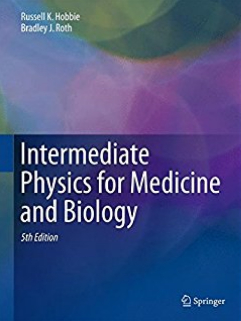
Diffusion tensor imaging is one of the topics that Russ Hobbie and I added to the 4th edition of Intermediate Physics for Medicine and Biology (see Chapter 18, Section 13). We also wrote a new homework problem that asked the student to show that the trace of the diffusion tensor is independent of fiber direction. We had trouble deciding if this problem belonged in Chapter 4 (on diffusion) or Chapter 18 (on magnetic resonance imaging), and we ended up putting the problem in both chapters (see Problems 4.22 and 18.40). Another homework problem featuring Peter’s work on cartilage appears in Chapter 5 (Problem 5.6).

The Office of NIH History has published an interview with Peter, in which he explains how he developed diffusion tensor imaging. Below is a brief excerpt of this interview, describing the moment Peter conceived the idea of DTI (I make a cameo):

Actually, the first exposure I had to diffusion imaging was a talk that Denis Le Bihan had given. He had recently come to the NIH from France and talked about how diffusion could be used—I think it was in stroke—and I thought it was very interesting, but I didn’t really initially make a connection to it. But in the early 1990s, Denis Le Bihan and, I believe it was Philippe Douek had a poster presentation at one of the NIH research festivals off in a corner in one of the white tents that they had constructed over here in the parking lot East of Building 30. They had done something very novel. They had shown that they could color code different parts of the brain according to what they thought was the orientation of diffusion. That was a poster that resulted in a paper, I think early in the next year, by Denis and Philippe. But I visited that poster and I was there with my friend and colleague, Brad Roth, the guy I was doing the magnetic stimulation with, and I realized that there was something really fundamentally wrong with the approach that Denis and Philippe were using.
The rest, as they say, is history. One of Peter's first papers on DTI (Basser PJ, Mattiello J, LeBihan D. MR Diffusion Tensor Spectroscopy and Imaging, Biophysical Journal, Volume 66, Pages 259–267, 1994) has been cited over 700 times according to the ISI Web of Knowledge. His coauthors were Denis Le Bihan (a previous ISMRM Gold Medal Winner) and James Mattiello (the first graduate of the Oakland University Medical Physics PhD Program). The technique is now widely used to map fiber orientation in the brain and the heart.

Congratulations Peter! 


 Listen to Peter Basser describe the invention and development of Diffusion Tensor Imaging.
https://www.youtube.com/watch?v=1_BeCeDak3w 
Friday, May 23, 2008

Should We Have a Molybdenum-99 Source in the United States?

In the December 14, 2007 entry to this blog, I discussed how the shutdown of a nuclear reactor at Chalk River, Ontario caused a shortage of an isotope of technetium (Tc-99m) used for medical imaging. Tc-99m is obtained from an isotope of molybdenum, Mo-99, that is produced at a handful of international facilities, including the Canadian reactor. The production and use of Tc-99m in nuclear medicine is discussed in Chapter 17 of the 4th edition of Intermediate Physics for Medicine and Biology.

The most recent (May 2008) issue of Physics Today contains an article by science writer Toni Feder that explores this topic further. Feder writes

The disruption late last year of Mo-99 production in Canada threw the nuclear medicine community into a panic. With a half-life of 66 hours, Mo-99 can’t be stockpiled. The Canadian reactor was down for maintenance, and its startup was delayed because of safety violations. Such was the upset in the nuclear medicine community that the Canadian government stepped in and ordered the reactor to start up despite some remaining safety concerns—and demoted the head of Canadas nuclear regulatory agency.
The article also discusses plans for the production of Mo-99 in the United States. The US producers will use “low-enriched uranium” (LEU) rather than the highly enriched uranium (HEU) used at other facilities, including the Chalk River plant. This would have advantages for nonproliferation, and could reduce the amount of HEU available for production of weapons of mass destruction by terrorist groups. Feder continues
Our program is to minimize civilian use of HEU, says Parrish Staples, manager of the National Nuclear Security Administrations program to convert reactors from HEU to LEU fuel. The only HEU the US is currently exporting is for production of Mo-99 in foreign production facilities. The US exports about 25 kg of HEU each year, or about half the total used for making Mo-99, he says. If the US stops exporting HEU, he adds, according to the IAEA [International Atomic Energy Agency] definition a weapons worth of material would be removed [from circulation each year].

At some point there will be an incident somewhere in the world that will cause the US to close its borders to radioactive materials for a day, a week, two or three weeks, whatever, says MURR [The University of Missouri-Columbia Research Reactor] director Ralph Butler. And when you think that there are tens of thousands of patients per day [in the US] utilizing this diagnostic tool [radioactive isotopes], thats a huge impact. The US does not have a Mo-99 source, he adds. There is a national need, and its an opportunity we [at MURR] can meet.

Friday, May 16, 2008

A Firm Foundation For Aspiring Biophysicists

In January 1989, John Wikswo of Vanderbilt University wrote a review in Physics Today (Volume 42, Pages 75–76) about the 2nd edition of Intermediate Physics for Medicine and Biology. His review began
In our introductory physics courses as well as in our daily use of physics, we regularly encounter the early work of Galileo, Isaac Newton, Luigi Galvani, Alessandro Volta, Thomas Young, Jean Poiseuille, Julius Mayer, Hermann von Helmholtz, William Gilbert and Jacques d’Arsonval. Many of us fail to recognize that the first four were physicists who in the course of their studies of physical systems made major contributions to the life sciences, while the remainder were physicians whose fundamental contributions to physics were largely motivated by their interest in biology and medicine. In the past 40 years, the Nobel Prize in Physiology or Medicine has been awarded to a remarkable number of physicists, including Hugo Theorell, Georg von Bekesy, Francis Crick, Maurice Wilkins, Alan Hodgkin, Andrew Huxley, Haldan Hartline, Max Delbruck, Rosalyn Yalow, Allan Cormack and Geoffrey Hounsfield. There must be a multitude of reasons why each of these modern-day physicists chose a career that spanned both physics and the life sciences, but it is unlikely that any single book, with the possible exception of Erwin Schrodinger’s What is Life?, could have been the stimulus. Why are there so few books that successfully span physics, medicine and biology?

While there are excellent texts, treatises and reviews of medical and radiological physics and biophysics, none of these provides the breadth and depth required of a guidebook for a physicist or biologist desiring to explore, possibly for the first time, the realm where physics joins medicine and biology. The problem in part is that such a book should develop simultaneously both the physics and the biology without assuming extensive prior knowledge of either, and yet should explore the subject with sophistication and quantitative rigor. In 1977, I was confronting the dilemma of finding no suitable text for the very first physics course I had been assigned to teach, an introductory medical physics course for undergraduate premedical students, when a friend of mine from the Mayo Clinic told me that Russell Hobbie of the University of Minnesota was writing just the book I needed. For two years my students and I learned from typed manuscripts kindly provided by Hobbie, and my colleagues and I have been using the first edition of Intermediate Physics for Medicine and Biology ever since then. This year, we can greet our students with the second edition.

Friday, May 9, 2008

See Russ Hobbie on YouTube!

In Chapter 15 of the 4th edition of Intermediate Physics for Medicine and Biology, Russ Hobbie and I discuss the interaction of radiation with matter, a topic that is crucial for understanding the medical use of X-rays. Twenty years ago, Russ wrote a computer program called MacDose that provides a two-dimensional simulation of the photoelectric effect, Compton scattering, and pair production; the primary mechanisms of X-ray interaction. MacDose runs on any Macintosh with OS-9 or earlier, including Classic in OS-X. You can download a copy of MacDose, including a student manual and instructors guide, at our book’s website. To learn more about MacDose, see Hobbie’s article in Computers in Physics (Volume 6, Pages 355–359, 1992).

You can also download a 26 minute Quicktime movie in which Russ demonstrates MacDose and explains various concepts related to the attenuation and absorption of X-rays (you can view the movie on either a PC or a Mac). With help from my daughter Stephanie, I have uploaded this movie onto Youtube. Because of a limit on the duration of Youtube videos, Stephanie had to split the movie into three parts. Search on YouTube for “MacDose” and you should find all three. Then pop some popcorn, pour yourself a drink, find a seat, and watch Hollywood
’s leading man Russ Hobbie explain how radiation interacts with matter.

 Russ Hobbie Demonstrates MacDose, Part 1

 Russ Hobbie Demonstrates MacDose, Part2

 Russ Hobbie Demonstrates MacDose, Part3
https://www.youtube.com/watch?v=hB4U7T0gH6M&t=2s
 

Friday, May 2, 2008

The Hodgkin and Huxley Model

In the 1940s and 50s, Alan Hodgkin and Andrew Huxley discovered the ionic basis for nerve conduction, work that resulted in their sharing the 1963 Nobel Prize in Physiology or Medicine. Chapter 6 in the 4th edition of Intermediate Physics for Medicine and Biology describes the Hodgkin-Huxley model in detail. Yet, no textbook can replace the experience of peering over Hodgkin's shoulder while he performs the voltage clamp experiments on a squid nerve axon that were crucial for their discoveries. Fortunately, a movie was made of these experiments, and clips from it can be found online, at a website for a neurophysiology class at Smith College. I particularly recommend the clip “Dissection and Anatomy” showing the dissection of the giant axon from a squid by J. Z. Young, and “Voltage Clamping” by P. F. Baker and Hodgkin himself.

When I teach Biological Physics at Oakland University, I like to have my students read Hodgkin and Huxley's classic paper
“A Quantitative Description of Membrane Current and its Application to Conduction and Excitation in Nerve” (Journal of Physiology, Volume 117, Pages 500–544, 1952). A pdf of this article is available online. However, if you encourage your students to read it, be sure to warn them that the definition of the transmembrane potential is different than is used now, with their definition being the outside minus the inside voltage, and zero being rest. (Nowadays, researchers typically use inside minus outside, with rest corresponding to -65 mV). Writing a program to simulate the Hodgkin and Huxley model is the best way to learn about it (we have a sample of such a program in Fig. 6.38 or our textbook), but those who are not programmers might want to try this applet that allows online simulation of a nerve action potential.

Alan Hodgkin and voltage clamping.
https://www.youtube.com/embed/Wd_gKJoo25Y

 John Young dissecting a squid giant axon.
https://www.youtube.com/embed/pw6_Si5jOpo