Friday, December 31, 2010

Brownian Motion

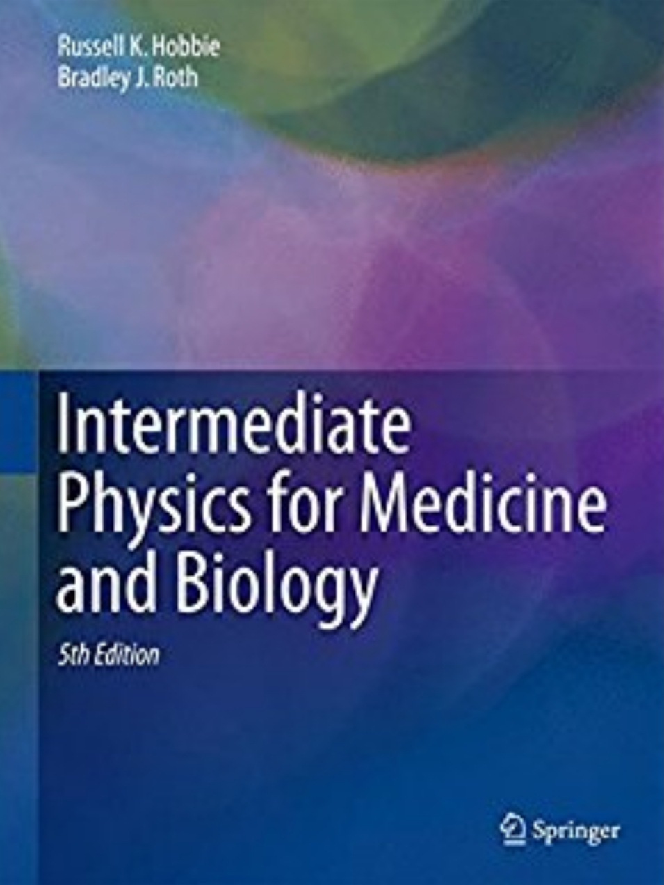
In the 4th edition of Intermediate Physics for Medicine and Biology, Russ Hobbie and I discuss Brownian motion. We first address this topic in Chapter 3 when deriving the equipartition of energy: the average thermal kinetic energy of an object at temperature T is 3kBT/2, where kB is Boltzmann’s constant.
This result is true for particles of any mass: atoms, molecules, pollen grains, and so forth. Heavier particles will have a smaller velocity but the same average kinetic energy. Even heavy particles are continually moving with this average kinetic energy. The random motion of pollen particles in water was first seen by a botanist, Robert Brown, in 1827. This Brownian motion is an important topic in the next chapter.
We next address this topic in Chapter 4, as we motivate the reader for a discussion of diffusion.
This movement of microscopic-sized particles, resulting from bombardment by much smaller invisible atoms, was first observed by the English botanist Robert Brown in 1827 and is called Brownian motion. Solute particles are also subject to this random motion. If the concentration of particles is not uniform, there will be more particles wandering from a region of high concentration to one of low concentration than vice versa. This motion is called diffusion.
As so often happens when you look deeply into a subject, the story is more complicated than can be described in an introductory (or even an intermediate) textbook. In the December 2010 issue of the American Journal of Physics, Philip Pearle and his colleagues published the fascinating article “What Brown Saw and You Can Too” (Volume 78, Pages 1278–1289).
A discussion of Robert Brown’s original observations of particles ejected by pollen of the plant Clarkia pulchella undergoing what is now called Brownian motion is given. We consider the nature of those particles and how he misinterpreted the Airy disk of the smallest particles to be universal organic building blocks. Relevant qualitative and quantitative investigations with a modern microscope and with a “homemade” single lens microscope similar to Brown’s are presented.
One interesting conclusion of their study is that Brown did not actually see pollen grains move.
We emphasize that Brown did not observe the pollen move. Instead, he observed the motion of much smaller objects that reside within the pollen.15 Nonetheless, statements that Brown saw the pollen move are common.16
Fortunately, reference 16 does not cite Intermediate Physics for Medicine and Biology. But it raises the question: Did Russ and I get it wrong? Our discussion in Chapter 4 seems safe. The text in Chapter 3 depends on if you interpret “pollen particles” as the entire “pollen grain” or “particles arising from pollen.” Russ may have been aware of this distinction when he wrote the original text, but I confess I was not. I always thought Brown saw the entire pollen grain move.

Pearle et al. show electron microscope pictures of pollen grains, which are 50–100 microns in diameter. I summarize their analysis about what Brown saw as a new homework problem for Chapter 4.
Problem 5 1/2. This problem looks at the original observations of Robert Brown that established Brownian motion.
(a) Combine Eqs. 4.23 and 4.71 to determine an expression for the average distance a particle of radius a will diffuse through a fluid of viscosity η in time t.
(b) Assume you observe a pollen grain with a radius of 50 microns in water at room temperature, and that your visual perception is particularly sensitive to motions occurring over a time of about one second. What is the average distance you observe the grain to move?
(c) Now assume your eye cannot see movements that occur over angles of less than 1 minute of arc, or 3 × 10⁻⁴ radians (in Chapter 14, we estimate 3 minutes of arc, but use 1 arc min to be conservative). Most eyes cannot focus on objects closer than 25 cm. Determine the smallest displacement you can observe with the naked eye.
(d) Robert Brown had a microscope that could magnify objects by a factor of about 370. What is the smallest displacement he could observe with his microscope? Is this larger or smaller than the displacement of a pollen grain in one second?
In fact, Brown did not observe the motion of entire pollen grains. He observed fat and starch particles about 2 microns in diameter that are released by pollen. For more on Brown’s original observations, see Pearle et al. (2010).
The authors also analyze the microscope that Brown used, and estimate the diffraction effects he had to contend with. Using an analysis similar to that presented in Section 13.7 (Medical Uses of Ultrasound) of Intermediate Physics for Medicine and Biology, they show that Brown probably could not resolve some of the smaller particles, but instead observed their diffraction pattern. As in Eq. 13.40 in our textbook, the diffraction pattern involves a Bessel function, and implies that the apparent size of an object is larger than the real size. The effect is minor for large objects but dominates for small objects.

I find the history and analysis of Brown’s original studies to be fascinating. For me, Pearle et al.’s paper reminds me that 1) the American Journal of Physics is still my favorite journal, and 2) physics has much to offer biology and medicine.
Friday, December 24, 2010

The littlest things can drive you nuts

I hope that our readers (and Russ Hobbie and I do value and appreciate all our dear readers) find the list of references at the end of each chapter in the 4th edition of Intermediate Physics for Medicine and Biology useful. We tried to include books and articles that you would enjoy, and that would help you understand the material in our textbook better. But, you may wonder, what do I see when I look at those lists of references? The first thing I see—the thing that jumps out of the page and screams at me—is that in each list, the first reference is not indented like the rest!!! As I recall, it is some issue in LaTex that is difficult to fix. I think it is related to the policy of not indenting the first paragraph of a section (a practice that I don’t care for).

I suppose what really should worry me are the errors that creep into the book. But at least we can correct those in the erratum, available at the book website. For some reason, I can live with those errors (que sera, sera) but the indentation issue is killing me. You can find a lot of other useful information at the book website, including an interview with Russ Hobbie published in the December 2006 issue of the American Physical Society Division of Biological Physics newsletter, a movie of Russ Hobbie explaining how radiation interacts with tissue based on his Mac Dose computer program, an American Journal of Physics resource letter that Russ and I published last year, and other supplementary material.

Let me use this post to update you on a few issues mentioned previously in this blog. In an October post, I talked about tanning and skin cancer. A recent article in the online newspaper MinnPost.com suggests that the problem is not getting any better, especially in the midwest, and that “people are still not recognizing that indoor tanning use is linked to skin cancer.” An article in medicalphysicsweb.com reports that “supply shortages of molybdenum-99 could become commonplace over the next decade unless longer-term actions are taken.” I discussed this issue several times before: here, here, and here. Felix Baumgartner’s attempt to jump out of a balloon at the edge of space and break the sound barrier in free fall has been put on hold, apparently because of a law suit over who owns the rights to this idea. Finally, you can watch online a series of lectures about the physics of hearing and cochlear implants delivered at the University of Michigan.

I wish you all a peaceful and happy Christmas Eve. If you are lucky, you will wake up tomorrow morning to find that Santa has left the 4th edition of Intermediate Physics for Medicine and Biology in your stocking. For those unfortunate few who received something else from Santa, I suggest amazon.com.

Merry Christmas!

Friday, December 17, 2010

Subtracting Large Numbers

One of the most notorious difficulties in numerical computations is the loss of precision when subtracting two similar, large numbers to obtain a smaller one. Russ Hobbie and I illustrate this hazard in Chapter 11 of the 4th edition of Intermediate Physics for Medicine and Biology. We begin this chapter with a discussion of the method of least squares, and we derive the formulas (Eqs. 11.5a and 11.5b) for fitting data to a straight line, y = ax + b. We then add “In doing computations where the range of data is small compared to the mean, better numerical accuracy can be obtained from…” and then present alternative formulas (Eqs. 11.5c, 11.5d, and 11.5e). Homework Problem 7 in Chapter 11 (one of the many new problems in the 4th edition) illustrates the advantage of the second set of equations.
Problem 7 Consider the data

   x       y
100   4004
101   4017
102   4039
103   4063

(a) Fit these data with a straight line y=ax+b using Eqs. 11.5a and 11.5b to find a.
(b) Use Eq. 11.5c to determine a. Your result should be the same as in part (a).
(c) Repeat parts (a) and (b) while rounding all the intermediate numbers to 4 significant figures. Do Eqs. 11.5a and 11.5b give the same result as Eq. 11.5c? If not, which is more accurate?
(Spoiler alert: Don’t continue reading if you want to solve the problem yourself first, as you should.) If you solve this problem, you will find that Eqs. 11.5a and 11.5b do not work very well at all for this problem. Their flaw is that they require you to subtract two really big numbers to get a much smaller one.

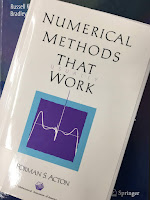
Numerical Methods That Work,  by Forman Acton, superimposed on Intermediate Physics for Medicine and Biology.
Numerical Methods That Work,
by Forman Acton.
A good discussion of this issue can be found in Forman Acton’s book Numerical Methods that Work.
The following problem often appears as a puzzle in Sunday Supplements. The difficulties are numerical rather than formulative and hence it is an especially appropriate challenge to the aspiring numerical analyst. We strongly urge that the reader solve it in his own way before turning to the “official” solution.

A railroad rail 1 mile long is firmly fixed at both ends. During the night some prankster cuts the rail and welds in an additional foot, causing the rail to bow up in the arc of a circle. The classical question concerns the maximum height this rail now achieves over its former position. To put it more precisely: We are faced…with the chord of a circle AB that is exactly 1 mile long and the corresponding arc AB that is 1 mile plus 1 foot and our question concerns the distance d between the chord and the arc at their midpoints. [See Acton’s book for the accompanying figure]

The relationships available are the simple ones from trigonometry involving the subtended half angle, θ, and the Pythagorean relationship. The student at this point should attempt to solve the problem before turning to the solution given in Chapter 2. He should attempt to find the distance d to an accuracy of three significant figures. In his effort he will probably be faced with subtracting two large and nearly equal numbers, which will cause a horrendous loss of significant figures. He can live with this process by shear brute force, but it will involve use eight-significant-figure trigonometric tables to preserve three figures in his answer. The point of the problem here is to find another method of calculating d, one that does not require such extreme measures. The three-figure answer can, indeed, be obtained rather easily using nothing more than pencil, paper, and a slide rule. The student should seek such a method.
If you find numerical methods interesting (as I do), you will love Acton’s delightfully written book. Originally published in 1970, it is all the more charming for its now-quaint references to slide rules and trigonometric tables. Yet, the concepts are not out-of-date. Even with powerful computers, errors can arise from subtracting nearly equal numbers. I’ve run into the issue myself when using the finite difference method and relaxation to solve Laplace’s equation with a fine grid and only single precision arithmetic.

Real Computing Made Real, by Forman Acton, superimposed on Intermediate Physics for Medicine and Biology.
Real Computing Made Real,
by Forman Acton.
Unfortunately, Acton’s book is not cited in the 4th edition of Intermediate Physics for Medicine and Biology (we’ll have to fix that in later editions), although I have mentioned it before in this blog. Acton is an emeritus professor in the Department of Computer Science at Princeton University (a department with an illustrious history). Also interesting is his more recent book Real Computing Made Real: Preventing Errors in Scientific and Engineering Calculations.

Friday, December 10, 2010

Robert Millikan

One fundamental constant that appears repeatedly in the 4th edition of Intermediate Physics for Medicine and Biology is the charge of the electron (the elementary charge, e), equal to 1.6 × 10⁻¹⁹ C. The first appearance of e that I can find is in Section 3.8 on the Nernst Equation. It appears in another context in Section 8.9, The Detection of Weak Magnetic Fields, when discussing Superconducting Quantum Interference Device (SQUID) magnetometers and the quantum of flux, equal to Planck’s constant divided by two times e. It shows up repeatedly in Chapter 9 on Electricity and Magnetism at the Cellular Level, and then again in Chapter 14 when discussing the energy levels of the hydrogen atom. It appears in Chapter 15 in the Klein-Nishina formula and in the expression for the classical radius of the electron.

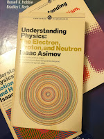
Understanding Physics: The Electron, Proton, and Neutron, by Isaac Asimov, suuperimposed on Intermediate Physics for Medicine and Biology.
Understanding Physics:
The Electron, Proton, and Neutron,
by Isaac Asimov.
How was the charge of the electron first measured? Isaac Asimov tells the story in Understanding Physics: The Electron, Proton, and Neutron.
The experiments that determined the size of the electric charge on the electron were conducted by the American physicist Robert Andrews Millikan (1868–1953) in 1911.

Millikan made use of two horizontal plates, separated by about 1.6 centimeters, in a closed vessel containing air at low pressure, The upper plate had a number of fine holes in it and was connected to a battery that could place a positive charge upon it. Millikan sprayed fine drops of nonvolatile oil into the closed vessel above the plates. Occasionally, one droplet would pass through one of the holes in the upper plate and would appear in the space between the plates. There it could be viewed through a magnifying lens because it was made to gleam like a star through its reflection of a powerful beam of light entering from one side.

Left to itself, the droplet of oil would fall slowly, under the influence of gravity. The rate of this fall in response to gravity, against the resistance of air (which is considerable for so small and light an object as an oil droplet), depends on the mass of the droplet. Making use of an equation first developed by the British physicist George Gabriel Stokes (1819–1903), Millikan could determine the mass of the oil droplets.

Millikan then exposed the container to the action of X rays. This produced ions in the atmosphere within (see page 110). Occasionally, one of these ions attached itself to the droplet. If it were a positive ion, the droplet, with a positive charge suddenly added, would be repelled by the positively-charged plate above, and would rush downward at a rate greater than could be accounted for by the action of gravity alone. If the ion were negative, the droplet would be attracted to the positively-charged plate and might even begin to rise in defiance of gravity.

The change in velocity of the droplet would depend on the intensity of the electric field (which Millikan knew) and the charge on the droplet, which he could now calculate.

Millikan found that the charge on the droplet varied according to the nature of the ion that was adsorbed and on the number of ions that were adsorbed. All the charges were, however, multiples of some minimum unit, and this minimum unit could reasonably be taken as the smallest possible charge on an ion and therefore, equal to the charge on the electron. Millikan's final determination of this minimum charge was quite close to the value now accepted, which is 4.80298 × 10⁻¹⁰ electrostatic units (“esu”), or 0.000000000480298 esu.
We don’t use electrostatic units in Intermediate Physics for Medicine and Biology (although they appear briefly in homework problem 3 in Chapter 6), but this is equivalent to 1.6 × 10⁻¹⁹ Coulombs.

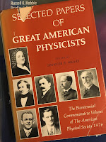
Selected Papers of Great American Physicists superimposed on Intermediate Physics for Medicine and Biology.
Selected Papers of
Great American Physicists.
I remember doing Millikan’s oil drop experiment as an undergraduate physics major at the University of Kansas. It required several hours in a dark room staring at small oil drops through a microscope. When in graduate school, I read one of Millikan’s papers in the book Selected Papers of Great American Physicists: The Bicentennial Commemorative Volume of The American Physical Society. I was particularly impressed by Millikan’s careful analysis of sources of systematic error in his experiment. In fact, I used that paper as a model for one of my few experimental papers: “The Magnetic Field of a Single Axon: A Comparison of Theory and Experiment” (Roth and Wikswo, Biophysical Journal, Volume 48, Pages 93–109, 1985). Some have claimed that Millikan committed scientific fraud by an improper selection of data to use in his analysis, but that claim has been debunked (see “Data Selection and Responsible Conduct: Was Millikan a Fraud?” by Richard Jennings, Science and Engineering Ethics, Volume 10, Pages 639–653, 2004).

I have a personal reason for being interested in the work of Robert Millikan. According to his Nobel Prize biography, he was born in Morrison Illinois, a small town 120 miles west of Chicago, about 15 miles from the Mississippi River. This is the town I grew up in, from an age of just a few months until I was 12 years old. At the time, I didn’t realize who Robert Millikan was, or that Morrison was the home to a Nobel Prize winning physicist. But over the years I have become a big fan of “Millikan from Morrison.” According to the Morrison chamber of commerce, there is now a downtown park named after Millikan. I must go visit.

Friday, December 3, 2010

Physical Biology of the Cell

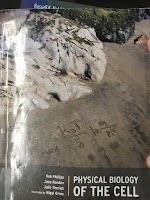
Physical Biology of the Cell, by Phillips, Kondev, and Theriot, superimposed on Intermediate Physics for Medicine and Biology.
Physical Biology of the Cell,
by Phillips, Kondev, and Theriot.
I spent some time this week looking over the recently published textbook Physical Biology of the Cell, by Rob Phillips, Jane Kondev, and Julie Theriot. In some ways this book is a competitor of the 4th edition of Intermediate Physics for Medicine and Biology (it is always good to know your competition). Bernard Chasan reviewed Physical Biology of the Cell in the November 2010 issue of the American Journal of Physics.
The authors of this book are, in a very real sense, missionaries. They want to convince a wide audience to share their enthusiasm for and commitment to a more quantitative and scientifically rigorous approach to cell biology than is normally encountered in the teaching literature.

To achieve this goal, they set out a program of quantitative model building based on physical principles…. What the authors describe (awkwardly but evocatively) as the mathematizing of the semiqualitative models of cell biology (referred to as “cartoons” in some circles) has now become central to cell biology—as evidenced by a half a dozen recent texts and the relatively new and thriving discipline of systems biology. The work being reviewed is the latest and most comprehensive attempt to foster and advocate for this approach…

At the center of their approach is the art of model making—well presented with the aid of some excellent figures, which show the choices needed to model proteins, as one example. The main point is that modeling requires a simplifying choice, which emphasizes one view of the protein and essentially ignores others. If it suits your purposes to model the protein as a collection of hydrophobic and hydrophilic amino acid residues—a good model for protein folding—then you cannot at the same time consider the protein as a two state system.
After skimming through Physical Biology of the Cell (I wish I had time to read it thoroughly), I have several observations.
  1. The second half of Intermediate Physics for Medicine and Biology (IPMB) is about clinical medical physics: imaging and therapy. None of this appears in Physical Biology of the Cell (PBC). Also, in IPMB Russ Hobbie and I steer clear of molecular biology, saying in the preface that “molecular biophysics has been almost completely ignored: excellent texts already exist, and this is not our area of expertise.” PBC is all molecular and cellular. The main overlap between the two books is several chapters in PBC that cover similar topics as are in the first half of IPMB. So, I guess IPMB and PBC are not really in direct competition. However, if I was Phil Nelson, author of Biological Physics: Energy, Information, Life, I might be concerned about market share.
  2. PBC is illustrated by Nigel Orme. Let me be frank; Orme’s drawings are much better than what we have in IPMB. One thing I like about PBC is that you can skip the text altogether and just look at the pictures, and still learn the gist of the subject. Figure 1.4 showing the genetic code reminds me of the sort of graphics that Edward Tufte promotes in The Visual Display of Quantitative Information. The authors of PBC state in the acknowledgments “this book would never have achieved its present incarnation without the close and expert collaboration of our gifted illustrator, Nigel Orme, who is responsible for the clarity and visual appeal of the more than 550 figures found in these pages, as well as the overall design of the book.” As generous as this tribute is, it may be an understatement. Then, just when I thought the artwork couldn’t get any better, I found that PBC contains several beautiful figures contributed by David Goodsell, author of The Machinery of Life.
  3. In the 4th edition of IPMB, Russ and I added an initial section exploring the relative size of biological objects. In PBC, a similar discussion fills the entire Chapter 2. There is lots of numerical estimating in this chapter, reminding me of the Bionumbers website. Chapter 3 looks at different temporal scales, which is more difficult to show visually than spatial scales (Russ and I didn’t try), although Orme’s drawings do a pretty good job. Chapter 4 of PBC looks at the many model systems used in biology, with an eye toward history (Mendel’s pea plants, hemoglobin and the structure of a protein, the bacteriophage in genetics, etc.). Great reading.
  4. Some subjects—such as diffusion, fluid dynamics, thermodynamics, and bioelectricity—are covered in both PBC and IPMB. Which book explains these topics better? Obviously I am biased, but I suggest that Russ and I develop the physics in a more detailed and systematic way, starting from the fundamentals, whereas Phillips, Kondev and Theriot present the physics rather quickly, and then apply it to many interesting biological applications. I would say that PBC does for molecular and cellular biology what Air and Water by Mark Denny does for physiology: use physics and math to explain biological concepts quantitatively. Russ and I, on the other hand, teach physics using biological examples. The difference is more about approach, tone, and point-of-view than about substance. The reader can look at both books and draw their own conclusions.
  5. PBC has a few nice homework problems, but I prefer IPMB’s more extensive collection. The student learns more by doing than by reading.
  6. The final chapter in PBC, “Wither Physical Biology,” is an excellent summary of the “the role of quantitative analysis in the study of living matter.” Anyone working at the interface between physics and biology must read these ten pages.
Phillips, Kondev, and Theriot ought to have the last word, so I will finish this blog entry by quoting PBC’s eloquent closing paragraph.
The act of writing this book has convinced each of us that the study of living matter is one of the most exciting frontiers in human thought. Just as the makings of the large scale universe are being revealed by ever more impressive telescopes, living matter is now being viewed in ways that were once as unimaginable as was going to the Moon. Despite the muscle-enhancing weight of this book, we feel that we have only scratched the surface of the rich and varied applications of physical reasoning to biological problems. Our overall goal has been to communicate a style of thinking about problems where we have done our best to illustrate the power of the style using examples chosen from biological systems that are well defined and usually well studied from a biological perspective. As science moves forward into the twenty-first century, it is our greatest hope that synthetic approaches for understanding the natural world from biological, physical, chemical, and mathematical perspectives simultaneously will enrich all of these fields and illuminate the world around us. We can only hope the reader has at least a fraction of the pleasure in answering that charge as we have had in attempting to describe the physical biology of the cell.