Showing posts with label obituary. Show all posts

Friday, February 9, 2024

Robert Kemp Adair (1924–2020)—Notes on a Friendship

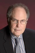
Robert Adair.
Robert Adair.
Photo credit: Michael Marsland/Yale University.

I try to write obituaries of scientists who appear in Intermediate Physics for Medicine and Biology, but for some reason I didn’t write about Robert Adair’s death in 2020. Perhaps the covid pandemic over-shadowed his demise. In Chapter 9 of IPMB, Russ Hobbie and I cite seven of his publications. He was a leader in studying the health effects (or, lack of heath effects) from electric and magnetic fields.

Recently, I read a charming article subtitled “Notes on a Friendship” about Adair, written by Geoffrey Kabat, the author of Getting Risk Right: Understanding the Science of Elusive Health Risks. I have Getting Risk Right on my to-read list. It sounds like my kind of book.

I admire Adair’s service in an infantry rifle platoon during World War II. I loved his book about baseball. I respect his independent assessment of the seriousness of climate change, although I don’t agree with all his conclusions. He certainly was a voice of reason in the debate about health risks of electric and magnetic fields. He led a long and useful life. We need more like him.

I will give Kabat the final word, quoting the last paragraph of his article.
In early October 2020, Bob’s daughter Margaret called me to tell me that Bob had died. I looked for an obituary in the New York Times, and was shocked when none appeared, likely due to the increased deaths from the pandemic. I wrote to an epidemiologist colleague and friend, who knew Bob’s work on ELF-EMF [extremely low frequency electromagnetic fields] and microwave energy, and who had served on a committee to assess possible health effects of the Pave Paws radar array on Cape Cod. My friend Bob Tarone wrote back, “Very sad to hear that. Adair was not directly involved in the Pave Paws study, but we relied heavily on his superb published papers on the biological effects of radio-frequency energy in our report. He and his wife were superb scientists. Losing too many who don’t seem to have competent replacements. Too bad honesty and truth are in such short supply in science today.” He concurred that there should have been an obituary in the Times.

Friday, January 26, 2024

Craig Henriquez (1959–2023)

I just learned that my friend Craig Henriquez passed away last summer. Craig earned his PhD at Duke University in their Department of Biomedical Engineering under the guidance of the renowned bioelectricity expert Robert Plonsey. His 1988 dissertation, titled “Structure and Volume Conductor Effects on Propagation in Cardiac Tissue,” was closely related to work I was doing at that time. Craig sent me a copy of his dissertation after he graduated. I really wanted to read it, but I was swamped with my my new job at the National Institutes of Health and helping care for my newborn daughter Stephanie. There wasn’t time to read it at work, and when I got home it was my turn to watch the baby, as my wife had been with her all day. The solution was to read Craig’s dissertation out loud to Stephanie as she crawled around in her play pen. She seemed to like the attention and I got to learn about Craig’s work.

Craig and I are nearly the same age. He was born in 1959 and I in 1960. Our careers progressed along parallel lines. After he graduated he stayed at Duke and joined the faculty. I recall he told me at the time that he didn’t know if he would make a career in academia, but he certainly did. He was on the Duke faculty for 35 years. In the early 1990s three young researchers at Duke—Craig, Natalia Trayanova, and Wanda Krassowska—were all from my generation. They were my friends, collaborators, and sometimes competitors as we worked to establish the bidomain model as the state-of-the-art representation of the electrical properties of cardiac tissue.

In my recent review about bidomain modeling (Biophysics Reviews, Volume 2, Article 041301, 2021) , I wrote (referring to myself in third person, as required by the journal; in the quotes below references are removed):

Roth’s calculation was not the first attempt to solve the active bidomain model using a numerical method. In 1984, Barr and Plonsey had developed a preliminary algorithm to calculate action potential propagation in a sheet of cardiac tissue. Simultaneous with Roth’s work, Henriquez and Plonsey were examining propagation in a perfused strand of cardiac tissue. For the next several years, Henriquez continued to improve bidomain computational methods with his collaborators and students at Duke. His 1993 article published in Critical Reviews of Biomedical Engineering remains the definitive summary of the bidomain model.
I’ve cited his 1993 review article (Crit. Rev. Biomed. Eng., Volume 21, Pages 1–77) many times, including in Intermediate Physics for Medicine and Biology. It’s a classic.

Craig and I were both interested in determining if Madison Spach’s electrical potential data from cardiac tissue samples should be interpreted as evidence of discontinuous propagation (Spach’s hypothesis) or a bath effect.
The original calculations of action potential propagation in a continuous bidomain strand perfused by a bath hinted at different interpretations of Spach’s data. As discussed earlier, the wave front is not one-dimensional because its profile varies with depth below the strand surface. The same effect occurs during propagation through a perfused planar slab, more closely resembling Spach’s experiment. The conductivity of the bath is higher than the conductivity of the interstitial space, so the wave front propagates ahead on the surface of the tissue and drags along the wave front deeper below the surface, resulting in a curved front. The extra electrotonic load experienced at the surface slows the rate of rise and the time constant of the action potential foot. Plonsey, Henriquez, and Trayanova analyzed this effect, and subsequently so did Henriquez and his collaborators and Roth.

Craig became an associate editor of the IEEE Transactions on Biomedical Engineering, and he would often send me papers to review. He was a big college basketball fan. We would email each other around March, when our alma maters—my Kansas Jayhawks and his Duke Blue Devils—would face off in the NCAA tournament. His research interests turned to nerves and the brain, and he co-directed a Center of Neuroengineering at Duke. He eventually chaired Duke’s biomedical engineering department, and at the time of his death he was an Associate Vice Provost.

I found out about Craig’s death when I was submitting a paper to a journal. This publication asks authors to suggest potential reviewers, and I was about to put Craig’s name down as a person who would give an honest and constructive assessment. I googled him to get his current email address, and discovered the horrible news. What a pity. I will miss him. 

Short bio published in the IEEE Transactions on Biomedical Engineering in January, 1990.

 Craig Henriquez talking about cardiac tissue and the bidomain model.

https://www.youtube.com/watch?v=OiSiLwP1ZPo

Friday, August 18, 2023

Paul Maccabee (1944–2023)

Paul Maccabee
Paul Maccabee (1944–2023).
Photo used with permission from the
Downstate Health Sciences University website.

My friend and collaborator Paul Maccabee died on July 24. Paul was a pioneer in the field of magnetic stimulation, a topic that Russ Hobbie and I discuss in Chapter 8 of Intermediate Physics for Medicine and Biology. Paul’s career and mine had many parallels. We both worked on magnetic stimulation in the late 1980s and early 1990s. We both collaborated with a leading neurophysiologist: Paul with Vahe Ammasian and me with Mark Hallett. We both recognized the importance of laboratory animal experiments for identifying physiological mechanisms. We both were comfortable working with biomedical engineers, I entered that field from physics and Paul from medicine. 

Paul was about 15 years older than me and I viewed him as a role model. I believe I first met him at the 1989 International Motor Evoked Potential Symposium in Chicago, a key early conference dedicated to magnetic stimulation. Our paths crossed at other scientific meetings and his research had a major impact on my own. For years I taught a graduate class on bioelectricity at Oakland University and I had my students read Paul’s 1993 Journal of Physiology paper (described below) which I assigned because it’s a classic example of a well-written scientific article. According to Google Scholar that paper has been cited 374 times, and it should be cited even more.

I wrote about Paul in my review of the development of transcranial magnetic stimulation (BOHR International Journal of Neurology and Neuroscience, Volume 1, Pages 8–20, 2022, https://doi.org/10.54646/bijnn.002). Below I quote part of that article. I put his name in bold so you can find it easily.

Although this experiment [performed by Jan Nilsson and Marcela Panizza at the National Institutes of Health, see reference 49] confirmed [Peter Basser and my] prediction [that neural excitation occurs where the gradient of the induced electric field is largest, see reference 58], there were nevertheless concerns because of the heterogeneous nature of the bones and muscles in the human arm. At about the same time Nilsson and Panizza were doing their experiment at NIH, Paul Maccabee was performing an even better experiment at the New York Downstate Medical Center in Brooklyn. Maccabee obtained his MD from Boston University and collaborated in Brooklyn with the internationally acclaimed neuroscientist Vahe Ammasian [1, 40–43]. This research culminated in their 1993 article in the Journal of Physiology, in which they examined magnetic stimulation of a peripheral nerve lying in a saline bath [44]. First, they measured the electric field Ey (they assumed the nerve would lie above the coil along the y-axis) and its derivative along the nerve produced by a figure-8 coil located under the bath (Figure 9). They found that the electric field was maximum directly under the center of the coil, but the magnitude of the gradient dEy/dy was maximum a couple centimeters either side of the center.
Figure 9. Contour plots of the electric field (Ey, red) and its spatial derivative (dEy/dy, blue) induced by a figure-eight coil (purple) placed under a tank filled with saline and measured using a bipolar recording electrode. The y direction is downward in the figure, parallel to the direction of the nerve (see Figure 10). Adapted from Figure 2 of Maccabee et al. [44].
Next they placed a bullfrog sciatic nerve in the dish and recorded the electrical response from one end (Figure 10). They found a 0.9 ms delay between the recorded action potentials when the polarity of a magnetic stimulus was reversed (the yellow and red traces on the right). Given a propagation speed of about 40 m/s, the shift in excitation position was about 3.6 cm, consistent with what Basser and I would predict.
Figure 10. Recordings from an electrode (black dot) at the distal end of a bullfrog sciatic nerve (green) that was immersed in a trough filled with saline (blue) and stimulated with a figure-8 coil (purple). The nerve emerged from the saline to rest on the recording electrode in air. The compound nerve action potentials were elicited by a stimulus of one polarity (orange), then the other (red). Adapted from Figure 3 of Maccabee et al. [44].
So far, their study was similar to what we performed at NIH in a human, but then they did an experiment that we could not do. To determine how a heterogeneity would impact their results, they placed two insulating cylinders on either side of the nerve (Figure 11). These cylinders modified the electric field, moving the negative and positive peaks of the activating function closer together. They observed a corresponding reduction in the latency shift. This experiment provided insight into what happens when a human nerve passes between two bones, or some similar heterogeneity.

Figure 11. Magnetic stimulation of a sheep phrenic nerve immersed in a homogeneous (left) and inhomogeneous (right) volume conductor. The figure-8 coil (purple) was positioned under the nerve (green). The yellow circles indicate the position of the insulating cylinders. The electric field Ex (red) and its gradient dEx/dx (blue) were measured along the nerve trajectory. The compound nerve action potentials at the recording electrode were measured for a magnetic stimulus of one polarity (orange) and then the other (green). Adapted from Figure 5 of Maccabee et al. [44].

Finally, they changed the experiment by bending the nerve and found that a bend caused a low threshold “hot spot,” and that excitation at that spot occurred where the electric field, not its gradient, was large. This result was consistent with Nagarajan and Durand’s analysis of excitation of truncated nerves [47].

In my opinion, Maccabee’s [44] article is the most impressive publication in the magnetic stimulation literature. Only Barker’s original demonstration of transcranial magnetic stimulation can compete with it [2].

Later in that review, I discussed a collaborative paper that Paul and I published.

One frustrating feature of the activating function approach is that excitation does not occur directly under the center of a figure-8 coil, where the electric field is largest, but off to one side, where the gradient peaks (Figure 9). Medical doctors do not want to guess how far from the coil center excitation occurs; they would prefer a coil for which “x” marks the spot. It occurred to me that such a coil could be designed using two adjacent figure-8 coils. I called this the four-leaf coil (Figure 12). John Cadwell from Cadwell Laboratories (Kennewick, Washington) built such a coil for me. Having seen the excellent results that Maccabee was obtaining using his nerve-in-a-dish setup, I sent the coil to him so he could test it. The resulting paper [65] showed that for one polarity of the stimulus the magnitude of the gradient of the electric field was largest directly under the coil center so the axons there were depolarized (“x” really did mark the spot of excitation). In addition, if the polarity of the stimulus was reversed, the magnitude of the gradient remained large under the coil center, but it now tended to hyperpolarize rather than depolarize the axons. Maccabee and I hoped that such hyperpolarization could be used to block action potential propagation, acting like an anesthetic. The Brooklyn experiments verified all the predictions of the activating function model for the four-leaf coil. Unfortunately, Maccabee never observed any action potential block. Perhaps, the hyperpolarization required for block was greater than the coil could produce.

Figure 12. A four-leaf coil (purple) used to stimulate a peripheral nerve (blue). Adapted from Figure 1 of Roth et al. [65].


[1] Amassian, V. E., Eberle, L., Maccabee, P. J., and Cracco, R. Q. (1992). Modelling magnetic coil excitation of human cerebral cortex witha peripheral nerve immersed in a brain-shaped volume conductor:The significance of fiber bending in excitation. Electroenceph. Clin. Neurophysiol., 85, 291–301.

[2] Barker, A. T., Jalinous, R., and Freeston, I. L. (1985). Non-invasive magnetic stimulation of human motor cortex. Lancet, 1, 1106–1107.

[40] Maccabee, P. J., Amassian, V. E., Cracco, R. Q., and Cadwell, J. A. (1988). An analysis of peripheral motor nerve stimulation in humans using the magnetic coil. Electroenceph. Clin. Neurophysiol., 70, 524–533.

[41] Maccabee, P. J., Eberle, L., Amassian, V. E., Cracco, R. Q., Rudell, A., and Jayachandra, M. (1990). Spatial distribution of the electric field induced in volume by round and figure ‘8’ magnetic coils: Relevance to activation of sensory nerve fibers. Electroenceph. Clin. Neurophysiol., 76, 131–141.

[42] Maccabee, P. J., Amassian, V. E., Cracco, R. Q., Cracco, J. B., Eberle, L., and Rudell, A. (1991a). Stimulation of the human nervous system using the magnetic coil. J. Clin. Neurophysiol., 8, 38–55.

[43] Maccabee, P. J., Amassian, V. E., Eberle, L. P., Rudell, A. P., Cracco, R. Q., Lai, K. S., and Somasundarum, M. (1991b). Measurement of the electric field induced into inhomogeneous volume conductors by magnetic coils: Application to human spinal neurogeometry. Electroenceph. Clin. Neurophysiol., 81, 224–237.

[44] Maccabee, P. J., Amassian, V. E., Eberle, L. P., and Cracco, R. Q. (1993). Magnetic coil stimulation of straight and bent amphibian and mammalian peripheral nerve in vitro: Locus of excitation. J. Physiol., 460, 201–219.

[47] Nagarajan, S. S., Durand, D. M., and Warman, E. N. (1993). Effects of induced electric fields on finite neuronal structures: A simulation study. IEEE Trans. Biomed. Eng., 40, 1175–1188.

[49] Nilsson, J., Panizza, M., Roth, B. J., Basser, P. J., Cohen, L. G., Caruso, G., and Hallett, M. (1992). Determining the site of stimulation during magnetic stimulation of a peripheral nerve. Electroenceph. Clin. Neurophysiol., 85, 253–264.

[58] Roth, B. J. and Basser, P. J. (1990). A model of the stimulation of a nerve fiber by electromagnetic induction. IEEE Trans. Biomed. Eng., 37, 588–597.

[65] Roth, B. J., Maccabee, P. J., Eberle, L., Amassian, V. E., Hallett, M., Cadwell, J., Anselmi, G. D., and Tatarian, G. T. (1994a). In-vitro evaluation of a four-leaf coil design for magnetic stimulation of peripheral nerve. Electroenceph. Clin. Neurophysiol., 93, 68–74.

Although my name was listed first on our joint 1994 article, Paul could easily have been the lead author. The coil shape was my idea but he performed all the experiments. I never set foot in Brooklyn; I just mailed the coil to him.

Paul was a giant in the field of magnetic stimulation. The articles I list above are only a few of the many he published. For a medical doctor he had a strong grasp of electricity and magnetism. I lost track of him over the years but had the good fortune to reconnect with him a few months ago by email.

I miss Paul Maccabee. Anyone who studies, uses, or benefits from magnetic stimulation owes him a debt of gratitude. I know I do.

Friday, July 28, 2023

John Moulder (1945–2022)

Photo of John Moulder.
John Moulder,
from Khurana et al. (2008) Med. Phys.,
35:5203, with permission from Wiley.
John Moulder, a leading expert in radiation biology, died about a year ago (on July 17, 2022; I wasn’t aware of his death until last week). When Russ Hobbie and I discuss the possible health risks of weak electric and magnetic fields in Intermediate Physics for Medicine and Biology, we cite a website about powerlines and cancer “that unfortunately no longer exists.” (However, in a previous blog post I found that is does still exist.) We also cite several papers that Moulder wrote with his collaborator Ken Foster about potential electromagnetic field hazards, including
Moulder JE, Foster KR (1995) Biological Effects of Power-Frequency Fields as they Relate to Carcinogenesis. Proceedings of the Society for Experimental Biology and Medicine Volume 209, Pages 309–324.

Moulder JE, Foster KR (1999) Is There a Link Between Exposure to Power-Frequency Electric Fields and Cancer? IEEE Engineering in Medicine and Biology Magazine, Volume 18, Pages 109–116.

Moulder JE, Foster KR, Erdreich LS, McNamee JP (2005) Mobile Phones, Mobile Phone Base Stations and Cancer: A Review. International Journal of Radiation Biology, Volume 81, Pages 189–203.

Foster KR, Moulder JE (2013) Wi-Fi and Health: Review of Current Status and Research. Health Physics, Volume 105, Pages 561–575.

Perhaps my favorite of Moulder’s publications is his Point/Counterpoint article in the journal Medical Physics.
Khurana VG, Moulder JE, Orton CG (2008) There is Currently Enough Evidence and Technology Available to Warrant Taking Immediate Steps to Reduce Exposure of Consumers to Cell-Phone-Related Electromagnetic Radiation. Medical Physics, Volume 35, Pages 5203–5206.
Here is how Moulder is introduced in that paper.
Dr. Moulder obtained his Ph.D. in Biology in 1972 from Yale University. Since 1978, he has served on the faculty of the Medical College of Wisconsin, where he directs the NIH-funded Center for Medical Countermeasures Against Radiological Terrorism. His major research interests include the biological basis for carcinogenesis and cancer therapy, biological aspects of human exposure to non-ionizing radiation, and the prevention and treatment of radiation-induced normal tissue injuries. He has served on a number of national advisory groups concerned with environmental health, non-ionizing radiation, and radiological terrorism; and he currently serves as a radiation biology consultant to NASA.

Are Electromagnetic Fields Making Me Ill? superimposed on Intermediate Physics for Medicine and Biology.
Are Electromagnetic Fields
Making Me Ill?

In my book Are Electromagnetic Fields Making Me Ill? I wrote:

Radiation biologist John Moulder, of the Medical College of Wisconsin, began maintaining a website titled “Power Lines and Cancer FAQs [frequently asked questions],” which exhaustively summarized the evidence pro and con. Although this website is no longer available online, an archived pdf of it is [13]. In a 1996 article published by IEEE Engineering in Medicine and Biology, Moulder reviewed dozens of studies, and concluded that:
Given the relative weakness of the epidemiology, combined with the extensive and unsupportive laboratory studies, and the biophysical implausibility of interactions at relevant field strengths, it is often difficult to see why there is still any scientific controversy over the issue of power-frequency fields and cancer. [14]

13. large.stanford.edu/publications/crime/references/moulder/moulder.pdf. Access date: January 12, 2022. 

14. Moulder JE (1996) Biological Studies of Power-Frequency Fields and Carcinogenesis. IEEE Engineering in Medicine and Biology Magazine, Volume 15, Pages 31–49.

In a special issue of the International Journal of Radiation Biology dedicated to Moulder, Andrea DiCarlo and her colleagues discussed his work on radiological terrorism.

Through his awarded research grant and cooperative agreements from the NIH and beyond, John leaves behind a legacy of excellent, rigorous, and robust scientific findings, research collaborators who benefited from his expertise and dedication, and a cadre of well-trained students. Although it is impossible to list here all the lives that were touched, and the careers that were impacted by John’s influence, the authors can state with certainty that the field of medical preparedness for a radiation public health emergency would not be where it is now without the steadying hand and role played by Dr. Moulder, both in the early days in the program and during his final years as an active researcher. We are grateful for his years of research and join the entire radiation community in mourning the loss of a great investigator and person.
John Moulder, you were a voice of reason in a crazy world. We’ll miss you.

To hear Moulder in his own words, go to times 4:40 and 5:05 in this video about Power Line Fears.

https://www.youtube.com/watch?v=kf7KWkod3Zw

Friday, February 17, 2023

Abraham Liboff (1927–2023)

Abe Liboff, in his office at Oakland University.
Abe Liboff, in his
office at Oakland University
Oakland University
physicist Abe Liboff died recently. A notice from President Ora Hirsch Pescovitz, published on the OU website, stated:
It is with deep sadness that I inform you of the death of Professor Emeritus Abraham Liboff who passed away on January 9, 2023. Dr. Liboff joined the Oakland University community in the Department of Physics on August 15, 1972, where he served until his retirement in August 2000.

During his tenure here at OU, Dr. Liboff was Chair of the Department of Physics. He is credited with 111 research publications, more than two dozen patents and nearly 3,400 scholarly citations during his career.

I arrived at OU in 1998, so his time at OU and mine overlapped by a couple years. I remember having a delightful breakfast with him during my job interview. He was one of the founders of OU’s medical physics PhD program that I directed for 15 years. His office was just a few doors down the hall from mine and he helped me get started at Oakland. I’ll miss him.

Although I loved the man, I didn’t love Abe’s cyclotron resonance theory of how magnetic fields interact with biological tissue. It’s difficult to reconcile admiration for a scientist with rejection of his scientific contributions. Rather than trying to explain Abe’s theory, I’ll quote the abstract from his article “Geomagnetic Cyclotron Resonance in Living Cells,” published in the Journal of Biological Physics (Volume 13, Pages 99–102, 1985).

Although considerable experimental evidence now exists to indicate that low-frequency magnetic fields influence living cells, the mode of coupling remains a mystery. We propose a radical new model for electromagnetic interactions with cells, one resulting from a cyclotron resonance mechanism attached to ions moving through transmembrane channels. It is shown that the cyclotron resonance condition on such ions readily leads to a predicted ELF-coupling at geomagnetic levels. This model quantitatively explains the results reported by Blackman et al. (1984), identifying the focus of magnetic interaction in these experiments as K+ charge carriers. The cyclotron resonance concept is consistent with recent indications showing that many membrane channels have helical configurations. This model is quite testable, can probably be applied to other circulating charge components within the cell and, most important, leads to the feasibility of direct resonant electromagnetic energy transfer to selected compartments of the cell.
In my book Are Electromagnetic Fields Making Me Ill? I didn’t have the heart to attack Abe in print. When discussing cyclotron resonance effects, I cited the work of Carl Blackman instead, who proposed a similar theory. What’s the problem with this idea? If you calculate the cyclotron frequency of a calcium ion in the earth’s magnetic field, you get about 23 Hz (see Eq. 8.5 in Intermediate Physics for Medicine and Biology). However, the thermal speed of a calcium ion at body temperature is about 440 m/s (Eq. 4.12 in IPMB). At that speed, the radius of the cyclotron orbit would be 3 meters (roughly ten feet)! The mean free path of a ion in water, however, is about an angstrom, which means the ion will suffer more than a billion collisions in one orbit; these interactions should swamp any cyclotron motion. Moreover, ion channels have a size of about 100 angstroms. In order to have a orbital radius similar to the size of a ion channel, the calcium ion would need to be moving extremely fast, which means it would have a kinetic energy vastly larger than the thermal energy. The theory just doesn’t work.

Since Liboff isn’t around to defend himself, I’ll let Louis Slesin—the editor and publisher of Microwave News—tell Abe’s side of the story. Read Slesin’s Reminiscence on the Occasion of Abe Liboff’s 90th Birthday. Although I don’t agree with Slesin on much, we both concur that Abe was a “wonderful and generous man.” If you want to hear about cyclotron resonance straight from the horse’s mouth, you can hear Abe talk about his career and work in a series of videos posted on the Seqex YouTube channel. (Seqex is a company that sells products based on Abe’s theories.) Below I link to the most interesting of these videos, in which Abe tells how he conceived of his cyclotron resonance idea.

Abe Liboff discussing the cyclotron resonance theory.
https://www.youtube.com/watch?v=YL-wqJ-PMAQ&list=PLCO-VktC6wofkMeEeZknT9Y4WhMnP76Ee&index=6

Friday, September 23, 2022

Michael Joy (1940–2020)

This week I belatedly learned that Mike Joy died. This was sad news indeed. Joy was a Canadian electrical engineer who measured current density in the body using magnetic resonance imaging. In Chapter 8 of Intermediate Physics for Medicine and Biology, Russ Hobbie and I include a homework problem in which the magnetic field throughout the body is known and the student’s task is to determine the current density.
Section 8.6 
Problem 24. The differential form of Ampere’s law, Eq. 8.24, provides a relationship between the current density j and the magnetic field B that allows you to measure biological current with magnetic resonance imaging (see, for example, Scott et al. (1991)). Suppose you use MRI and find the distribution of magnetic field to be 
Bx = C(yz2 − yx2
By = C(xz2 − xy2) 
Bz = C4xyz 
where C is a constant with the units of T m3. Determine the current density. Assume the current varies slowly enough that the displacement current can be neglected.
To solve this homework problem, calculate the curl of the magnetic field to get, within a proportionality constant, the current density. By the way, the problem doesn’t ask you to do this, but you might want to verify that the divergence of B is zero as it must be according to Maxwell’s equations, and that the divergence of j is zero (conservation of current).
Scott et al. (1991) IEEE Trans Med Imaging 10:362–374, superimposed on Intermediate Physics for Medicine and Biology.
Scott et al. (1991)
IEEE Trans Med Imaging

10:362–374.

The article we cite in IPMB is a beautiful paper by Greig Scott, Robin Armstrong, Mark Henkelman, and Joy. At that time Scott was Joy’s graduate student at the University of Toronto.
Scott GC, Joy MLG, Armstrong RL, Henkelman RM (1991) Measurement of nonuniform current density by magnetic resonance. IEEE Transactions on Medical Imaging Volume 10, Pages 362–374.
Using MRI to measure current density was one of those ideas I wish I’d thought of, but I didn’t. When Peter Basser and I wrote a paper analyzing an alternative (and less successful) method to detect action currents using MRI, we cited four of Joy’s articles in our very first sentence! I first met Joy when we co-chaired a session at the 2009 IEEE Engineering in Medicine and Biology Society Conference in Minneapolis. I had the honor of being the external examiner for one of Joy’s graduate students, Nahla Elsaid, at her 2016 dissertation defense. Joy was a delightful guy, and a joy to work with. I’ll miss him.

Below is Joy’s obituary.
MICHAEL LAWRENCE GRAHAME JOY (July 31, 1940–July 5, 2020) was born in Toronto and died at Drynoch Farm in Caledon, on his own terms, in his own time. He was predeceased by his wife Jane (née Andras) and will be dearly missed by his wife Carol Fanning, his son Rob, his daughters Gwen and Ellen, their partners, his grandchildren (Asha, Nel, Tallulah, Freya, Kelvin, and Skyler) and generations of nieces, nephews, cousins, former students, friends and colleagues.

Mike was professor emeritus at the University of Toronto; Institute of Biomaterials & Biomedical Engineering; Department of Electrical & Computer Engineering. He was a pioneer in the development of Magnetic Resonance and Electric Current Density Imaging and earned numerous significant grants, awards and citations.

Mike, (Muncle Ike, Zeepa) was truly a unique individual. He was a man of many interests who always had time for the numerous children who would follow him like shadows as he puttered on his latest amazing project. He could turn the most mundane chore into both an adventure and a learning experience. He imparted his love of nature, enquiry and adventure on his young assistants, whether tinkering on his jet boat Feeble, constructing a zip line, building model rockets, fishing, or going on long walks where “getting lost” was all part of the fun.

Mike enjoyed being surrounded by those he loved. His birthday parties at the Bay were the highlight of the summer while the Christmas tree parties at the Farm kicked off the festive season. Whether at summer picnics, Church, dinners, gatherings, bridge games, visiting family at Nares Inlet or summer afternoons on the side porch, he was always at the center of things with his distinctive laugh and quick sense of humour.

Mike left his imprint on so many. His was a life well lived and well loved. In lieu of flowers, please consider a donation to the Georgian Bay Land Trust, one of the many conservation projects Mike supported.

Friday, January 21, 2022

Howard Berg (1934–2021)

Random Walks in Biology,
by Howard Berg.
Look up at the picture of books at the top of this blog, showing my ideal bookshelf. You see Intermediate Physics for Medicine and Biology towering in the center. The small volume two books to the right of IPMB is Random Walks in Biology, by Howard Berg.

Berg died on December 30, 2021. He was the Herchel Smith Professor of Physics in the Rowland Institute at Harvard University. He was known for his studies of flagellar motility and sensory transduction in bacteria, as described in his 2004 book E. coli in Motion.

Berg obtained his bachelor’s degree from Cal Tech, and a PhD from Harvard. He was on the faculty at the University of Colorado, then at Cal Tech, and finally at Harvard. He was a fellow of the American Physical Society and a member of the National Academy of Sciences. In 1984 he and Edward Purcell received the Max Delbrück Prize in Biological Physics from the American Physical Society “for the elucidation of complex biological phenomena, in particular chemotaxis and bacterial locomotion, through simple but penetrating physical theories and brilliant experiments.”

I had the pleasure of meeting Berg at a 2014 Gordon Research Conference about Physics Research and Education: The Complex Intersection of Biology and Physics, held at Mount Holyoke College in South Hadley, Massachusetts. He was a quiet, thoughtful, kind man. I wish I knew him better.

Purcell mentioned Berg in his influential article “Life at Low Reynolds Number” (American Journal of Physics, Volume 45, Pages 3–11, 1977).
I might say what got me into this. To introduce something that will come later, I’m going to talk partly about how microorganisms swim. That will not, however, turn out to be the only important question about them. I got into this through the work of a former colleague of mine at Harvard, Howard Berg. Berg got his Ph.D. with Norman Ramsey, working on a hydrogen maser, and then he went back into biology, which had been his early love, and into cellular physiology. He is now at the University of Colorado at Boulder, and has recently participated in what seems to me one of the most astonishing discoveries about the questions we're going to talk about. So it was partly Howard's work, tracking E. coli and finding out this strange thing about them, that got me thinking about this elementary physics stuff.
Section 4.10 of Intermediate Physics for Medicine and Biology analyzes chemotaxis, and cites Berg’s 1977 paper with Purcell “Physics of Chemoreception” (Biophysical Journal, Volume 20, Pages 119–136). Below is the abstract.
Statistical fluctuations limit the precision with which a microorganism can, in a given time T, determine the concentration of a chemoattractant in the surrounding medium. The best a cell can do is to monitor continually the state of occupation of receptors distributed over its surface. For nearly optimum performance only a small fraction of the surface need be specifically adsorbing. The probability that a molecule that has collided with the cell will find a receptor is Ns/(Ns + Ï€a), if N receptors, each with a binding site of radius s, are evenly distributed over a cell of radius a. There is ample room for many independent systems of specific receptors. The adsorption rate for molecules of moderate size cannot be significantly enhanced by motion of the cell or by stirring of the medium by the cell. The least fractional error attainable in the determination of a concentration c is approximately (TcaD)−1/2, where D is the diffusion constant of the attractant. The number of specific receptors needed to attain such precision is about a/s. Data on bacteriophage adsorption, bacterial chemotaxis, and chemotaxis in a cellular slime mold are evaluated. The chemotactic sensitivity of Escherichia coli approaches that of the cell of optimum design.

To learn more about Berg's life, education, and career, read his interview with Current Biology.

I will end with Berg’s introduction to his masterpiece Random Walks in Biology. If you want to learn about diffusion, start with Berg’s book.
Biology is wet and dynamic. Molecules, subcellular organelles, and cells, immersed in an aqueous environment, are in continuous riotous motion. Alive or not, everything is subject to thermal fluctuations. What is this microscopic world like? How does one describe the motile behavior of such particles? How much do they move on the average? Questions of this kind can be answered only with an intuition about statistics that very few biologists have. This book is intended to sharpen that intuition. It is meant to illuminate both the dynamics of living systems and the methods used for their study. It is not a rigorous treatment intended for the expert but rather an introduction for students who have little experience with statistical concepts.

The emphasis is on physics, not mathematics, using the kinds of calculations that one can do on the back of an envelope. Whenever practical, results are derived from first principles. No reference is made to the equations of thermodynamics. The focus is on individual particles, not moles of particles. The units are centimeters (cm), grams (g), and seconds (sec).

Topics range from the one-dimensional random walk to the motile behavior of bacteria. There are discussions of Boltzmann’s law, the importance of kT, diffusion to multiple receptors, sedimentation, electrophoresis, and chromatography. One appendix provides an introduction to the theory of probability. Another is a primer on differential equations. A third lists some constants and formulas worth committing to memory. Appendix A should be consulted while reading Chapter 1 and Appendix B while reading Chapter 2. A detailed understanding of differential equations or the methods used for their solution is not required for an appreciation of the main theme of this book.


Howard Berg. Marvels of Bacterial Behavior. Part 1.

Howard Berg. Marvels of Bacterial Behavior. Part 2.

Friday, December 31, 2021

Adrianus Kalmijn (1933–2021)

Adrianus Kalmijn, a biophysicist known for his studies of electroreception in sharks, died December 7, 2021, at the age of 88.

In Chapter 9 of Intermediate Physics for Medicine and Biology, Russ Hobbie and I discuss Kalmijn’s work.

Kalmijn et al. discovered that the ocean flounder generates a current dipole of 3 × 10⁻⁷ A m. Sea water with resistivity of 0.23 Ω m gives an electric field of 2 × 10⁻⁵ V m−1 at a distance 10 cm in front of the flounder. They were able to show in a beautiful series of behavioral experiments that dogfish (a small shark) could detect the electric field 0.4 m from a current dipole of 4 × 10⁻⁷ A m, corresponding to an electric field of 5 × 10⁻⁷ V m−1. The fish would bite at the electrodes, ignoring a nearby odor source. A field of 10−4 V m−1 would elicit the startle response. A field ⅒ as large caused a physiologic response. 
The first page of Kalmijn, A. J. (1977) The electric and magnetic sense of sharks, skates, and rays. Oceanus 20:45–52, superimposed on Intermediate Physics for Medicine and Biology.
Kalmijn, A. J. (1977) The electric and
magnetic sense of sharks, skates,
and rays. Oceanus 20:45–52.

 My favorite paper by Kalmijn is

Kalmijn, A. J. (1977) The electric and magnetic sense of sharks, skates, and rays. Oceanus 20:45–52.
Below is an excerpt.
During the summer of 1976, we learned from longline fishing off Cape Cod that the smooth dogfish Mustelus regularly frequents the shallow, inshore waters of Vineyard Sound on its nightly feeding excursions. This predatory shark is a warm-season visitor, arriving at Woods Hole in May and leaving for the South again in late October or shortly thereafter. It is an active bottom hunter, preying on small fish as well as crustaceans and other invertebrate animals. The females reach an average length of 115 centimeters; the males are slightly smaller. The smooth dogfish is truly live-bearing; the new-born measure 29 to 37 centimeters.

To observe the sharks’ feeding behavior, we worked from an inflatable rubber raft (Zodiac Mark II) free of any metal under the waterline. On station in 2.5 to 3.0-meter-deep water over a sand patch devoid of seaweed, we attracted the sharks by squeezing liquified herring through a long Tygon tube that ran from the raft to the bottom of the sea. The Tygon chumming tube was attached to a polypropylene line, suspended from a Styrofoam float and stretched over the ocean floor between two polyvinyl pipes anchored in low-profile cinder blocks (Figure 3). Starting after dark, we illuminated the area with a 100-watt, battery-operated underwater light. To break the water surface, we used a glass-bottom viewing box secured behind the stern of the raft. 
Figure 3 from Kalmijn, A. J. (1977) Oceanus 20:45–52.
With permission from the Woods Hole Oceanographic Institution.
Two pairs of agar-filled, salt-bridge electrodes were tied to the polypropylene line and positioned on the sand, one on either side of the odor source and 30 centimeters from it. Mekka underwater plugs with stainless steel pins and integral cables connected the thin, 30 to 90-centimeter-long Silastic salt-bridge tubes to the electrical equipment set up in the rubber raft. The use of a constant-current source virtually eliminated the adverse effects of polarization at the stainless steel/seawater interfaces. From the raft, we could conveniently vary the strength of the field and select the pair of electrodes to be energized, the other pair functioning as the control. The applied direct-current dipole moments ranged from 1 to 8 microamperes × 5 centimeters (dipole current × distance between electrodes), roughly corresponding to the bioelectric fields of small prey at a seawater resistivity of 20.0 to 20.5 ohm·centimeters and a temperature of 19 to 22 degrees Celsius.
After entering the area, the smooth dogfish began frantically searching over the sand, apparently trying to locate the odor source. Both young and mature sharks were observed, sometimes alone, sometimes in groups of two to five. Neither the raft nor the underwater light appeared to bother them. Most interestingly, when nearing the odor source, the animals did not bite at the opening of the chumming tube but from distances up to 25 centimeters turned sharply to the current-electrodes, viciously attacking the electrically simulated prey. After snapping the line with their teeth right at the position of the electrodes, the sharks usually attempted to rip them apart—and one night they succeeded. When the current was switched to the other pair of electrodes, the animals let go, circled around for awhile, and attacked again, but at the electrodes on the other side of the odor source. At the lower current levels, the sharks kept responding, though from increasingly shorter distances.

These observations convincingly demonstrate that odor-motivated sharks are capable of detecting and taking prey by the exclusive use of their electric sense, not only under well-controlled laboratory conditions, but also in their electrically more noisy, ocean habitat.
You can find an obituary of Kalmijn at the Scripps Institution of Oceanography website. It states
Family members remember Kalmijn as a renaissance man and a maverick. His work was his passion. He set a very high standard of integrity in his work and sought truth, accuracy, and scientific insight.

As a scientist, you can’t ask for more than that.

Friday, December 17, 2021

Russell Hobbie (1934–2021)

Russell Hobbie
Russell Hobbie
I am heartbroken to have to tell you that Russ Hobbie, my coauthor and friend, passed away this week, after a long battle with Parkinson’s disease. Russ was the sole author on the first three editions of Intermediate Physics for Medicine and Biology, and was the senior author with me on the fourth and fifth editions.

Below are excerpts from Russ’s preface to the first edition, describing how he came to write IPMB.
Between 1971 and 1973 I audited all of the courses medical students take in their first two years at the University of Minnesota. I was amazed at the amount of physics I found in those courses and how little of it is discussed in the general physics course.

I found a great discrepancy between the physics in some papers in the biological research literature and what I knew to be the level of understanding of most biology majors who had taken a year of physics. It was clear that an intermediate level physics course would help these students. It would provide the physics they needed and would relate it directly to the biological problems where it is useful. Making the connection with biology is something that we tend to leave for the student. When we do that, I think we overestimate the ability of most students to see the application and work out the details. Seeing few applications is also a powerful motivation for mastering difficult material.

This book is the result of my having taught such a course since 1973. It is intended to serve as a text for an intermediate course taught in a physics department and taken by a variety of majors… 

Here is a list from Google Scholar of some of his publications.

First ten listings in the Google Scholar entry for Russ Hobbie.

His most highly cited publication, by far, was IPMB (all editions are combined into one citation score). His most highly cited research article was a 2010 paper that he coauthored with his daughter Sarah. Some of the figures in that paper found their way, with slight modifications, into the 5th edition of IPMB: exponential decay assuming constant error bars (Fig. 2.6 in IPMB) or a constant percentage error bars (Fig. 2.7). He has articles going back to 1960, and the most recent edition of IPMB was in 2015, implying an amazing 55 year publication history. His earliest papers in nuclear physics were published during his time at the Harvard cyclotron laboratory

You can learn more about Russ by reading the transcript of his 1994 interview as part of an oral history project at the University of Minnesota. Some excerpts:

I grew up as a college brat. My parents both taught at Skidmore College in upstate New York. I was born in 1934. One of my earliest recollections is at age three or four falling in the college fishpond and being fished out by some of the Skidmore students. My father taught physics there...
[My high school had] a standard college preparatory course except that they had machine shop and mechanical drawing. I don’t know how I decided it, but I decided that I wanted to go to college at MIT [Massachusetts Institute of Technology], which I did and I thoroughly enjoyed that experience. I liked Cambridge so much that I wanted to stay in Cambridge. Everybody told me that I ought to change schools for graduate school; so, I went up the river to Harvard as a graduate student...
[At Harvard] my TA [teaching assistant] assignment was to work with Ed[ward Mills] Purcell, who is a Nobel Prize winning physicist, a very wonderful and humble person, redesigning the junior electricity and magnetism lab. That was a very great experience...

I became an RA [research assistant] at the Harvard cyclotron and drifted into doing my thesis in experimental nuclear physics on the cyclotron. I took my final Ph.D. oral exam on April Fool's Day in 1960, which means that I had done my Ph.D. in slightly under four years because I graduated from college in 1956...

I had met my wife [Cynthia], a public health nurse, in Boston. She had grown up in Iowa, had gone to the University of Iowa, and had then come out to work for the Visiting Nurses’ Association. She had some interest in moving closer to her home and I had seen this about Minnesota...
I got out here [an interview at the University of Minnesota] and discovered that I was actually being interviewed by Morris Blair to be a post-doc on the old Van de Graaff generator. I spent the day with him. About two o’clock that afternoon, he offered me a job and I accepted. All of this is a little bit different from the way things are done now...

I was attracted by the fact that it appeared to me at that time that at the University of Minnesota, research was important. It was a research university. Having grown up seeing a small college, I thought that that was a bit stifling and I didn’t want that. I thought that one could combine teaching and a research career here...
I also saw coming up the fact that nuclear physics was going to change and that the experiments were going to have to be done at national laboratories because individual universities could not afford to keep these things going; so, I made a conscious decision about that time—I was now an associate professor—that I wanted not to continue in nuclear physics. The department was fairly supportive of that and I became the director of undergraduate studies in physics for a few years...

I’d come out here [Minnesota] and happened, at some point along here, to be invited to the neighbors for a dinner party at which I met a pathologist named Richard Riess [spelled Reece], who was a pathologist at, then, St. Barnabas Hospital, a principal in Lufkin Medical Laboratories, and who was interested in using computers for interpreting lab test results...
So, Riess was making things that he called diagnotes, which were just lists of what could cause an elevated uric acid, or a low calcium, or a high calcium, and so on. At that dinner party, he started asking, "Was there any way that one could computerize this?" Having just put in this online computer at the Tandem Lab, I started using it to try to do some pattern matching. This, then, led to, for several years, my working with Riess as a collaborator and Lufkin Medical Labs having a research contract with the University of Minnesota that supported a couple of students over the years. We did a lot of work on developing automatic interpretation of the clinical laboratory results and published several papers in this area. I can remember still being the director of undergraduate studies in Physics and Mort[on] Hamennesh, who was the department head, coming in one day to tell me that they were promoting me to full professor based on the work that I had done at the Tandem in nuclear physics and the online computer...
[Reece] got me to thinking that it might be interesting to put some [medical] examples in the pre-med physics course; so, I wrote Al Sullivan who was the assistant dean of the Medical School asking if it was possible to snoop around over there. Al asked me to have lunch with him one day—it was in October—and said, "What you really ought to do is to attend Medical School." I said, "I can’t. I’m director of undergraduate studies in Physics. I’m teaching a full load, which is a course each quarter. There’s just no time to do that." He said, "You could just audit things and skip the labs." So, for two years, I did that....

Probably around 1972 or 1973, I started teaching that course, developing it as I went. That turned into a book [Intermediate Physics/or Medicine and Biology] that was published by Wiley in 1978 with a second edition about 1988. I’m trying, without much success, to do a third edition right now [Russ was ultimately successful]....

Included in the interview was a story about Russ’s daughter Ann.

It is now time to do the second edition of my book. I have the solutions manual and the new problems, I have already put into the computer. The new problems are there, but the old ones aren’t; so, Wiley agreed to hire my youngest daughter, Anne [Ann], to type all of the old solutions manual into the computer. She thought she had a summer job. She was done in two and half weeks.

If you still want to know more about Russ, check out the December 2006 issue of The Biological Physicist, the newsletter of the Division of Biological Physics in the American Physical Society. Russ and I (mostly Russ) answered questions about IPMB. Here is what I said about becoming Russ’s coauthor.

THE BIOLOGICAL PHYSICIST (to Brad Roth): Tell us a little about how you first became acquainted with Hobbie’s text, and how you see it has having influenced the field. 

Brad Roth: I used the first edition of Hobbie’s book for a class taught by John Wikswo when I was a graduate student at Vanderbilt University. This was a very crucial time in my education, when I was changing from a physics undergraduate student to a biological physics graduate student. The book had a huge impact on me and my career.

When visitors come by my office at OaklandUniversity, they sit politely and listen to me describe my research. Then I mention “Oh, by the way, I am also going to be second author on the 4th edition of Hobbie’s book Intermediate Physics in Medicine and Biology.” At this point, their eyes usually light up and they say, almost with disbelief, “Really? I know that book.”

At the end of the preface to the first edition of IPMB, Russ wrote

Every list of acknowledgments seems to close with thanks to a long-suffering family. I never knew what those words really meant, nor how deep the indebtedness, until I wrote this book.

 Below is the obituary prepared by his family.

Russell Klyver Hobbie, 87, died peacefully at home surrounded with love on December 16. Russ was born on November 3, 1934. His parents were Eulin Klyver and John Remington Hobbie. He grew up in Saratoga Springs, NY and Springfield, MA. He graduated from MIT with a BS in physics, and earned a PhD in physics from Harvard University. In 1960, he and his wife Cynthia moved with their young family to Minneapolis, where he joined the faculty at the University of Minnesota. Over his 38-year career, he was a wonderful professor and stalwart advocate for students. 
After auditing two years of medical school, Russ changed his specialty from nuclear physics to biophysics. He developed a new medical physics course which led to the first edition of his text book, Intermediate Physics for Medicine and Biology. For 12 years, Russ served as Associate Dean of Student Affairs in the Institute of Technology. After retirement, despite having been diagnosed with Parkinson’s disease, Russ completed the fourth and fifth edition of the book. 
Russell had many interests, among them canoeing and fishing in the BWCA, the Quetico and the Canadian Arctic. He enjoyed working on genealogy, and developed his skills as a woodworker, making beautiful furniture for his children and grandchildren. He and Cynthia took many trips in the US and abroad and loved spending time at the family cabin on Burntside Lake. They enjoyed theater, music, and visual arts in the Twin Cities. 
Russ is survived by his wife of 64 years, and his children, Lynn (Kevin Little), Erik (Pam Gahr), Sarah (Jacques Finlay) and Ann (Jeff Benjamin), and by his six grandchildren, Henry Benjamin, Grace Little, William Benjamin, Owen Finlay, Phoebe Finlay, and Rosie Hobbie. His sister Jane Bacon, two nieces and several grand nieces and nephews also survive him. 
Russ was a most wonderful and kind person and we will miss him very much. There will be a Memorial Service over Zoom from First Congregational Church in SE Minneapolis on Sunday January 2 at 2PM. We invite people to join us on-line. In lieu of flowers, Memorials may be directed to Save the Boundary Waters or Friends of the Boundary Waters.

Russ, I will miss you. Your work left a legacy that will influence how physics is taught to medical and biology students for years to come.

Russ Hobbie demonstratg the computer program MacDose. Part 1.
https://www.youtube.com/watch?v=eZGfHBYMuHg

Russ Hobbie demonstrating the computer program MacDose. Part 2.
https://www.youtube.com/watch?v=Rwp8eeKFycQ

Russ Hobbie demonstrating the computer program MacDose. Part 3.

Friday, November 15, 2019

Thomas Tenforde (1940-2019) and The Wonders of Magnetism

A photograph of Thomas Tenforde (1940-2019).
Thomas Tenforde (1940-2019).
Photo used with permission of the
Health Physics Society.
Thomas Tenforde—an expert on the interaction of magnetic fields with biological tissue—died this fall.

When I was looking for employment in the late 1980s just after getting my PhD, I had a fellowship opportunity at the Pacific Northwest National Laboratory in Richland, Washington, where Tom Tenforde worked. At that time a debate raged about the health risks of 60-Hz power-line fields, and I recall having much respect for Tenforde’s research on this topic; his work seemed more rigorous and physics-based than many other studies. I ended up taking a position at the National Institutes of Health, but I seriously considered working for Tenforde.

When Russ Hobbie and I discuss power-line electric and magnetic fields in Intermediate Physics for Medicine and Biology, we cite a review by Tenforde.
9.10.2  Power Frequency (50-60 Hz) Fields
9.10.2.1 Fields in Homes are Weak

Much weaker fields in homes are produced by power lines, house wiring, and electrical appliances. Barnes (1995) found average electric fields in air next to the body of about 7 V m−1, with peak values of 200 V m−1. (We will find that since the body is a conductor, the fields within the body are much less.) Average residential magnetic fields are about 0.1 μT, with peaks up to four times as large. Within the body they are about the same. Tenforde (1995) reviews both power-line and radio-frequency field intensities.
Electromagnetic Fields: Biological Interactions and Mechanisms, edited by Martin Blank, superimposed on Intermediate Physics for Medicine and Biology.
Electromagnetic Fields:
Biological Interactions and Mechanisms
,
edited by Martin Blank.
The reference is to
Tenforde TS (1995) Spectrum and intensity of environmental electromagnetic fields from natural and man-made sources. In: Blank M (ed.) Electromagnetic Fields: Biological Interactions and Mechanisms. American Chemical Society, Washington, DC, pp 13–35.
An obituary for Tenforde published online by the Health Physics Society begins
Thomas S. Tenforde died on 6 September 2019 in Berkeley, California, at the age of 78. He was born in Middletown, Ohio, on 15 December 1940. He received his BA degree from Harvard University with a major in physics. He earned a PhD in biophysics at the University of California at Berkeley. He was a senior scientist at the Lawrence Berkeley National Laboratory from 1969 to 1988. He moved to Richland, Washington, where he was a fellow at Battelle's Pacific Northwest National Laboratory from 1988 to 2002. His special areas of research included health effects of nonionizing radiation and the role of radionuclides for medical applications. He received the dArsonval Medal from the Bioelectromagnetics Society in 2001. He also received awards from the US Department of Energy in 2000 and the Federal Laboratory Consortium in 2001 for leading the development of 90Y as a therapeutic medical isotope that is used worldwide for the treatment of cancer and other medical disorders...
A photograph of Jacques-Arsène d'Arsonval (1851-1940).
Jacques-Arsène dArsonval
(1851-1940).
Photo from Wikipedia.
The d’Arsonval Medal is the most prestigious award bestowed by the Bioelectromagnetics Society. It’s named after French physician and physicist Jacques-Arsène d’Arsonval, a pioneer in electrophysiology. (Two scientists mentioned in last week's postEleanor Adair and Ken Foster—also were awarded the d'Arsonval Medal.) Tenforde’s award announcement states
Thomas S. Tenforde, senior chief scientist in the environmental technology division, Pacific Northwest National Laboratory, Richland, Wash., will receive the eighth dArsonval Award, presented by the Bioelectromagnetics Society to recognize “extraordinary accomplishment within the discipline of bioelectromagnetics.” This award recognizes Tenforde’s extensive research on dosimetry and biophysical interaction of static and low-frequency electric and magnetic fields with living systems….
Tenforde’s strong interest in bioelectromagnetics began with the use of static electric fields for single-cell micro-electrophoresis during his doctoral thesis work. In the 1970s and 1980s at Lawrence Berkeley National Laboratory he conducted a broad range of biological studies on static and ELF [extremely low frequency] magnetic fields. Tom and his colleagues at the Donner Laboratory at the University of California developed what soon became the foremost program investigating the biological effects of strong static magnetic fields. These studies looked at the cardiovascular system, the nervous system, thermoregulation, circadian rhythmicity, lipid bilayer membrane permeability, and animal behavior.
This work initially began because of concerns about human exposure to strong magnetic fields near thermonuclear fusion reactors, magneto-hydrodynamic power systems, and high-energy physics facilities such as cyclotrons and bubble chambers. Tom and his colleagues played a key role in the evaluation of potential risks to patients and workers from MRI facilities…

As manager of PNNL’s Hanford Radioisotopes Program, Tenforde supervised work which produced the medical isotope yttrium-90, which is now being used worldwide to treat cancer…
Tenforde provides yet another example of a scientist who made the transition from physics to biology. His career indicates how working at this interface can lead to new insights and great accomplishments. His interest in how magnetic fields affect animals overlaps with my work on the magnetic field of a nerve axon, magnetic stimulation and magnetoacoustic imaging. I think he would have enjoyed Chapter 8 of Intermediate Physics for Medicine and Biology, about biomagnetism.

I will let Tenforde have the last word. In an article based on his speech accepting the d’Arsonval Medal (Bioelectromagnetics, Volume 24, Pages 3-11, 2003), he wrote
A recurring theme of my work during the past 25 years has been the beneficial uses of magnetism in advancing our scientific knowledge of living systems, and hence I have chosen the title, “The Wonders of Magnetism.”