Showing posts with label obituary. Show all posts

Friday, May 24, 2013

Eleanor Adair (1926-2013)

Eleanor Adair, who studied the health risks of microwave radiation, died on April 20 in Hamden, Connecticut at the age of 86. A 2001 interview with Adair, published in the New York Times, began
Eleanor R. Adair wants to tell the world what she sees as the truth about microwave radiation.

New widely reported studies have failed to find that cellular phones, which use microwaves to transmit signals, cause cancer. And most academic scientists say the microwave radiation that people are exposed to with devices like cell phones is harmless. But still, Dr. Adair knows that many people deeply fear these invisible rays.

She knows that many people hear the word “radiation” and assume that all radiation is dangerous, equating microwaves to the very different X-rays.

Microwaves, she points out, are at the other end of the electromagnetic spectrum from high energy radiation like X-rays and gamma rays. And unlike gamma rays and X-rays, which can break chemical bonds and injure cells, even causing cancer, microwaves, she says, can only heat cells. Of course, if cells get hot enough, they can die, but the heat level has to be closer to that in an oven than the extremely low level from cell phones.
The interview ends with this exchange:
Q. If I were to say to people, “Hey there’s this really cool idea: Why heat your whole house when you could use microwaves to heat yourself?” they would say: “You’ve got to be kidding. Don’t you know that microwaves are dangerous? They can even cause cancer.” What do you say to people who respond like that?

A. I try to educate them in exactly what these fields are. That they are part of the full electromagnetic spectrum that goes all the way from the radio frequency and microwave bands, through infrared, ultraviolet, the gamma rays and all that.

And the difference between the ionizing X-ray, gamma ray region and the microwave frequency is in the quantum energy. The lower you get in frequency the lower you get in quantum energy and the less it can do to the cells in your body.

If you have a really high quantum energy such as your X-rays and ionizing-radiation region of the spectrum, this energy is high enough that it can bump electrons out of the orbit in your cells and it can create serious changes in the cells of your body such that they can turn into cancers and various other things that are not good for you.

But down where we are working, in the microwave band, you are millions of times lower in frequency and there the quantum energy is so low that they can’t do any damage to the cells whatsoever. And most people don’t realize this.

Somehow, something is missing in their basic science education, which is something I keep trying to push. Learn the spectrum. Learn that you’re in far worse shape if you lie out on the beach in the middle of summer and you soak up that ultraviolet radiation than you are if you use your cell phone.

Q. Some people say that with the ever-increasing exposure of the population to microwaves—cell phones have really taken off in the past few years—we need to redouble our research efforts to look for dangerous effects of microwaves on cells and human tissues. Do you agree?

A. No. All the emphasis that we need more research on power line fields, cell phones, police radar—this involves billions of dollars that could be much better spent on other health problems. Because there is really nothing there.
We don’t cite Adair’s research in the 4th edition of Intermediate Physics for Medicine and Biology, but we do cover the interaction of electromagnetic fields with tissue in Chapter 9. Much of our discussion is about powerline (60 Hz) fields, but many of the same considerations apply to microwaves. In our discussion, we do cite Robert Adair, Eleanor’s husband and an emeritus professor of physics at Yale, who shares his wife’s interest in the health effect of microwave radiation.

Adair won the d’Arsonval Award, presented by the Bioelectromagnetics Society, to recognize her accomplishments in the field of bioelectromagnetics. In an editorial announcing the award, Ben Greenebaum writes (Bioelectromagnetics, Volume 29, Page 585, 2008)
It gives me great pleasure to introduce Dr. Eleanor R. Adair, the recipient of the Bioelectromagnetics Society’s 2007 D’Arsonval Award, as she presents her Award Lecture (Fig. 1). Dr. Adair is being honored by the Society for her body of work investigating physiological thermoregulatory responses to radio frequency and microwave fields. Her bioelectromagnetic career began with extensive experimental studies of electromagnetic radiation-induced thermophysiological responses in monkeys and concluded with experiments that accomplished the critical extrapolation of the earlier findings to humans. I believe that this body of work constitutes a majority of the literature on the latter topic.

She spent most of her career as a research scientist at the John B. Pierce Foundation Laboratory at Yale University, but finished it as a scientist at the US Air Force’s Brooks City Base in San Antonio, Texas. As she notes in her D’Arsonval address [Adair, 2008], she took her undergraduate degree at Mount Holyoke College in 1948 and her doctorate in psychology at the University of Wisconsin-Madison in 1955. Interspersed among her academic accomplishments in Madison were others—marriage to Robert Adair and children. We should not forget that combining a research career and family at that time was much rarer and required overcoming greater difficulties than those still encountered today. Those of us who have interacted with Dr. Adair over the years know that she has determination in plenty.

Dr. Adair was a charter member of the Society and was its Secretary-Treasurer (1983–1986) during a difficult time, when the Society decided to replace its first Executive Director with Bill Wisecup. She has also been active outside the Society, both with groups concerned with research into bioelectromagnetic effects and with groups concerned with the implications of these results.

However, it is for her overall scientific contributions to bioelectromagnetics that she is being presented the D’Arsonval Award. The criteria for the Award state that “. . . the D’Arsonval Medal is to recognize outstanding achievement in research in the field of Bioelectromagnetics.” And that is the topic that she will address today in her presentation entitled, “Reminiscences of a Journeyman Scientist.”
For those who want to read Adair's own words, you can find her presentation at:
Adair. E. R. (2008) “Reminiscences of a journeyman scientist: Studies of thermoregulation in non-human primates and humans,”  Bioelectromagnetics  Volume 29, Pages 586–597.
Friday, March 22, 2013

Barouh Berkovits (1926-2012)

When my March 2013 issue of the journal Heart Rhythm arrived this week, I found in it an obituary for Barouh Berkovits, who died last year.
Barouh Vojtec Berkovits passed away on October 23, 2012, at the age of 86 years. Berkovits was a master of science and an electrical engineer. Born in 1926 in Lucenec, Czechoslovakia (today Czech Republic), he worked as a technician behind the enemy lines. He escaped the Holocaust, but his parents and sister Eva perished in Auschwitz, Poland. In 1949 he immigrated to Israel and in 1956 to the United States… Berkovits invented and patented the first demand pacemaker capable of sensing the R wave…For his contributions to the treatment of cardiac arrhythmias, Berkovits received the “Distinguished Scientist Award” in 1982 by the Heart Rhythm Society.
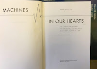
Machines in Our Hearts, by Kirk Jeffrey, superimposed on Intermediate Physics for Medicine and Biology.
Machines in Our Hearts,
by Kirk Jeffrey.
The story of how Berkovits invented the demand pacemaker is told in Machines in Our Hearts, by Kirk Jeffrey.
Barouh V. Berkovits (b. 1924), an engineer at the American Optical Company, was already well known as the inventor of the DC defibrillator and the cardioverter, a device that interrupts a rapid heart rate (tachycardia) with low-energy shocks. He knew that when the cardioverter discharged randomly into the tachycardia, it would “occasionally not only not stop the tachyarrhythmia…but would produce ventricular fibrillation.” Cardioversion has to be synchronized to fall within the QRS complex and avoid the vulnerable period of the heartbeat. In 1963, Berkovits applied this principle to cardiac pacing. To solve the problem of competition [between the SA node and the artificial pacemaker], Berkovits in 1963 designed a sensing capability into the pacemaker. His invention behaved exactly like an asynchronous pacer until it detected a naturally occurring R wave, the indication of a ventricular contraction. This event would reset the timing circuit of the pacemaker, and the countdown to the next stimulus would begin anew. Thus the pacer stimulated the heart only when the ventricles failed to contract. It worked only “on demand.” As an added benefit, non-competitive pacing extended the life of the battery.
The 4th edition of Intermediate Physics for Medicine and Biology does not mention Berkovits by name, but Homework Problem 45 in Chapter 7 does analyze the demand pacemaker.
Problem 45 A patient with “intermittent heart block” has an AV node which functions normally most of the time with occasional episodes of block, lasting perhaps several hours. Design a pacemaker to treat the patient. Ideally, your design will not stimulate the heart when it is functioning normally. Describe
(a) whether you will stimulate the atria or ventricles
(b) which chambers you will monitor with a recording electrode
(c) what logic your pacemaker will use to determine when to stimulate. Your design may be similar to a “demand pacemaker” described in Jeffrey (2001), p. 132.
Of course, the reference is to Machines in Our Hearts. Berkovits’s phenomenal career is yet another example of how knowledge of engineering and physics can allow you to contribute to medicine and biology.

Friday, June 1, 2012

Andrew Huxley (1917-2012)

Andrew Huxley, the greatest mathematical biologist of the 20th century, died on Wednesday, May 30. Huxley won the Nobel Prize for his groundbreaking work with Alan Hodgkin that explained electrical transmission in nerves.

In Chapter 6 of the 4th edition of Intermediate Physics for Medicine and Biology, Russ Hobbie and I describe the Hodgkin-Huxley model of membrane current in a nerve axon.
Considerable work was done on nerve conduction in the late 1940s, culminating in a model that relates the propagation of the action potential to the changes in membrane permeability that accompany a change in voltage. The model [Hodgkin and Huxley (1952)] does not explain why the membrane permeability changes; it relates the shape and conduction speed of the impulse to the observed changes in membrane permeability. Nor does it explain all the changes in current…Nonetheless, the work was a triumph that led to the Nobel Prize for Alan Hodgkin and Andrew Huxley.
The paper we cite (“A Quantitative Description of Membrane Current and its Application to Conduction and Excitation in Nerve,” Journal of Physiology, Volume 117, Pages 500–544) is one of my favorites. Whenever I teach biological physics, I assign this paper to my students as an example of mathematical modeling in biology at its best. In 1981 Hodgkin and Huxley wrote a “citation classic” article about their paper, which has now been cited over 9300 times. They concluded
Another reason why our paper has been widely read may be that it shows how a wide range of well-known, complicated, and variable phenomena in many excitable tissues can be explained quantitatively by a few fairly simple relations between membrane potential and changes of ion permeability—processes that are several steps away from the phenomena that are usually observed, so that the connections between them are too complex to be appreciated, intuitively. There now seems little doubt that the main outlines of our explanation are correct, but we have always felt that our equations should be regarded only as a first approximation that needs to be refined and extended in many ways in the search for the actual mechanism of the permeability change’s on the molecular scale.
As one who does mathematical modeling of bioelectric phenomena for a living, I can think of no better way to honor Huxley than to show you his equations.


This set of four nonlinear ordinary differential equations, plus six expressions relating how the ion channel rate constants depend on voltage, not only describes the membrane of the squid giant nerve axon, but also is the starting point for models of all electrically active tissue. Russ and I consider this model to be so important that we dedicate six pages to exploring it, and present in our Fig. 6.38 a computer program to solve the equations. For anyone interested in electrophysiology, becoming familiar with the Hodgkin-Huxley model is job one, just as analyzing the Bohr model for hydrogen is the starting point for someone interested in atomic structure. Remarkably, 60 years ago Huxley solved these differential equations numerically using only a hand-crank adding machine.

How can your learn more about this great man? First, the Nobel Prize website contains his biography, a transcript of his Nobel lecture, and a video of an interview. Another recent, more detailed interview is available on Youtube in two parts, part1 and part 2. Huxley wrote a fascinating description of the many false leads during their nerve studies in a commemorative article celebrating the 50th anniversary of his famous paper. Finally, the Guardian published an obituary of Huxley yesterday.

An interview with Andrew Huxley, Part 1.
https://www.youtube.com/watch?v=WdL-81i3Qg4

An interview with Andrew Huxley, Part 2.
https://www.youtube.com/watch?v=qL3aTfljBXE

I will conclude by quoting the summary at the end of Hodgkin and Huxley’s 1952 paper, which was the last of a series of five articles describing their voltage clamp experiments on a squid axon.
SUMMARY
1. The voltage clamp data obtained previously are used to find equations which describe the changes in sodium and potassium conductance associated with an alteration of membrane potential. The parameters in these equations were determined by fitting solutions to the experimental curves relating sodium or potassium conductance to time at various membrane potentials.
2. The equations, given on pp. 518–19, were used to predict the quantitative behaviour of a model nerve under a variety of conditions which corresponded to those in actual experiments. Good agreement was obtained in the cases:
(a) The form, amplitude and threshold of an action potential under zero membrane current at two temperatures.
(b) The form, amplitude and velocity of a propagated action potential.
(c) The form and amplitude of the impedance changes associated with an action potential.
(d) The total inward movement of sodium ions and the total outward movement of potassium ions associated with an impulse.
(e) The threshold and response during the refractory period.
(f) The existence and form of subthreshold responses.
(g) The existence and form of an anode break response.
(h) The properties of the subthreshold oscillations seen in cephalopod axons.
3. The theory also predicts that a direct current will not excite if it rises sufficiently slowly.
4. Of the minor defects the only one for which there is no fairly simple explanation is that the calculated exchange of potassium ions is higher than that found in Sepia axons.
5. It is concluded that the responses of an isolated giant axon of Loligo to electrical stimuli are due to reversible alterations in sodium and potassium permeability arising from changes in membrane potential.

Friday, September 30, 2011

Wilson Greatbatch (1919-2011)

This week we lost a giant of engineering: Wilson Greatbatch, inventor of the implantable cardiac pacemaker.

The cardiac pacemaker represents one of the most important contributions of physics and engineering to medicine. In Chapter 7 of the 4th edition of Intermediate Physics for Medicine and Biology, Russ Hobbie and I describe the pacemaker.
Cardiac pacemakers are a useful treatment for certain heart diseases [Jeffrey (2001); Moses et al. (2000); Barold (1985)]. The most frequent are an abnormally slow pulse rate (bradycardia) associated with symptoms such as dizziness, fainting (syncope), or heart failure. These may arise from a problem with the SA node (sick sinus syndrome) or with the conduction system (heart block)….

A pacemaker can be used temporarily or permanently. The pacing electrode can be threaded through a vein from the shoulder to the right ventricle (transvenous pacing, Fig. 7.31) or placed directly in the myocardium during heart surgery.
Several years ago, I taught a class about pacemakers and defibrillators as part of Oakland University’s honors college. The class was designed to challenge our top undergraduates, but not necessarily those majoring in science. Among the readings for the class was a profile in the March 1995 issue of IEEE Spectrum about Wilson Greatbatch (Volume 32, Pages 56-61). The article tells the story of Greatbatch’s first implantable pacemaker:
Greatbatch was on one team that had been summoned by William C. Chardack, chief of surgery at Buffalo’s Veteran’s Administration Hospital, to deal with a blood oximeter. The engineers could not help with that problem, but the meeting for the inventor was momentous: finally, after many previous attempts, he had met a surgeon who was enthusiastic about prospects for an implantable pacemaker. The surgeon estimated such a device might save 10000 lives a year.

Three weeks later, on May 7, 1958, the engineer brought what would become the worlds first implantable cardiac pacemaker to the animal lab at Chardack’s hospital. There Chardack and another surgeon, Andrew Gage, exposed the heart of a dog, to which Greatbatch touched the two pacemaker wires. The heart proceeded to beat in synchrony with the device, made with two Texas Instruments 910 transistors. Chardack looked at the oscilloscope, looked back at the animal, and said, “Well, I’ll be damned.”
Machines in Our Hearts, by Kirk Jeffrey, superimposed on Intermediate Physics for Medicine and Biology.
Machines in Our Hearts,
by Kirk Jeffrey.
Another source the honors college students studied from was Kirk Jeffrey’s excellent book Machines in Our Hearts: The Cardiac Pacemaker, the Implantable Defibrillator, and American Health Care. Jeffrey tells the long history of how pacemakers and defibrillators were developed. In a chapter titled Multiple Invention of Implantable Pacemakers he describes Greatbatch’s contributions as well as others, including Elmqvist and Senning in Sweden. Jeffrey writes
If theirs [Chardack and Greatbatch] was not the only pacemaker of the 1950s, it appears to be the only one that survives today in the collective memory of the community of physicians, engineers, and businesspeople whose careers are tied to the pacemaker… The Chardack-Greatbatch pacamaker stood out from other prototype implantables of the late 1950s not because it was first or clearly a better design, but because it succeeded in the U.S. market as did no other device.
Jeffrey also discusses at length Greatbatch’s contributions to developing the lithium battery.
Because of his prestige in the pacing community and his effectiveness as a champion of technology be believed in, Greatbatch was able almost single-handedly to turn the industry to lithium; in fact by 1978, a survey of pacing practices indicated that only 5 percent of newly implanted pulse generators still used mercury-zinc batteries.
Greatbatch was inducted into the National Inventor’s Hall of Fame in 1986. His citation says
Wilson Greatbatch invented the cardiac pacemaker, an innovation selected in 1983 by the National Society of Professional Engineers as one of the two major engineering contributions to society during the previous 50 years. Greatbatch has established a series of companies to manufacture or license his inventions, including Greatbatch Enterprises, which produces most of the world's pacemaker batteries.

Invention Impact

His original pacemaker patent resulted in the first implantable cardiac pacemaker, which has led to heart patient survival rates comparable to that of a healthy population of similar age.

Inventor Bio

Born in Buffalo, New York, Greatbatch received his preliminary education at public schools in West Seneca, New York. In 1936 he entered military service and served in the Atlantic and Pacific theaters during World War II. He was honorably discharged with the rating of aviation chief radioman in 1945. He attended Cornell University and graduated with a B.E.E. in electrical engineering in 1950. Greatbatch received a master's from the State University of New York at Buffalo in 1957 and was awarded honorary doctor's degrees from Houghton College in 1970 and State University of New York at Buffalo in 1984. Although trained as an electrical engineer, Greatbatch has primarily studied interdisciplinary areas combining engineering with medical electronics, agricultural genetics, the electrochemistry of pacemaker batteries, and the electrochemical polarization of physiological electrodes.
Below are some links related to Wilson Greatbatch that you might find useful.
An article about Greatbatch published by the Lemelson Center for the Study of Invention and Innovation: http://invention.smithsonian.org/centerpieces/ilives/lecture09.html

A video about Greatbatch produced by the Vega Science Trust: http://www.vega.org.uk/video/programme/248

Biography of Wilson Greatbatch on the Heart Rhythm Society website:
http://www.hrsonline.org/News/ep-history/notable-figures/wilsongreatbatch.cfm

New York Times obituary: http://www.nytimes.com/2011/09/28/business/wilson-greatbatch-pacemaker-inventor-dies-at-92.html

BBC obituary: http://www.bbc.co.uk/news/world-us-canada-15085056

A video honoring Wilson Greatbatch, the 1996 Lemelson-MIT Lifetime Achievement Award Winner.
Learn about Wilson Greatbatch, 1996 Lemelson-MIT Lifetime Achievement Award Winner.
https://www.youtube.com/embed/WLZBl118Ads

Friday, February 4, 2011

Britton Chance (1913-2010)

Britton Chance died late last year. The website www.brittonchance.org states that
Britton Chance, M.D., Ph.D., D.Sc., for more than 50 years one of the giants of biochemistry and biophysics and a world leader in transforming theoretical science into useful biomedical and clinical applications, died on November 16, 2010, at age 97 in Philadelphia, PA. Dr. Chance had the rare distinction of being the recipient of a National Medal of Science (1974), a Gold Medal in the Olympics (1952, Sailing, Men’s 5.5 Meter Class), and a Certificate of Merit for his sensitive work during World War II.
His obituary in the New York Times describes his early work.
Over a lifetime of research, Dr. Chance focused on the observation and measurement of chemical reactions within cells, tissue and the body. But unlike most researchers, he also had expertise in mechanics, electronics and optics, and a great facility in instrument-building. His innovations helped transform theoretical science into biochemical and biophysical principles, the stuff of textbooks, and useful biomedical and clinical applications.

Early in his career he invented a tool, known as a stopped-flow apparatus, for measuring chemical reactions involving enzymes; it led to the establishment of a fundamental principle of enzyme kinetics, known as the enzyme-substrate complex.
Another obituary, in the December 17 issue of Science magazine, observed that
In his mid-70s, Chance (then emeritus) launched a new field of optical diagnostics that rests on the physics of light diffusion through scattering materials such as living tissue. He showed that scattered near-infrared light pulses could not only measure the dynamics of oxy- and deoxyhemoglobin levels in performing muscles, but also reveal and locate tumors and cancerous tissue in muscles and breast as well as injury in the brain. Because changing patterns of oxy- and deoxyhemoglobin in the brain reflect cognitive activity, the applications of this diagnostic approach widened to include assessing neuronal connectivity in premature babies.
Chance appears in the 4th edition of Intermediate Physics for Medicine and Biology because of his research on light diffusion. In Section 14.4 (Scattering and Absorption of Radiation), Russ Hobbie and I analyze the absorption and scattering coefficients of infrared light, and then give typical values that “are eyeballed from data from various tissues reported in the article by Yodh and Chance (1995),” with the reference being to Yodh, A. and B. Chance (1995) “Spectroscopy and Imaging with Diffusing Light,” Physics Today, March, Pages 34–40.

Then in Sec. 14.5 (The Diffusion Approximation to Photon Transport), we analyze pulsed measurements of infrared light.
A technique made possible by ultrashort light pulses from a laser is time-dependent diffusion. It allows determination of both [the scattering coefficient] and [the absorption coefficient]. A very short (150-ps) pulse of light strikes a small region on the surface of the tissue. A detector placed on the surface about 4 cm away records the multiply-scattered photons. A typical plot of the detected photon fluence rate is shown in Fig. 14.13.
Figure 14.13 is a figure from Patterson, M. S., B. Chance, and B. C. Wilson (1989) “Time Resolved Reflectance and Transmittance for the Noninvasive Measurement of Tissue Optical Properties,” Applied Optics, Volume 28, Pages 2331–2336, which has been cited over 1000 times in the scientific literature.

Finally, in Sec. 14.6 (Biological Applications of Infrared Scattering), we reproduce a figure from the Physics Today article by Yodh and Chance, which shows the absorption coefficient for water, oxyhemoglobin and deoxyhemoglobin.
The greater absorption of blue light in oxygenated hemoglobin makes oxygenated blood red…The wavelength 800 nm at which both forms of hemoglobin have the same absorption is called the isosbestic point. Measurements of oxygenation are made by comparing the absorption at two wavelengths on either side of this point.
This property of infrared absorption of light is the basis for pulse oximeters that measure oxygenation. Not all measurements of blood oxygen use pulsed light. Russ and I cite one of Chance’s papers that uses a continuous source: Liu, H., D. A. Boas, Y. Zhang, A. G. Yodh, and B. Chance (1995) “Determination of Optical Properties and Blood Oxygenation in Tissue Using Continuous NIR Light,” Physics in Medicine and Biology, Volume 40, Pages 1983–1993. A fourth of Chance’s paper that we include in our references is Sevick, E. M., B. Chance, J. Leigh, S. Nioka, and M. Maris (1991) “Quantitation of Time- and Frequency-Resolved Optical Spectra for the Determination of Tissue Oxygenation,” Analytical Biochemistry, Volume 195, Pages 330–351.

In 1987, Chance won the Biological Physics Prize (now known as the Max Delbruck Prize in Biological Physics) from the American Physical Society
for pioneering application of physical tools to the understanding of Biological phenomena. The early applications ranged from novel spectrometry that elucidated electron transfer processes in living systems to analog computation of nonlinear processes. Later contributions have been equally at the forefront.

Friday, June 5, 2009

Ichiji Tasaki (1910-2009)

Ichiji Tasaki (1910–2009) died January 4 in Bethesda, Maryland. Tasaki was known for his discovery in 1939 of saltatory conduction of action potentials in a myelinated nerve axon. You can learn more about myelinated fibers and saltatory conduction in the 4th edition of Intermediate Physics for Medicine and Biology.

Tasaki had a long and fascinating career in science. His life is described in an obituary published in the May 2009 issue of Neuroscience Research. He is also featured in an article of the NIH Record, the weekly newsletter for employees of the National Institutes of Health.

I knew Tasaki when I was working at NIH in the 1990s. Late in his career he worked with my friend Peter Basser in the National Institute of Child Health and Human Development. I recall him working every day in his laboratory, despite being in his 80s, with his wife as his assistant. He led a fascinating life. His best known research on saltatory conduction was performed in Japan just before and during World War II. After the war, he spent over 50 years at NIH.

Basser describes Tasaki as “a scientist’s scientist, never afraid to question current dogma, always digging deeper to discover the truth.” Congressman Chris van Hollen of Maryland paid tribute to Tasaki a few months before he died, beginning
Madam Speaker, I rise today to recognize the outstanding achievements of my constituent Dr. Ichiji Tasaki. Dr. Tasaki has worked at the National Institutes of Health for 54 years, since November 1953, and has made invaluable contributions to the scientific community.

Friday, November 28, 2008

Adrian Kantrowitz (1918-2008)

Last week heart surgeon and pacemaker pioneer Andrian Kantrowitz died in Ann Arbor, Michigan. Among his many roles, Kantrowitz was an Adjunct Professor in the Department of Physics here at Oakland University where I work. Soon after I arrived at OU in 1998, Emeritus Professor Norm Tepley and I visited Kantrowitz’s company L.VAD Technology in Detroit, which makes a left ventricular assist device that helps the heart pump blood. In February 2005 I invited Kantrowitz to give our weekly physics colloquium. At the time his health was already fragile and he gave his lecture sitting down. But it was an excellent talk to one of the largest crowds we ever had at our colloquium series.
Machines in Our Hearts: The Cardiac Pacemaker, the Implantable Defibrillator, and American Health Care, by Kirk Jeffrey, superimposed on Intermediate Physics for Medicine and Biology.
Machines in Our Hearts: The Cardiac Pacemaker,
the Implantable Defibrillator, and American Health Care,
by Kirk Jeffrey.

Kantrowitz had an inspirational life story. As a young man, he served as a battalion surgeon in World War Two. He later performed the first heart transplant in the United States. He also played a role in the early development of the pacemaker, a topic discussed in Chapter 7 of the 4th edition of Intermediate Physics for Medicine and Biology. Kirk Jeffrey, in his book 
Machines in Our Hearts: The Cardiac Pacemaker, the Implantable Defibrillator, and American Health Care, wrote
GE [General Electric] had developed an implantable pacemaker in its electronics laboratory in cooperation with heart surgeon Adrian Kantrowitz of Maimonides Hospital in Brooklyn. This project began in 1960, apparently in response to the announcement of the Chardack-Greatbatch pacemaker. The initial model was implanted in May 1961 and, as was common with these early devices, the designers made improvements based on the experience of the early patients.

The GE pacemakers had one remarkable technological feature—an external control unit that communicated with the implanted generator by magnetic induction. When taped to the skin on the patient's abdomen, the controller enabled the physician to set the pacing rate anywhere between 64 and 120 beats per minute. Kantrowitz viewed rate control as a means to safeguard the elderly patient.
You can learn more about Adrian Kantrowitz from obituaries in the New York Times, the Washington Post and the Los Angeles Times.