Friday, July 8, 2022

The Physics of Radiology

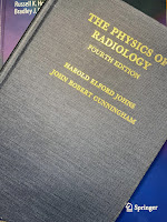
The Physics of Radiology, by Johns and Cunningham, superimposed on Intermediate Physics for Medicine and Biology.
The Physics of Radiology,
by Johns and Cunningham.
In last week’s blog post, I reviewed the recently published book True Tales of Medical Physics. A point I didn’t mention in my review was the central role of one textbook in the education of many of the authors who described their life story in True Tales. The Physics of Radiology was written by Harold Johns and John Cunningham. The first edition was published in 1953, but I have access through the Oakland University library to the fourth edition from 1983. This iconic book defined the field of medical physics and radiology in the second half of the twentieth century. Chapters 15–17 in Intermediate Physics for Medicine and Biology summarize material covered in more detail in The Physics of Radiology.

In True Tales, Jacob Van Dyk wrote
In 1971, I was hired by Professor Harold Elford Johns to work as a medical physicist at the Princess Margaret Hospital (PMH) in Toronto. Professor Jack Cunningham was my immediate boss. Professor Johns was considered the guru of Medical Physics with a world-renowned reputation for being a great scientist, a feared graduate student supervisor, and humanitarian. Over the years, he received multiple awards including five honorary doctorate degrees, and Officer of the Order of Canada. He was the first medical physicist to be inducted into the Canadian Medical Hall of Fame. Jack Cunningham also received the Officer of the Order of Canada along with multiple other awards, largely for his work on software development for computerized radiation treatment planning systems. Johns and Cunningham were the authors of The Physics of Radiology, the textbook which gave me my medical physics grounding as it did for all other young, aspiring medical physicists at that time….

[When Van Dyk was studying for his medical physics certification exams], Professors Johns and Cunningham were working on a draft of the fourth edition of their book, The Physics of Radiology. In January, I asked Jack Cunningham if I could review the available draft of this fourth edition. Considering that they would be contributors to the certification examination questions, my guess was that some, if not all, of the questions could be answered if I knew everything in this new edition. So, I went through this draft of the book from cover to cover and I solved (at least I worked on) every problem that was posed at the end of each chapter. As part of this process, I provided Jack with some comments on some questionable things that I found in the draft. As a result, my name was listed, along with others, in the acknowledgments when the book was published in 1983.
In his chapter of True Tales, Terry Peters wrote
During my early years at McGill, I became involved in the activities of the newly formed Canadian College of Physicists in Medicine (CCPM), which had begun the process of credentialling Medical Physicists in Canada. While I had Engineering, rather than Physics, training, I felt that my background had prepared me well for the roles I was playing in Diagnostic Radiology at the Neuro. Nevertheless, I felt it would do no harm to formally study radiation physics and its practical implementation in medicine, so in 1983 I embarked on a mission to devour The Physics of Radiology, by Johns and Cunningham, in preparation for the CCPM Fellowship exams in 1984. The examination process had evolved into an oral session, and a closed book examination—where three questions were selected from a previously published catalogue of questions covering all aspects of Medical Physics. Every Friday afternoon for almost a year I studied “Johns and Cunningham” with Gino Fallone, then a physicist at the Montreal General Hospital, who had also decided to take the certification examination. A gruelling process, but finally successful—we both became CCPM fellows that year.
Martin Yaffe's contribution to True Tales described a bit about Johns background and career.
Dr. Johns had been born in Chengdu, China to Canadian church missionary parents and he had spent his early years there, roaming about small communities in the mountains of Szechuan province with his father, a no-nonsense disciplinarian who believed strongly in devotion to duty and hard work. He learned to be focussed and driven to succeed at whatever was his mission. When the family eventually returned to Canada, he brought that to his graduate work in physics and later to the University of Saskatchewan where he built a strong medical physics research group, concentrating on developing and refining radiation therapy. There he developed the first (or possibly the second—there is some debate as his unit and a competitor, built by Eldorado Mining and Refining Ltd., were used to treat patients within a week or two of each other) cobalt-60 radiation treatment system and carried out pioneering work on radiation dosimetry and treatment planning. Dr. Johns and his work in Saskatchewan were actually mentioned in the film First Man, about the astronaut Neil Armstrong whose daughter had suffered from a brain tumour. Later, he began work on The Physics of Radiology, a textbook which he referred to jokingly (I think) as “The Bible”. This book truly became a guide to those working in radiation oncology all over the world and was published in multiple languages. While the book and its various editions consumed many of his evenings after a hard day at the lab, Johns reverse bragged that he earned about two cents per hour on his textbook writing efforts.

I once estimated that I make about ten cents an hour for my work on IPMB. I guess the difference represents inflation.

Yaffe also reminisced on Johns’ personality and mentoring technique.

Johns had an abrupt nature, not hesitating to poke you emphatically when he felt that you needed to think harder. Often, he would read your carefully written document, hold it up between you and slowly rip it to shreds before filing it in the trash bin. If it was late in the day, he would tell you to meet him the next morning at eight to re-write.

What I learned in those sessions was how to sharply focus your thinking on a problem and how to persist until you had a workable solution. Dr. Johns had two more senior students at the time—Aaron Fenster… and Don Plewes. These two and the lab technician, Dan Ostler, more or less adopted me and provided mentoring to prepare me for my sessions with Johns. Also, Johns used to invite me into his office when he was working on a paper with Aaron or Don and let me watch. While he was more respectful toward them, it was not uncommon for him to fix one of them with his laser-like glare which he held on them for what seemed like minutes and then say something like: “Plews” (he never pronounced the “e” that made it rhyme with Lewis; instead he made it rhyme with “news”), “If you sent that to a journal, they’d crap all over you”. Or, as he slowly ripped up a piece of writing that Aaron had proudly submitted, he’d say at a similar slow pace, “well (rrrrip) Fenster (rrrrip), your (rrrrip) writing (rrrrip) is improving.” So, rather than feel discriminated against, I simply realized that the standards were high, and I’d have to present my best game at all times.
The Physics of Radiology is one of those landmark textbooks (like Jackson’s Classical Electrodynamics in physics) that is a rite of passage for a student in that a field of study. As a coauthor on IPMB, I know what an honor it would be for your book to make that sort of impact. Johns and Cunningham was cited in the second edition of IPMB, but not in earlier or later editions.

The fourth edition is the last that Johns and Cunningham published. However, just last year an updated fifth edition was prepared by a team of five authors.

Friday, July 1, 2022

True Tales of Medical Physics: Insights into a Life-Saving Specialty

True Tales of Medical Physics: Insights into a Live-Saving Speciality, by Jacob Van Dyk, superimposed on Intermediate Physics for Medicine and Biology.
True Tales of Medical Physics:
Insights into a Live-Saving Specialty
,
by Jacob Van Dyk.
I’ve found the perfect book for readers of Intermediate Physics for Medicine and Biology who are fascinated by medical physics but who don’t want a lot of technical details and math. Jacob Van Dyk recently published True Tales of Medical Physics: Insights into a Life-Saving Specialty. The book consists of 22 chapters written by leading medical physicists, in which they each discuss their career, focusing on interesting anecdotes and life lessons. If I were a young student pondering what career to pursue, this is a book I’d want to read.

Van Dyk’s instructions to the contributors were
to communicate what medical physics is and what medical physicists do to a broad audience including science students, graduate students and residents, experienced medical physicists and their family members, and the general public who are wondering about medical physics. The book will consist of a series of short stories written by award-winning medical physicists—stories that are of personal interest as it relates to their careers. Each story will be unique to the author and could serve any one or more of the following purposes:
  1. Be an inspiration to young people searching for career directions, as well as more experienced physicists who are seeking direction on leadership development. 
  2. Provide an overview of what medical physicists do with a level of description that is understandable by the non-medical physicist.
  3. Provide lessons on life’s experiences from high-profile medical physicists who have significant experience and who are clearly at the top of the field as shown by the awards that they have won. 
  4. Be entertaining for those working in the field as well as others.
You can look at this book as a Plutarchian collection of comparative biographies, or you can focus on cross-cutting lessons that appear again and again in the various chapters. Here are some of the lessons I noticed.
  • The critical role of mentoring. All the authors stressed the importance of having supportive, inspirational mentors early in their career, and the satisfaction of mentoring their own students.
  • Crucial advances grow out of discussions at scientific meetings. Clearly the opportunity to travel and attend meetings is a part of a scientist’s career that’s highly valued, and often leads to new research directions and collaborations.
  • The division of their duties into three parts: research, teaching, and clinical work. The variety that arises from having these three different tasks keeps a medical physicist’s job from ever becoming dull or routine.
  • Leading scientific societies. The American Association of Physicists in Medicine (AAPM), the American Society for Radiation Oncology (ASTRO), the International Organization for Medical Physics (IOMP), and others professional groups are mentioned over and over by these authors.
  • The challenge of the American Board of Radiology (ABR) exams. Today, these certification exams act as a gateway to a career in clinical medical physics.
  • The interdisciplinary nature of medical physics. Many of these authors brought expertise from one field (say, computer programming) and integrated that knowledge with other fields (say, anatomy or medical imaging). 
  • Failures are opportunities. These scientists had their share of setbacks, but managed to overcome them and use them as springboards to success. They persisted.
  • The role of industry in medical physics research. Many authors tell stories of interacting with for-profit companies making imaging or therapy devices. Working with industry can be complicated and aggravating, but when successful the resulting products can have a huge impact on medical practice.
  • Science is an international activity. Many authors had collaborators and students from all over the world, leading to lifelong friendships.
True Tales of Medical Physics illustrates what a career in medical physics is like better than any textbook can (even IPMB!). Some of the authors worked on old, now obsolete devices—colbalt-60 radiation therapy, the betatron, film/screen cassettes—but they also helped develop today’s cutting edge technologies: computed tomography (CT), intensity-modulated radiation therapy (IMRT), magnetic resonance imaging (MRI), and medical ultrasound (US). These authors worked at leading medical institutions, such as the Memorial Sloan Kettering Cancer Center and the MD Anderson Cancer Center. Many were included in the IOMP’s 50th anniversary list of 50 medical physicists who have made an outstanding contribution to the advancement of medical physics over the last 50 years (I can’t help but compare this to the list of the 50 greatest basketball players prepared by the National Basketball Association on their 50th anniversary).

While I enjoyed all the chapters in True Tales, my favorite was by Marcel van Herk, a Steve Wozniak-like Dutch electronics guru. He started out as a 12-year-old hobbyist who built his own computer. Van Herk writes “One of the first things I did was to design and build a completely functional relay-based full adder (a circuit that can add two 4-bit binary numbers), soldered together while listening to Black Sabbath’s Iron Man in the living room, not totally to my mother’s liking due to the music and the spilled solder on the carpet. The parts I used were small electromechanical relays from 1950 punched card sorting machines, acquired cheaply.” He ended up developing software for the Elekta cone-beam CT imaging guidance system integrated with a medical linear accelerator. While his story is fascinating, it’s not uncommon; many of these scientists traveled individual, meandering paths into medical physics, taking them from a clueless neophyte to a giant in their field. A key lesson for students is that there’s no single route to success in science, and certainly not in medical physics.

If you are considering possible careers, I urge you to read True Tale of Medical Physics. It may change your life. You may change medicine.

Friday, June 24, 2022

Specific Heat Capacity

One of the most important parameters in thermodynamics is the specific heat capacity. In Chapter 3 of Intermediate Physics for Medicine and Biology, Russ Hobbie and I define the specific heat capacity in this way:
Consider a system into which a small amount of heat Q flows. In many cases the temperature of the system rises [by an amount ΔT]… The heat capacity C of the system is defined as

                                        C = Q/ΔT .                 (3.39)

Heat capacity has units of J K-1. It depends on the size of the object and the substance it is made of. The specific heat capacity, c, is the heat capacity per unit mass (J K−1 kg−1).
Air and Water, by Mark Denny, superimposed on Intermediate Physics for Medicine and Biology.
Air and Water,
by Mark Denny.
In Air and Water: The Biology and Physics of Life's Media, Mark Denny compares the specific heat capacity of life’s two most important substances.
The specific heat of air is 1006 J kg−1 K−1, a typical value for a gas… Water, however, … has a very high specific heat for a liquid… about 4200 J kg−1 K−1… It thus takes about four times as much heat to raise the temperature of a kilogram of water one degree as it does to raise the temperature of an equal mass of air.
A factor of four is significant, but frankly I would have expected an even bigger difference. The reason for the somewhat similar values of the specific heat capacity for air and water is that we are comparing heat capacity per unit mass. It may be more intuitive to compare heat capacity per unit volume. To convert from per kilogram to per cubic meter you must multiply the specific heat capacity per unit mass by the mass per unit volume, which is just the density, ρ. The density of air and water are very different. Air has a density of about 1.2 kg m−3, whereas water has a density of 1000 kg m−3.

We can express the specific heat capacity per unit volume as the product c times ρ. For air cρ is 1207 J K−1 m−3 but for water cρ is 4,200,000 J K−1 m−3. So water has a vastly higher specific heat capacity compared to air when expressed as per unit volume.

Denny says the same thing this way:
The relative similarity of specific heat between air and water can be misleading, however, because specific heat is measured on per-mass basis. One cubic meter of air weighs only about 1.2 kg, while a cubic meter of water weighs 1000 kg. It thus takes about 3500 times as much heat to raise the temperature of a given volume of water one degree as it does to raise the temperature of the same volume of air.
For similar volumes of air and water in thermal equilibrium, the heat stored in the air is negligible compared to that stored in the water. Biological tissue is mostly water, so that means air holds a lot less heat than tissue, per unit volume. This has implications for biological processes, such as heat exchange between air and tissue in the lungs.

Friday, June 17, 2022

Limping

In Chapter 1 of Intermediate Physics for Medicine and Biology, Russ Hobbie and I discuss biomechanics. One of our most important examples is the force on the hip.
The forces in the hip joint can be several times a person’s weight, and the use of a cane can be very effective in reducing them.
Indeed, a cane is very useful, as Russ and I show in Section 1.8 of IPMB. But what if you don’t have a cane handy, or if you prefer not to use one? You limp. In this post, we examine the biomechanics of limping.

When you limp, you lean toward the injured side to reduce the forces on the hip. The reader can analyze limping in this new homework problem.
Section 1.8

Problem 11 ½. The left side of the illustration below analyzes normal walking and reproduces Figures 1.11 and 1.12. The right side shows what happens when you walk with a limp. By leaning toward the injured side you reduce the distance between the hip joint and the body’s center of gravity, and your leg is more vertical than in the normal case.
Pertinent features of the anatomy of the leg: normal (left) and limping (right), based on figures 1.11 and 1.12 in Intermediate Physics for Medicine and Biology.
Pertinent features of the anatomy of the leg: normal (left) and limping (right).

(a) Reproduce the analysis of Section 1.7 to calculate of the forces on the hip during normal walking using the illustration on the left. Begin by making a free-body diagram of the forces acting on the leg like in Figure 1.13. Then solve the three equations for equilibrium: one for the vertical forces, one for the horizontal forces, and one for the torques. Verify that the magnitude of the force on the hip joint is 2.4 times the weight of the body. 
(b) Reanalyze the forces on the hip when limping. Use the geometry and data shown in the illustration on the right. Assume that any information missing from the diagram is the same as for the case of normal walking; For example, the abductor muscle makes an angle of 70° with the horizontal for both the normal and limping cases. Draw a free-body diagram and determine the magnitude of the force on the hip joint in terms of the weight of the body.

I won’t solve the entire problem for you, but I’ll tell you this: limping reduces the force on the hip from 2.4 times the body weight in the normal case to 1.2 times the body weight in the case of limping. No wonder we limp! 

The main reason for the lower force when limping is the smaller moment arm. If we calculate torques about the head of the femur, then in the normal case the moment arm for the force that the ground exerts on the foot is 18 – 7 = 11 cm. When limping, this moment arm reduces to 9 – 7 = 2 cm. The moment arm for the abductor muscles (the gluteus minimus and gluteus medius) is the same in the two cases. Therefore, rotational equilibrium can be satisfied with a small muscle force when limping, although a large muscle force is required normally. The torque is a critical concept for understanding biomechanics.

What do you do if both hips are injured? When walking, you first lean to one side and then the other; you waddle. This reduces the forces on the hip, but results in a lot of swinging from side to side as you walk.

If you are having trouble solving this new homework problem, contact me and I’ll send you the solution. 

Enjoy!


Friday, June 10, 2022

The Genetics of Cystic Fibrosis

In Appendix H of Intermediate Physics for Medicine and Biology, Russ Hobbie and I briefly mention the severe lung disease cystic fibrosis. Analyzing this disease provides an opportunity to examine the prevalence of a genetic disorder. I’ll do this by creating a new homework problem.
Appendix H

Problem 5 ½. About 1 in every 2500 people is born with cystic fibrosis, an autosomal recessive disorder. What is the probability of the gene responsible for cystic fibrosis in the population? What fraction of the population are carriers of the disease?

To answer these questions, first we must know that an “autosomal recessive disorder” is one in which you only get the disease if you have two copies of a recessive gene. To a first approximation, there are often two variants (or alleles) of a gene governing a particular protein: dominant (A) and recessive (a). In order to have cystic fibrosis, you must have two copies of the recessive allele (aa). If you have only one copy (Aa), you are healthy but are a carrier for the disease: your children could potentially get the disease if your mate is also a carrier. If you have no copies of the recessive allele (AA) then you’re healthy and your children will also be healthy.

Let’s assume the probability of the dominant allele is p, and the probability of the recessive allele is q. Since we assume there are only two possibilities, we know that p + q = 1. Our goal is to find q, the probability of the gene responsible for cystic fibrosis in the population.

When two people mate, they each pass on to their offspring one of their two copies of the gene. The probability that both parents are dominant (AA), so the child is normal, is p2. The probability that both parents are recessive (aa), so the child has the disease, is q2. There are two ways for the child to be a carrier: A from dad and a from mom, or a from dad and A from mom. So, the probability of a child being a carrier (Aa) is 2pq. There are only three possibilities or genotypes: AA, Aa, and aa. The sum of their probabilities must equal one: p2 + 2pq + q2 = 1. But this expression is equivalent to (p + q)2 = 1, and we already knew that p + q = 1, so the result isn’t surprising.

The only people that suffer from cystic fibrosis have the genotype aa, so q2 is equal to the fraction of people with the disease. The problem states that this fraction is 1/2500 (0.04%). So, q is the square root of 1/2500, or 1/50 (2%; wasn’t that nice of me to make the fraction be the reciprocal of a perfect square?). One out of every fifty copies of the gene governing cystic fibrosis is defective (that is, it is the recessive version that can potentially lead to the disease). If q is 1/50, then p is 49/50 (98%). The fraction of carriers is 2pq, or 3.92%. The only reason this result is not exactly 4% is that we don’t count someone with the disease (aa) as a carrier, even though they could pass the disease to their children (a carrier by definition has the genotype Aa). If we are rounding off our result to the nearest percent, then 1 out of every 25 people (4% of the population) are carriers.

This calculation is based on several assumptions: no natural selection, no inbreeding, and no selection of embryos based on genetic testing. Cystic fibrosis is such a severe disease that often victims don’t survive long enough to have children (modern medicine is making this less true). The untreated disease is so lethal that one wonders why natural selection didn’t eliminate it from our gene pool long ago. One possible reason is that carriers of cystic fibrosis might be better able to resist other diseases—such as cholera, typhoid fever, or tuberculosis—than are normal people. 

A nice discussion of cystic fibrosis.
https://www.youtube.com/watch?v=QfjIGXNey3g


This video from the Cystic Fibrosis Foundation suggests that gene editing may cure cystic fibrosis. https://www.youtube.com/watch?v=nWj7Be6PSS4

Friday, June 3, 2022

Roger Bacon, Biological Physicist

The Story of Civilization, by Will and Ariel Durant, surrounding a copy of Intermediate Physics for Medicine and Biology.
The Story of Civilization,
by Will and Ariel Durant.
About a year ago I began reading the eleven-volume series The Story of Civilization by Will and Ariel Durant. I just finished Volume 4, The Age of Faith. A History of Medieval Civilization—Christian, Islamic, and Judaic—from Constantine to Dante: A.D. 325–1300. Of course, I’m always on the lookout for how a book overlaps with Intermediate Physics for Medicine and Biology. In The Age of Faith I found a scholar from the Middle Ages who might qualify as a biological physicist: Roger Bacon. Durant writes (citations removed)
VII. ROGER BACON: c. 1214–92

The most famous of medieval scientists was born in Somerset about 1214. We know that he lived till 1292, and that in 1267 he called himself an old man. He studied at Oxford under Grosseteste, and caught from the great polymath a fascination for science; already in that circle of Oxford Franciscans the English spirit of empiricism and utilitarianism was taking form. He went to Paris about 1240, but did not find there the stimulation that Oxford had given him…
Bacon is known for his support of the role of experiment in science. So much of medieval thought was based on religion and mysticism, and an emphasis on science and experiment is refreshing.
We must not think of him [Bacon] as a lone originator, a scientific voice crying out in the scholastic wilderness. In every field he was indebted to his predecessors, and his originality was the forceful summation of a long development. Alexander Neckham, Bartholomew the Englishman, Robert Grosseteste, and Adam Marsh had established a scientific tradition at Oxford; Bacon inherited it, and proclaimed it to the world. He acknowledged his indebtedness, and gave his predecessors unmeasured praise. He recognized also his debt—and the debt of Christendom—to Islamic science and philosophy, and through these to the Greeks

Like Russ Hobbie and I, Bacon appreciated the role of math in science. Durant summarized Bacon’s view as “though science must use experiment as its method, it does not become fully scientific until it can reduce its conclusions to mathematical form.”

Bacon’s work on optics and vision overlaps with topics in IPMB. Durant notes that “one result of these studies in optics [performed by Bacon and others] was the invention of spectacles.” I can hardly think of a better example of physics interacting with physiology than eyeglasses. Durant concludes:

Experimenting with lenses and mirrors, Bacon sought to formulate the laws of refraction, reflection, magnification, and microscopy. Recalling the power of a convex lens to concentrate many rays of the sun at one burning point, and to spread the rays beyond that point to form a magnified image, he wrote:
We can so shape transparent bodies [lenses], and arrange them in such a way with respect to our sight and the objects of vision, that the rays will be refracted and bent in any direction we desire; and under any angle we wish we shall see the object near or at a distance. Thus from an incredible distance we might read the smallest letters…
These are brilliant passages. Almost every element in their theory can be found before Bacon, and above all in al-Haitham [an Arab scientist also known as Alhazen]; but the material was brought together in a practical and revolutionary vision that in time transformed the world. It was these passages that led Leonard Digges (d. c. 1571) to formulate the theory of which the telescope was invented.

I enjoy reading the Durants’ books. They contain not only the usual political and military history of the world, but also the history of science, art history, music history, comparative religion, linguistics, the history of medicine, philosophy, and literature. While The Story of Civilization may not be the definitive source on any of these topics, it is the best integration of all of them into one work that I am aware of. Had the Durants lived longer, future volumes (which they tentatively titled The Age of Darwin and The Age of Einstein) might have focused even more on the role of science in civilization. 

I won’t finish The Story of Civilization anytime soon; I still have seven volumes to go. The series runs to over ten thousand pages, single-spaced, small font (I had to buy more powerful reading glasses for this project). I’ll continue to search for discussions of medical physics and biological physics throughout.

Now, on to The Renaissance

The Story of Civilization. 1. Our Oriental Heritage, 2. The Life of Greece, 3. Caesar and Christ, 4. The Age of Faith, 5. The Renaissance, 6. The Reformation, 7. The Age of Reason Begins, 8. The Age of Louis XIV, 9. The Age of Voltaire, 10. Rousseau and Revolution, and 11. The Age of Napoleon.


In Our Time: Season 19/Episode 30, Roger Bacon (April 20, 2017)

https://www.youtube.com/watch?v=i3riF-F7hGY


The Durants—Will & Ariel Durant: The Story of Civilization Documentary.

https://www.youtube.com/watch?v=Jc8aFTuCAu0

Friday, May 27, 2022

Radiology in Our Changing Climate: A Call to Action

Schoen et al., (2021) “Radiology in Our Changing Climate: A Call to Action” JACR, 18:1041–1043, superimposed on Intermediate Physics for Medicine and Biology.
Schoen et al. (2021)
“Radiology in Our Changing Climate:
A Call to Action,”
JACR, 18:1041–1043.
Lately I’ve been thinking more and more about the importance of combating climate change, which may be the most urgent technological challenge of our time. But you haven’t seen much about it in this blog, because climate change doesn’t have much to do with Intermediate Physics for Medicine and Biology. Or does it? Last year, Julia Schoen, Geraldine McGinty, and Cody Quirk published an opinion piece in the Journal of the American College of Radiology titled “Radiology in Our Changing Climate: A Call to Action” (Volume 18, Pages 1041–1043). It’s short, clear, and well worth reading. The introduction begins:
Just as early radiologists did not understand the dangers of high radiation doses, today we are naive to imaging’s carbon footprint and its implications for public health. The world’s temperature has already risen more than 1 °C from preindustrial levels. We see the effects of climate change across the world, from extreme wildfires and stronger storms to rising sea levels and ocean acidification. If we continue with “business as usual,” children born today will experience a planet that is 4 °C warmer than in preindustrial times and the associated health consequences. These consequences are disproportionately felt by children, the elderly, those with preexisting conditions, and outdoor workers. As our climate crisis worsens, radiologists must urgently consider our role in climate change.
According to Schoen et al., the health care system may be responsible for nearly ten percent of American’s greenhouse gas emissions. TEN PERCENT! Yikes. They suggest that radiology departments are “likely a major contributor to energy use within hospital systems.” They identify four ways to address the energy use in radiology.

Imaging Exams

Schoen et al. claim that “over a year, the energy use of one CT [computed tomography] scanner was comparable with that of 5 four-person households, and the energy use of one MR [magnetic resonance] scanner was close to that of 26 four-person households.” I always thought MRI was the ideal imaging method, but it turns out it’s an energy hog, contributing significantly to radiology’s carbon footprint. There are few easy ways to reduce energy use; perhaps use ultrasound more when appropriate and adopt new technologies that shorten imaging time.

Scanners in the Off State

Imaging systems use a lot of energy even in standby mode. You must keep the superconducting coil of a MRI scanner cold all the time, not just when it’s imaging. Solutions are not simple. Schoen et al. suggest using scanners 24 hours a day (patients won’t like that) and working with manufacturers to find ways of reducing energy use when a scanner is not operating.

Wasteful Habits

We have to cut the waste in radiology departments. Simple improvements would be to turn off computers and picture archiving and communication systems (PACSs) at night or when not in use, and reducing excess packaging. I support these easy changes, but wonder if they’ll have a major impact on our carbon footprint.

Energy Sources

Alternative energy sources—including ones like wind, solar, and nuclear—will reduce greenhouse gas emissions. This is something individual radiologists, or even radiology departments, have little control over, but if major health care systems demand cleaner energy sources they might be able to influence regional utilities and politicians.

Conclusion

Schoen, McGinty, and Quirk discuss an important issue, and I thank them for raising it. Their call to action must be addressed by radiologists in collaboration with hospital administrators, academic researchers, and medical device companies. All of us—including the past, present, and future patients needing radiological services—must advocate for reducing our impact on the climate.

I’ll give Schoen et al. the last word by quoting the eloquent final paragraph of their publication.

Radiology faces many challenges, from improving diversity to changes in reimbursement in a budget-neutral system. Addressing climate change is an opportunity to protect vulnerable populations and increase our value in the health care system. Initiatives to address climate change align with the ACR’s [American College of Radiology’s] core purpose of serving both patients and society. Our field has made great strides in patient safety by decreasing radiation doses. Similarly, through our technological expertise and awareness, we can decrease our carbon footprint, with the ultimate goal of mitigating climate change and preventing a looming public health crisis.


Listen to a podcast of Julia Schoen discussing sustainability and radiology.

https://www.youtube.com/watch?v=_66M2PgnTxA


The group “Radiologists for a Sustainable Future” is on Twitter. Follow them at @Rads4SF.

Friday, May 20, 2022

Using the Mechanical Bidomain Model to Analyze the Biomechanical Behavior of Cardiomyocytes

During the decade of 2010–2020, my research shifted from bioelectricity and biomagnetism to biomechanics and mechanotransduction. I took the bidomain model of cardiac electrophysiology—described in Chapter 7 of Intermediate Physics for Medicine and Biology— and adapted it to describe growth and remodeling in response to mechanical forces. In other words, I traded resistors for springs. This effort was not entirely successful, but I think it provided some useful insights.

In 2015 I described the mechanical bidomain model in a chapter of Cardiomyocytes: Methods and Protocols. This book was part of the series Methods in Molecular Biology, and each chapter had a unusual format. The research was outlined, with the details relegated to an extensive collection of endnotes. A second edition of the book was proposed, and I dutifully submitted an updated chapter. However, the new edition never come to pass. Rather than see my chapter go to waste, I offer it to you, dear reader. You can download a draft of my chapter for the second edition here. For those of you who have time only for a summary, below is the abstract.

The mechanical bidomain model provides a macroscopic description of cardiac tissue biomechanics, and also predicts the microscopic coupling between the extracellular matrix and the intracellular cytoskeleton of cardiomyocytes. The goal of this chapter is to introduce the mechanical bidomain model, to describe the mathematical methods required for solving the model equations, to predict where the membrane forces acting on integrin proteins coupling the intracellular and extracellular spaces are large, and to suggest experiments to test the model predictions.

The main difference between the chapter in the first edition and the one submitted for the second was a new section called “Experiments to Test the Mechanical Bidomain Model.” There I describe how the model can reproduce data obtained when studying colonies of embryonic stem cells, sheets of engineered heart tissue, and border zones between normal and ischemic regions in the heart. The chapter ends with this observation:

The most important contribution of mathematical modeling in biology is to make predictions that can be tested experimentally. The mechanical bidomain model makes many predictions, in diverse areas such as development, tissue engineering, and hypertrophy.
I particularly like a new figure in the second edition. It’s a revision of a figure created by Xavier Trepat and Jeffrey Fredberg that compares mechanobiology to a game of tug-of-war. I added the elastic properties of the extracellular space (the green arrows), saying “It is as if the game of tug-of-war is played on a flexible surface, such as a flat elastic sheet.” In other words, tug-of-war on a trampoline

Enjoy!

The “tug-of-war” model of tissue biomechanics, adapted from an illustration by Trepat and Fredberg.
The “tug-of-war” model of tissue biomechanics, adapted from an illustrationby Trepat and Fredberg. Top: the intracellular (yellow), extracellular (green) and integrin (blue) forces acting on a monolayer of cells. Middle: The analogous forces among the players of a game of tug-of-war. This figure is extended beyond that of Trepat and Fredberg by allowing both the intracellular and extracellular spaces to move. Bottom: Representation of the mechanical bidomain model by a ladder of springs.

Friday, May 13, 2022

Aquaporins and Peter Agre

In Chapter 5 of Intermediate Physics for Medicine and Biology, Russ Hobbie and I mention aquaporins, a type of membrane channel. In a footnote, we write
Some aquaporins are permeable only to water, and not to any other small molecules or ions, even hydrogen ions (Preston et al. 1992). Aquaporins are formed by proteins that span the cell membrane. Their structure has been determined by x-ray crystallography (Murara et al. 2000). Their selectivity arises from a narrowing of the channel to about 0.3 nm, about the size of a single water molecule. Aquaporins allow water to cross cell membranes at a much higher rate than it could diffuse through. Genetically defective aquaporins may be responsible for some clinical diseases, such as nephrogenic diabetes insipidus and congenital cataracts (Agre et al. 2002).

In 2003, Peter Agre was awarded the Nobel Prize in Chemistry for the discovery of aquaporins. My goal in this post is to provide a bit more detail about aquaporins and Agre

To learn more about these water channels, let’s begin with this simple, fun Claymation video.

Claymation video about aquaporins by Sophia Dudte.
https://www.youtube.com/watch?v=7EGPtMqZ7pY

Next is a more rigorous simulation of an aquaporin

A simulation of a water channel in a cell membrane, performed by The Theoretical and Biophysics Group at the NIH Center for Macromolecular Modeling and Bioinformatics.
https://www.youtube.com/watch?v=GSi5-y6NHjY

Russ and I cite the paper by Murara et al. (2000). The full citation is

Murata K, Mitsuoka K, Hirai T, Walz T, Agre P, Heymann JB, Engel A, Fujiyoshi Y (2000) Structural determinants of water permeation through aquaporin-1. Nature, Volume 407, Pages 599–605.
The abstract is listed below.
Human red cell AQP1 is the first functionally defined member of the aquaporin family of membrane water channels. Here we describe an atomic model of AQP1 at 3.8 Å resolution from electron crystallographic data. Multiple highly conserved amino-acid residues stabilize the novel fold of AQP1. The aqueous pathway is lined with conserved hydrophobic residues that permit rapid water transport, whereas the water selectivity is due to a constriction of the pore diameter to about 3 Å over a span of one residue. The atomic model provides a possible molecular explanation to a longstanding puzzle in physiology—how membranes can be freely permeable to water but impermeable to protons.
Below is a illustration of the aquaporin molecule. The view is perpendicular to the membrane, and the little hole in the middle is the pore. 


Illustration of an aquaporin molecule. Drawn by David Goodsell.

Next is the introduction to the Wikipedia article about Agre (references removed).

Peter Agre /ˈɑːɡriː/ (born January 30, 1949) is an American physician, Nobel Laureate, and molecular biologist, Bloomberg Distinguished Professor at the Johns Hopkins Bloomberg School of Public Health and Johns Hopkins School of Medicine, and director of the Johns Hopkins Malaria Research Institute. In 2003, Agre and Roderick MacKinnon shared the 2003 Nobel Prize in Chemistry for "discoveries concerning channels in cell membranes." Agre was recognized for his discovery of aquaporin water channels. Aquaporins are water-channel proteins that move water molecules through the cell membrane. In 2009, Agre was elected president of the American Association for the Advancement of Science (AAAS) and became active in science diplomacy.
You can learn more about Agre in the videos below.

Peter Agre talking about aquaporin channels at the National Institutes of Health. https://www.youtube.com/watch?v=L1TyWo86w4Q 


Peter Agre answering questions about his life and research 


Peter Agre giving a TED Talk (2011)
https://www.youtube.com/watch?v=-eq5tfU1kZY

Friday, May 6, 2022

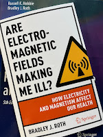
Are Electromagnetic Fields Making Me Ill? How Electricity and Magnetism Affect Our Health

Are Electromagnetic Fields Making Me Ill? by Brad Roth superimposed on Intermediate Physics for Medicine and Biology.
Are Electromagnetic Fields Making Me Ill?
How Electricity and Magnetism Affect Our Health
,
by Brad Roth
Big News! This week Springer published my new book: Are Electromagnetic Fields Making Me Ill? How Electricity and Magnetism Affect Our Health. This book is different from Intermediate Physics for Medicine and Biology: it’s short (122 pages), uses no math, and is aimed at a general audience. Readers of this blog may find parts of the book familiar; over the last couple years I’ve written posts that served as first drafts of some sections. Below is an excerpt from the Introduction.
This book is about electric and magnetic fields, and their effect on your body. We will examine the use of magnets for pain relief, the risk of power line magnetic fields, the safety of cell phones, and the possibility that microwave weapons are responsible for the Havana syndrome. Many medical treatments are based on electromagnetism, including well established ones like heart pacemakers and neural prostheses, and more questionable ones such as bone healing, transcutaneous electrical nerve stimulation, and transcranial direct current stimulation. Innumerable books and articles have been written about each of these topics; my goal in this book is to examine them together, to get the big picture.

This book is not a research monograph. It presents no original discoveries and makes no attempt to be comprehensive. Moreover, it omits numerous details and technicalities that experts often argue about. It does, however, try to offer an overall view of the field that is accurate.

My target readers are nonscientists: journalists, politicians, teachers, students, and anyone who has heard about electric and magnetic fields interacting with biological tissue and wants to learn more. I use no mathematics, avoid jargon, and employ abbreviations only when repeating the same mouthful of words over and over again becomes tedious. I tried my best to make the book understandable to a wide audience….

Sometimes the effect of electric and magnetic fields is controversial. For any debate, I have tried to present both sides. Nevertheless, readers will soon catch on that I’m a skeptic. Each chapter title is a question, of which my answer is usually “probably not” or “no.”
Here is the Table of Contents.
  1. Introduction 
  2. Can Magnets Cure All Your Ills? 
  3. Can a 9-Volt Battery Make You Smarter? 
  4. Do Power Lines Cause Cancer? 
  5. Will Electrical Stimulation Help Your Aching Back? 
  6. Is Your Cell Phone Killing You? 
  7. Did 5G Cell Phone Radiation Cause Covid-19? 
  8. Did Cuba Attack America with Microwaves? 
  9. Is That Airport Security Scanner Dangerous? 
  10. Conclusion

Although Russ Hobbie is not a coauthor on my new book, readers familiar with IPMB will see his influence on each page. In one of our last email exchanges before he passed away, I sent Russ an early draft of the book and he claimed to love it (that may have been Russ being kind, as he always was).

Enjoy!

Listen to me read the final chapter of Are Electromagnetic Fields Making Me Ill?

https://www.youtube.com/watch?v=5jJLkBsU4V0