Friday, January 17, 2020

Leonardo Da Vinci, Biological Physicist

Leonardo da Vinci, by Walter Isaacson, superimposed upon Intermediate Physics for Medicine and Biology.
Leonardo da Vinci,
by Walter Isaacson.
Leonardo da Vinci (1452 – 1519) is never mentioned in Intermediate Physics for Medicine and Biology, but his presence can be felt throughout. Over the Christmas break I listened to Walter Isaacson’s biography of da Vinci. He’s best known for his famous paintings such as The Last Supper and Mona Lisa. Yet, his accomplishments as a scientist are what tie him to IPMB.

I’m a big fan of Isaacson, and I enjoyed his biographies of Einstein and Jobs (his book about Franklin is on my to-read list). In his introduction, Isaacson describes why he chose to write about da Vinci.
I embarked on this book because Leonardo da Vinci is the ultimate example of the main theme of my previous biographies: how the ability to make connections across disciplines—arts and sciences, humanities and technology—is a key to innovation, imagination, and genius. Benjamin Franklin, a previous subject of mine, was a Leonardo of his era: with no formal education, he taught himself to become an imaginative polymath who was Enlightenment America’s best scientist, inventor, diplomat, writer, and business strategist… Albert Einstein, when he was stymied in his pursuit of his theory of relativity, would pull out his violin and play MozartAda Lovelace, whom I profiled in a book on innovators, combined the poetic sensibility of her father, Lord Byron, with her mother’s love of the beauty of math to envision a general-purpose computer. And Steve Jobs climaxed his product launches with an image of street signs showing the intersection of the liberal arts and technology. Leonardo was his hero.
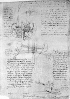
Drawings of blood vessels, by Leonardo da Vinci
Drawings of blood vessels,
by Leonardo da Vinci.
Credit: Wellcome Collection,
CC BY.
In Chapter 14 of IPMB, Russ Hobbie and I describe the many different medical imaging techniques used to study atherosclerosis: the narrowing of an artery. I learned from Isaacson that da Vinci was the first to understand this deadly disease. He figured it out during his autopsy of a man who claimed, just before died, that he was over one hundred years old.
In his quest to figure out how the centenarian died, Leonardo made a significant scientific discovery: he documented the process that leads to arteriosclerosis, in which the walls of arteries are thickened and stiffened by the accumulation of plaque-like substances. “I made an autopsy in order to ascertain the cause of so peaceful a death, and found that it proceeded from weakness through the failure of blood and of the artery that feeds the heart and the other lower members, which I found to be very dry, shrunken, and withered,” he wrote. Next to a drawing of the veins in the right arm, he compared the centenarian’s blood vessels to those of a two-year-old boy who also died at the hospital. He found those of the boy to be supple and unconstricted, “contrary to what I found in the old man.” Using his skill of thinking and describing through analogies, he concluded, “The network of vessels behaves in man as in oranges, in which the peel becomes tougher and the pulp diminishes the older they become.”
A photograph of the American Horse, inspired by da Vinci’s unfinished Horse sculpture, at Meijer Gardens in Grand Rapids, Michigan.
I’m standing in front of The American Horse,
inspired by da Vinci’s unfinished Horse sculpture,
at Meijer Gardens in Grand Rapids, Michigan.
One aspect of da Vinci’s career that I hadn’t appreciated before was how many of his projects were unfinished, including paintings such as the Adoration of the Magi and the Battle of Anghiari, as well as his Horse sculpture. Much of his scientific work was incomplete, or at least unpublished. An example was his collaborative research with Marcantonio della Torre, an anatomy professor at the University of Pavia.
Marcantonio died in 1511 of the plague that was devastating Italy that year. It is enticing to imagine what he and Leonardo could have accomplished. One of the things that could have most benefited Leonardo in his career was a partner who would help him follow through and publish his brilliant work. Together he and Marcantonio could have produced a groundbreaking illustrated treatise on anatomy that would have transformed a field still dominated by scholars who mainly regurgitated the notions of the second-century Greek physician Galen. Instead, Leonardo’s anatomy studies became another example of how he was disadvantaged by having few rigorous and disciplined collaborators along the lines of Luca Pacioli, whose text on geometric proportions Leonardo had illustrated. With Marcantonio dead, Leonardo retreated to the country villa of Francesco Melzi’s family to ride out the plague.
I think Isaacson lets da Vinci off the hook too easily. Leonardo needed some of Michael Faraday’s discipline to “Work, Finish, Publish.”

A drawing of the heart, by Leonardo da Vinci.
A drawing of the heart, by
Leonardo da Vinci.
Much of Chapter 7 in IPMB is about the heart. da Vinci contributed much to our understanding the heart’s anatomy.
Leonardo’s studies of the human heart, conducted as part of his overall anatomical and dissection work, were the most sustained and successful of his scientific endeavors. Informed by his love of hydraulic engineering and his fascination with the flow of liquids, he made discoveries that were not fully appreciated for centuries…

Leonardo was among the first to fully appreciate that the heart, not the liver, was the center of the blood system. “All the veins and arteries arise from the heart,” he wrote on the page that includes the drawings comparing the branches and roots of a seed with the veins and arteries emanating from the heart. He proved this by showing, in both words and a detailed drawing, “that the largest veins and arteries are found where they join with the heart, and the further they are removed from the heart, the finer they become, dividing into very small branches.” He became the first to analyze how the size of the branches diminish with each split, and he traced them down to tiny capillaries that were almost invisible. To those who would respond that the veins are rooted in the liver the way a plant is rooted in the soil, he pointed out that a plant’s roots and branches emanate from a central seed, which is analogous to the heart.

Leonardo was also able to show, contrary to Galen, that the heart is simply a muscle rather than some form of special vital tissue. Like all the muscles, the heart has its own blood supply and nerves. “It is nourished by an artery and veins, as are other muscles,” he found.
Self portrait, by Leonardo da Vinci.
Self portrait,
by Leonardo da Vinci.
One of the greatest contributions of physics and engineering to medicine is artificial heart valves. Again, this work builds on da Vinci’s discoveries, including his research on biomechanics and hydrodynamics.
Leonardo’s greatest achievement in his heart studies, and indeed in all of his anatomical work, was his discovery of the way the aortic valve works, a triumph that was confirmed only in modern times. It was birthed by his understanding, indeed love, of spiral flows. For his entire career, Leonardo was fascinated by the swirls of water eddies, wind currents, and hair curls cascading down a neck. He applied this knowledge to determining how the spiral flow of blood through a part of the aorta known as the sinus of Valsalva creates eddies and swirls that serve to close the valve of a beating heart…

Leonardo’s breakthroughs on heart valves were followed, however, by a failure: not discovering that the blood in the body circulates. His understanding of one-way valves should have made him realize the flaw in the Galenic theory, universally accepted during his time, that the blood is pulsed back and forth by the heart, moving to-and-fro. But Leonardo, somewhat unusually, was blinded by book learning. The “unlettered” man who disdained those who relied on received wisdom and vowed to make experiment his mistress failed to do so in this case. His genius and creativity had always come from proceeding without preconceptions. His study of blood flow, however, was one of the rare cases where he had acquired enough textbooks and expert tutors that he failed to think differently. A full explanation of blood circulation in the human body would have to wait for William Harvey a century later.
Vitruvian Man, by Leonardo da Vinci.
Vitruvian Man,
by Leonardo da Vinci.
I’ll let Isaacson sum up the moral of his story. It’s a lesson that is relevant for interdisciplinary scientists working at the intersection between physics and physiology, who draw connections between mathematics and medicine.
The fifteenth century of Leonardo and Columbus and Gutenberg was a time of inventions, exploration, and the spread of knowledge by new technologies. In short, it was a time like our own. That is why we have much to learn from Leonardo. His ability to combine art, science, technology, the humanities, and the imagination remains an enduring recipe for creativity. So, too, was his ease at being a bit of a misfit: illegitimate, gay, vegetarian, left-handed, easily distracted, and at times heretical. Florence flourished in the fifteenth century because it was comfortable with such people. Above all, Leonardo’s relentless curiosity and experimentation should remind us of the importance of instilling, in both ourselves and our children, not just received knowledge but a willingness to question it—to be imaginative and, like talented misfits and rebels in any era, to think different.
The Last Supper, by Leonardo da Vinci.
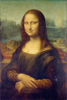
Mona Lisa, by Leonardo da Vinci.


Friday, January 10, 2020

Significant Advances in Computed Tomography

The journal Medical Physics recently published a virtual issue about “Significant Advances in Computed Tomography.” It’s accessible to all for free and is a wonderful resource for an instructor teaching a class based on Intermediate Physics for Medicine and Biology. Marc Kachelrieß, curator of the virtual issue, writes
It is now 40 years since Allan M. Cormack and Godfrey N. Hounsfield were jointly awarded the Nobel Prize in Physiology or Medicine for the development of computer assisted tomography, today known as computed tomography or simply as CT. Since its introduction in 1972 CT has become the most widespread and the most important tomographic medical imaging modality.

This inaugural virtual issue of the journal Medical Physics was created in honor of the 40th anniversary of Cormack and Hounsfield’s 1979 Nobel Prize. It is a compilation of the most significant original scientific papers on advances in CT that have been published in our journal. These papers have been selected among the most cited CT articles published in our journal so far, with a focus on clinical relevance. CAD [coronary artery disease] papers were not considered. If there were two or more papers on a similar topic that met all selection criteria the one that was published first was chosen.

This compilation reflects many important CT developments starting with Hounsfield’s Nobel award address on “Computed Medical Imaging” [cited in IPMB]. Some of the topics that are covered include basic image reconstruction technologies, spiral CT, cardiac CT, CBCT [cone beam CT], tube current modulation, 4D respiratory CT, dual-source dual-energy CT, and new technologies such as iterative image reconstruction as well as the future technology of photon counting detector CT.

Thus, this virtual issue provides the reader with an opportunity to reflect on the historical developments of CT and also to gain insights into the hot CT topics of today and of the near future.
Table of Contents:
These papers support and expand the discussion of computed tomography in Section 16.8 of Intermediate Physics for Medicine and Biology.

To learn more about this virtual issue, and about the history of computed tomography, listen to two videos by Cynthia McCollough, the president of the American Association of Physicists in Medicine


Cynthia McCollough introduces the virtual issue about 
“Significant Advances in Computed Tomography,
published by the journal Medical Physics

A video about the history of CT technology.

Friday, January 3, 2020

The Isaac Winners

Yesterday was the 100th anniversary of Isaac Asimov’s birth. Regular readers of this blog know that Asimov had a huge impact on my decision to become a scientist. Although his name never appears in Intermediate Physics for Medicine and Biology, his influence is on every page.

Adding a Dimension, by Isaac Asimov, superimposed on Intermediate Physics for Medicine and Biology.
Adding a Dimension,
by Isaac Asimov.
From 1959 to 1992, Asimov wrote a monthly essay for The Magazine of Fantasy & Science Fiction. Of all his writings, this series of essays was his favorite (and mine too). Each time he completed seventeen essays he would collect them in a book. One of these collections, Adding a Dimension, ended with an essay about his list of the ten greatest scientists.
The only scientist who, it seemed to me, indubitably belonged to the list and who would, without a doubt, be on such a list prepared by anyone but a consummate idiot, was Isaac Newton.

But how to choose the other nine?
Asimov needed a name for these awards.
I would be false to current American culture if I did not give the ten winners a named award… To go along with the Oscar, Emmy, Edgar, and Hugo, let us have the Isaac.
In a footnote, he added
If anyone has some wild theory that the choice of the name derives from any source other than Newton, let him try to prove it.
Below I list the Isaac winners in alphabetical order, and note which appear in Intermediate Physics for Medicine and Biology.
Half of the Isaac Award winners appear in Intermediate Physics for Medicine and Biology. Not bad.

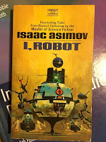
I, Robot, by Isaac Asimov, superimposed on Intermediate Physics for Medicine and Biology.
I, Robot, by Isaac Asimov.
In honor of Asimov’s centenary, yesterday I reread I, Robot, one of his best science fiction books. Delightful. I’ve read The Foundation Trilogy several times, and I’ve enjoyed his many short stories such as the classic “Nightfall.” I’m not sure how many Asimov books I’ve read, but probably on the order of a hundred.

If you want to learn more about Asimov, read the essay “Asimov at 100: From Epic Space Operas to Rules for Robots, the Prolific Author's Literary Legacy Endures,” by James Gunn. When I was an undergraduate at the University of Kansas, I took a science fiction class taught by Gunn; the topic of my term paper was Asimov’s future history.

I’ll close with the description of Asimov’s birth from In Memory Yet Green: The Autobiography of Isaac Asimov (his 200th book).
In Memory Yet Green: The Autobiography of Isaac Asimov, superimposed on Intermediate Physics for Medicine and Biology.
In Memory Yet Green:
The Autobiography of Isaac Asimov.
When my mother went into labor, there was no one to help her, therefore, but a midwife, and the process took three days and two nights, during much of which she walked the floor, leaning on my father. The result of all that was myself, and I was named Isaac after my mother’s dead father. (A Jewish child is, by tradition, named after a dead relative.)
The date of my birth, as I celebrate it, was January 2, 1920. It could not have been later than that. It might, however, have been earlier. Allowing for the uncertainties of the times, of the lack of records, of the Jewish and Julian calendars, it might have been as early as October 4, 1919. There is, however, no way of finding out. My parents were always uncertain and it really doesn’t matter.

I celebrate January 2, 1920, so let it be.
 Happy birthday, Isaac Asimov.

Listen to “Nightfall,” a short story by Isaac Asimov.

Friday, December 27, 2019

The Magnetic Field of an Axon: Ampere versus Biot-Savart

In Homework Problem 14 of Chapter 8 in Intermediate Physics for Medicine and Biology, Russ Hobbie and I ask the reader to calculate the magnetic field produced by the action current in a nerve axon using the law of Biot and Savart, and to compare it with the result found using Ampere’s law. This is a useful exercise, but I’ve always been uncomfortable with one aspect of the calculation. I’ll explain what I mean in today’s post.

In the homework problem you assume the intracellular current is uniform along one section of the axon, and is zero elsewhere (this is a big assumption, but it lets you derive an analytical solution). When you calculate the magnetic field using the law of Biot and Savart, you get a smooth, continuous function valid for any position along the axon. However, when you use Ampere’s law the result seems like it should be discontinuous. For some positions the intracellular current contributes to the current enclosed by the Amperian loop, but for other positions the intracellular current is zero and contributes nothing. How can the magnetic field be smooth and continuous if the intracellular current is discontinuous?

Below I’ll show you an elegant way to resolve this paradox. The bottom line is that the magnetic field you calculate using Ampere’s law is the same continuous function that you’d get using the law of Biot and Savart. I’ll change the details so that you don’t solve the homework problem in the book exactly, but the fundamental idea works for the book’s problem too.

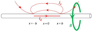
A uniform intracellular current I0 extending from x = -b to x = b, in a nerve axon.

Let the intracellular current be I0 for −b < x < b, and zero elsewhere, where x is the position along the axon (see the figure above). The axon is surrounded by saline with conductivity σ. The calculation consists of four steps: First calculate the extracellular voltage Ve in the saline, then differentiate Ve to find the x-component of the extracellular current density Jx, next integrate the current density across the area of the Amperian loop to get the return current Iret (that part of the extracellular current that passes through the loop), and finally determine the net current enclosed by the loop and calculate the magnetic field B.

Case 1: x > b

The current density and magnetic field surrounding a nerve axon; x>b.
Begin by calculating the magnetic field at point (x,y) where x > b, so you’re in the region where there is no intracellular current threading the Amperian loop (the green circle in the figure above, having radius r). To determine the extracellular voltage, realize that current crosses the membrane at only two locations: x = b (a positive point source when viewed from the extracellular space) and x = −b (a negative point source). The voltage produced by a point source is inversely proportional to the distance, so
A mathematical expression for the voltage in the saline surrounding a nerve axon.
(If you don’t follow how I derived this expression, see Section 7.1 in IPMB.)

To find the x-component of the current density, differentiate Ve with respect to x, multiple by σ, and add a minus sign.
A mathematical expression for the current density in the saline surrounding a nerve axon.
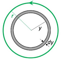
A drawing showing how to integrate the current density over the area of the Amperian loop to get the return current.

The most difficult part of the calculation is integrating the current density over the area enclosed by the loop to find the return current. This is a two-dimensional integral, with an area element of 2πy dy and limits of the integration from 0 to r (see the figure on the right).

You can look up the needed integral in an integral table, evaluate it at the limits, and fill in any missing steps. The result is

A mathematical expression for the return current through the Amperian loop.

The second term in the brackets is equal to minus one and the fourth term is equal to plus one, which cancel. There is no intracellular current for x > b, so the current enclosed by the loop is just the return current. The magnetic field is
A mathematical expression for the magnetic field produced by a nerve axon.
(I switched the order of the two surviving terms and brought the minus sign inside the bracket.) This is exactly the solution you get using the law of Biot and Savart; if you don’t believe me, calculate it yourself.

Case 2: −b < x < b

The current density and magnetic field surrounding a nerve axon; -b<x<b.
The calculation for the extracellular potential, current density, and return current in the region −b < x < b is exactly as before

A mathematical expression for the return current through the Amperian loop.

Now comes the interesting part. The second term in the brackets is not equal to minus one as it was earlier. Because x < b the numerator is negative, but the denominator is squared inside the square root so it is positive; the term becomes plus one. Because x > −b the fourth term is also equal to plus one, as before (both the numerator and denominator are positive). These two terms no longer cancel, so the return current becomes

A mathematical expression for the return current inside the Amperian loop. Because -b<x<b, the second and fourth terms no longer cancel, and the expression inside the brackets contains an extra term "+2".

This is different than we found for x > b. Don’t panic; remember that the total current enclosed by the loop is the return current plus the intracellular current. In this case, the intracellular current I0 exactly cancels the +2 term inside the brackets in the expression for Iret (remember, there is a minus one half in front of the brackets), so the enclosed current is just what we had for the x > b case, and the magnetic field is again

A mathematical expression for the magnetic field produced by a nerve axon.
The equation for the magnetic field is the same for any value of x (you can check the x < −b case yourself; you’ll get the same equation). The “magic” comes from the term (xb)/√(xb)2 switching from negative to positive, which is exactly what it had to do to cancel the intracellular current. The enclosed current is continuous even though the intracellular and return currents are not. The magnetic field calculated using Ampere’s law is a smooth function for all x, and is equivalent to the result obtained using the law of Biot and Savart (as it must be). Nice!

One limitation of this calculation is that the action potential has intracellular current only in one direction; a dipole. For an action potential propagating down an axon, the intracellular current first goes in one direction and then in another as the membrane depolarizes and then repolarizes; a quadrupole.

Two oppositely oriented dipoles of current along a nerve axon.
I’ll leave the calculation for this more complicated current distribution as an exercise for you. I suggest you do the calculation using both the Biot-Savart law and Ampere’s law. Enjoy!

Friday, December 20, 2019

This and That

Most of my blog posts are about a single topic related to Intermediate Physics for Medicine and Biology, but today’s post consists of a dozen brief notes. Read to the end for your Christmas gift.
  1. Previously in this blog, I’ve mentioned the website medicalphysicsweb.com. That site no longer exists, but was replaced by a page dedicated to medical physics on the Physics World website. Former medicalphysicsweb editor Tami Freeman is still in charge, and the new site is useful for instructors and students using IPMB. I get updates by email.
  2. I taught my Biological Physics class (PHY 3250) this fall at Oakland University, and videos of the class meetings are posted on Youtube. The quality is poor; often the blackboard is difficult to read. But if you want to see how I teach the first half of IPMB, take a look.
  3. On the Wednesday before Thanksgiving, my class played Trivial Pursuit IPMB. The students had fun and earned extra credit. Earlier this year my wife and I bought two Trivial Pursuit games—complete with game boards and pieces—at a garage sale for a couple dollars, so I was able to accommodate twelve students. You can download the questions at the IPMB homepage
  4. In 2012 I wrote about the website iBioMagazine. I no longer can find it, but I believe the website iBiology is related to it. I recommend iBiology for physics students trying to improve their knowledge of biology.
  5. Today The Rise of Skywalker opens. It’s the final episode in the Star Wars trilogy of trilogies. I remember watching the first Star Wars movie as a teenager in 1977; I can’t wait to see the latest.
  6. From the Oakland University campus you can see the Headquarters and Tech Center of Fiat Chrysler Automobiles. This fall the folks at Chrysler introduced a new advertising blitz called the Dodge Horsepower Challenge. Each week they presented a new physics problem about cars, and those who answered correctly were entered in a drawing for a 2019 Dodge Challenger SRT Hellcat Redeye. They needed a physicist to review their problems and solutions, and somehow I got the job. You can find the problems on Youtube, presented by a colorful wrestler named Goldberg.
  7. Lately I’ve been republishing these blog posts on medium.com. Oddly, among my most popular stories on Medium is the one about the Fourier series of the cotangent. It has over 160 reads while others that I think are better have just a handful.
  8. The Blogger software keeps its own statistics, and claims that my most popular post is about Frank Netter, with over 6000 page views. I think its popularity has to do with Search Engine Optimization.
  9. The IPMB Facebook page now has over 200 members. Thanks everyone, and let’s try to finish 2020 with 220.
  10. Regular readers know that my two favorite authors are Isaac Asimov and Charles Dickens. Recently I’ve discovered another: P. G. Wodehouse. His books about Bertie Wooster and Jeeves are hilarious, and a joy to read.
  11. If you want to know what books I’m reading, you can follow my Goodreads account. Often books in the category Read More Science become subjects of blog posts.
  12. Finally, here’s your Christmas present. Last year Oakland University Professor Andrea Eis organized an event—called Encountering the Rare Book—to highlight the OU Kresge Library’s special collections. I was one of the faculty members Andrea asked to select a book from the collection and write a brief essay about it. I chose A Christmas Carol and my essay is below. A Merry Christmas to you all.
I read A Christmas Carol every December, so I was delighted to find a first edition of Charles Dickens’ classic novella in the Rare Book Collection of Kresge Library. I never tire of Dickens’ “ghostly little book.” I love his language, humor, and wonderfully drawn characters.

I enjoy the Ghosts of Christmas Past and Present best; the Ghost of Christmas Yet to Come frightens me. One of my favorite scenes is when Scrooge’s nephew Fred and the Ghost of Christmas Present collude with Topper to catch Fred’s sister-in-law (the plump one with the lace tucker) during a game of blind man’s bluff. I’m a cheapskate focused on my work, so I have a certain sympathy for Ebenezer. I read the book each year as a reminder to not become a “tight-fisted hand at the grindstone.”

All of us in higher education ought to recall the words of the Ghost of Christmas Present at the end of Stave 3, as he revealed two wretched children hidden in his robes: “This boy is Ignorance. This girl is Want. Beware them both…but most of all beware this boy.”

I sometimes wonder if I should have been born a Victorian. I love their physics—Faraday, Maxwell, and Kelvin are my heroes—as well as their literature. A Christmas Carol was published in 1843, the same year that James Joule measured the mechanical equivalent of heat, George Stokes analyzed incompressible fluids, and Ada Lovelace wrote the first computer program. Holding a first edition in your hands connects you to that time; as if Dickens, like Marley’s Ghost, “sat invisible beside you.” The library’s copy has lovely illustrations, which at that time had to be painstakingly hand-colored.

I intend to continue reading A Christmas Carol each year, with the hope that I, like Scrooge, can “become as good a friend, as good a master, and as good a man, as the good old city knew.”
Ecountering the Rare Book, an exhibition celebrating the Special Collections in Kresge Library at Oakland University, organized by Andrea Eis, superimposed on Intermediate Physics for Medicine and Biology.
Encountering the Rare Book, an exhibition celebrating
the Special Collections in Kresge Library at
Oakland University, organized by Andrea Eis.

Friday, December 13, 2019

How Russ Hobbie Came to Write Intermediate Physics for Medicine and Biology

In the preface of Intermediate Physics for Medicine and Biology, Russ Hobbie writes
Between 1971 and 1973 I audited all the courses medical students take in their first 2 years at the University of Minnesota.

I was amazed at the amount of physics I found in these courses and how little of it is discussed in the general physics course. I found a great discrepancy between the physics in some papers in the biological research literature and what I knew to be the level of understanding of most biology majors or premed students who have taken a year of physics. It was clear that an intermediate level physics course would help these students. It would provide the physics they need and would relate it directly to the biological problems where it is useful.

This book is the result of my having taught such a course since 1973…
Want to hear more about how Russ came to write IPMB? You can! Russ was interviewed for the University of Minnesota Oral History Project. Below is an excerpt about the origin of the book.
Interview with Russell Hobbie
Interviewed by Professor Clarke A. Chambers. University of Minnesota
Interviewed on September 29, 1994. University of Minnesota Campus

…I wrote Al Sullivan who was the assistant dean of the Medical School asking if it was possible to snoop around over there. Al asked me to have lunch with him one day—it was in October—and said, “What you really ought to do is to attend Medical School.” I said, “I can’t. I’m director of undergraduate studies in Physics. I’m teaching a full load, which is a course each quarter. There’s just no time to do that.” He said, “You could just audit things and skip the labs.” So, for two years, I did that….

I sat through the remainder of the year in embryology, and biochemistry, and anatomy, and pathology, and physiology, and then, in the second year, the organ systems, the neuro psych, the cardiovascular, the pulmonary, the renal, the dermatology, the bones, the GI [gastrointestinal] ….

I really got a fairly good knowledge there and found that there was just too much physics ever to fit it into the pre-med physics course. I also found that there was a tremendous gap between what we teach the pre-meds, who will take one year of physics and that’s it, and what you found in the physiology and biophysics research literature. I convinced the Physics Department that I ought to try teaching a course to try to fill that gap, a 5000 level course that has a year of general physics and a year of calculus as a prereq[uisite] that would appeal to the physiologists and so on. Probably around 1972 or 1973, I started teaching that course, developing it as I went. That turned into a book [Intermediate Physics for Medicine and Biology] that was published by Wiley in 1978 with a second edition about 1988. I’m trying, without much success, to do a third edition right now….

…after I started teaching the course, I can remember Professor Jack Johnson from Physiology wanted to come and sit it in; and I was quite nervous about this because I was afraid I might get some of the physiology wrong. He reminded me, in no uncertain terms, that I’d been sitting through his course and turnabout really was fair play…

I think that, as I look back at my own career, the thing that I think that was most important, that has certainly given me the greatest intellectual satisfaction is the book.
If you can’t get enough of Russ, watch him in this video about his computer program MacDose.

Russell Hobbie demonstrates MacDose, part 1.

Russell Hobbie demonstrates MacDose, part 2.

Russell Hobbie demonstrates MacDose, part 3.

Friday, December 6, 2019

The Dimensionality of Color Vision in Carriers of Anomalous Trichromacy

Russ Hobbie and I discuss color vision in Chapter 14 of Intermediate Physics for Medicine and Biology.
14.15 Color Vision
The eye can detect color because there are three types of cones in the retina, each of which responds to a different wavelength of light (trichromate vision): red, green, and blue, the primary colors...
From Photon to Neuron: Light, Imaging, Vision, by Philip Nelson, superimposed on Intermediate Physics for Medicine and Biology.
From Photon to Neuron:
Light, Imaging, Vision,
by Philip Nelson.
Imagine my shock when I read about possible tetrachromate vision in Philip Nelson’s book From Photon to Neuron. I downloaded the article Phil cited—“The Dimensionality of Color Vision in Carriers of Anomalous Trichromacy,” by Gabriele Jordan, Samir Deeb, Jenny Bosten, and John Mollon, Journal of Vision, Volume 10, doi:10.1167/10.8.12, 2010—and quote the abstract below.
Some 12% of women are carriers of the mild, X-linked forms of color vision deficiencies called “anomalous trichromacy.” Owing to random X chromosome inactivation, their retinae must contain four classes of cone rather than the normal three; and it has previously been speculated that these female carriers might be tetrachromatic, capable of discriminating spectral stimuli that are indistinguishable to the normal trichromat. However, the existing evidence is sparse and inconclusive. Here, we address the question using (a) a forced-choice version of the Rayleigh test, (b) a test using multidimensional scaling to reveal directly the dimensionality of the participants' color space, and (c) molecular genetic analyses to estimate the X-linked cone peak sensitivities of a selected sample of strong candidates for tetrachromacy. Our results suggest that most carriers of color anomaly do not exhibit four-dimensional color vision, and so we believe that anomalous trichromacy is unlikely to be maintained by an advantage to the carriers in discriminating colors. However, 1 of 24 obligate carriers of deuteranomaly exhibited tetrachromatic behavior on all our tests; this participant has three well-separated cone photopigments in the long-wave spectral region in addition to her short-wave cone. We assess the likelihood that behavioral tetrachromacy exists in the human population.
Flatland: A Romance of Many Dimensions, by Edwin A. Abbott, superimposed on Intermediate Physics for Medicine and Biology.
Flatland: A Romance of Many Dimensions,
by Edwin A. Abbott. If you haven’t
read Flatland, ask Santa for a copy
this Christmas (or click on this link).
Wow! IPMB claims that “other animals...[can] have more than three types [of cones]” but offers no hint that people can. How cool is that? This is like finding someone who lives in a four-dimensional world. Would a tetrachromat explaining color to me (a trichromat) be like Square in Flatland describing a sphere to the Triangles? (Square was thrown in prison for that!) What would life be like with four color receptors (red, green, blue, and orange) instead of three? Would you perceive a fundamentally different world, or would any difference be subtle? Could we use CRISPR or some other gene editing tool to expand our color vision? (I’ll take a dozen different cones, please.) Is it fair that only women can be tetrachromats? (No, but let’s not go there.) Is tetrachromacy a superpower?

San Diego woman Concetta Antico diagnosed with “super vision.”
I don’t know how accurate this news story is, but it’s interesting.

Friday, November 29, 2019

The Rayl

Section 13.3 of Intermediate Physics for Medicine and Biology discusses acoustic impedance. For an ultrasonic wave the acoustic impedance is the pressure divided by the tissue velocity, so it has units of Pa/(m/s). In terms of kilograms, meters, and seconds, the units of acoustic impedance are kg/(m2 s).

Acoustic impedance is analogous to electrical impedance. Voltage over current is like pressure over velocity. Electromagnetic waves propagating through a transmission line reflect when the electrical impedance changes, just as ultrasonic waves propagating through tissue reflect when the acoustic impedance changes. The unit for electrical impedance is the ohm, and the unit for acoustic impedance is the…

Wait! What is the name of the unit for acoustic impedance? According to IPMB the units of acoustic impedance are kg/(m2 s). It has no name. The newton is a kg m/s2, the joule is a kg m2/s2, the pascal is a kg/(m s2), and the watt is a kg m2/s3. Why is there no name for the kg/(m2 s)?

There is a name. A kg/(m2 s) is called a rayl. It’s pronounced “rail.” I quote the last lines of Section 13.3.1 in IPMB, but using the rayl.
The quantity Z = ρ0c = √ρ0 κ is called the acoustic impedance of the medium [ρ0 is the density, c is the speed of sound, and κ is the compressibility]. The acoustic impedance of water is about (103 kg m-3)(1400 m s-1) = 1.4 × 10⁶ rayl, or 1.4 Mrayl. The acoustic impedance of air is about 400 rayl, so Zair is much less than Zwater (Denny 1993).
The rayl is also the name for a g/(cm2 s). But the rayl in the meter-kilogram-second system of units isn’t the same as the rayl in the centimeter-gram-second system. A CGS rayl is equal to ten MKS rayls. Maybe all this confusion is why no one uses the rayl (including IPMB). My vote is to abolish the CGS rayl. Erase it from history. Make it taboo. Let’s restrict the use of the term rayl to the MKS rayl.

Theory of Sound, by Lord Rayleigh, superimposed on Intermediate Physics for Medicine and Biology.
The Theory of Sound,
by Lord Rayleigh.
The unit is named after John William Strutt, better known as Lord Rayleigh (pronounced ray-lee). He is known for Rayleigh waves, Rayleigh scattering, Rayleigh-Benard convection, the Rayleigh criterion, the Rayleigh-Taylor instability, the Rayleigh-Ritz method, and the Rayleigh-Jeans law. His influential textbook The Theory of Sound makes him the logical choice for the unit of acoustic impedance. He won the Nobel Prize in 1904 “for his investigations of the densities of the most important gases and for his discovery of argon in connection with these studies.”

Below are excerpts from Rayleigh’s Nobel biography.
John William Strutt, third Baron Rayleigh, was born on November 12, 1842 at Langford Grove, Maldon, Essex

Throughout his infancy and youth he was of frail physique; his education was repeatedly interrupted by ill-health, and his prospects of attaining maturity appeared precarious. After a short spell at Eton at the age of 10, mainly spent in the school sanatorium, three years in a private school at Wimbledon, and another short stay at Harrow, he finally spent four years with the Rev. George Townsend Warner (1857) who took pupils at Torquay.

In 1861 he entered Trinity College, Cambridge, where he commenced reading mathematics, not at first equal in attainments to the best of his contemporaries, but his exceptional abilities soon enabled him to overtake his competitors. He graduated in the Mathematical Tripos in 1865 as Senior Wrangler and Smith’s Prizeman. In 1866 he obtained a fellowship at Trinity which he held until 1871, the year of his marriage.

A severe attack of rheumatic fever in 1872 made him spend the winter in Egypt and Greece. Shortly after his return his father died (1873) and he succeeded to the barony, taking up residence in the family seat, Terling Place, at Witham, Essex… In 1876 he left the entire management of the land to his younger brother.

From then on, he could devote his full time to science again. In 1879 he was appointed to follow James Clerk Maxwell as Professor of Experimental Physics and Head of the Cavendish Laboratory at Cambridge. In 1884 he left Cambridge to continue his experimental work at his country seat at Terling, Essex, and from 1887 to 1905 he was Professor of Natural Philosophy in the Royal Institution of Great Britain, being successor of Tyndall

Lord Rayleigh’s first researches were mainly mathematical, concerning optics and vibrating systems, but his later work ranged over almost the whole field of physics, covering sound, wave theory, colour vision, electrodynamics, electromagnetism, light scattering, flow of liquids, hydrodynamics, density of gases, viscosity, capillarity, elasticity, and photography… His Theory of Sound was published in two volumes during 1877-1878, and his other extensive studies are reported in his Scientific Papers – six volumes issued during 1889-1920…

He had a fine sense of literary style; every paper he wrote, even on the most abstruse subject, is a model of clearness and simplicity of diction. The 446 papers reprinted in his collected works clearly show his capacity for understanding everything just a little more deeply than anyone else…

Lord Rayleigh… was a Fellow of the Royal Society (1873) and served as Secretary from 1885 to 1896, and as President from 1905 to 1908. He was an original recipient of the Order of Merit (1902), and in 1905 he was made a Privy Councillor. He was awarded the Copley, Royal, and Rumford Medals of the Royal Society, and the Nobel Prize for 1904…

Lord Rayleigh died on June 30, 1919, at Witham, Essex.
Rayleigh has always been a hero of mine. The breadth of his contributions, the quality of his writing, the ability to excel at both theory and experiment, and the service he contributed to his profession amaze me. I’m glad Russ Hobbie and I discuss some of his contributions in Intermediate Physics for Medicine and Biology.

John William Strutt, Lord Rayleigh (1842-1919).


Lord Rayleigh by Peter Wells, Cardiff University

Friday, November 22, 2019

Pocket Ultrasound

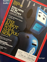
The November 4, 2019 issue of TIME magazine, about health innovation, superimposed on Intermediate Physics for Medicine and Biology.
The November 4, 2019 issue of
TIME magazine, about health innovation.

For decades I’ve been a loyal subscriber to TIME magazine. I read it—more or less cover-to-cover—every week. Most issues have little overlap with Intermediate Physics for Medicine and Biology, but the November 4, 2019 issue is an exception; it focuses on health innovation.

My favorite of the featured innovators is Jonathan Rothberg, who’s developed a handheld ultrasound imager. His device looks like an electric shaver and would fit in your pocket. Don Steinberg writes in TIME 
Jonathan Rothberg, a Yale genetics researcher and serial entrepreneur, figured out how to put ultrasound technology on a chip, so instead of a $100,000 machine in a hospital, it’s a $2,000 go-anywhere gadget that connects to an iPhone app.
On his website, Rothberg says his aim is to make healthcare accessible to everyone around the world. A noble goal, but what’s unique about the physics of Rothberg’s invention? In Chapter 13 of Intermediate Physics for Medicine and Biology, Russ Hobbie and I discuss traditional ultrasound transducers.
Ultrasound is typically produced using a piezoelectric transducer. A piezoelectric material converts a stress (or pressure) into an electric field, and vice versa. A high-frequency oscillating voltage applied across a piezoelectric material creates a sound wave at the same frequency. Conversely, an oscillating pressure applied to a piezoelectric material creates an oscillating voltage across it. Measurement of this voltage provides a way to record ultrasonic waves. Thus, the same piezoelectric material can serve as both source and detector.
According to an article by Eliza Strickland in IEEE Spectrum, Rothberg’s ultrasound device, called the Butterfly iQ, does away with the piezoelectricity.
Today’s ultrasound systems use piezoelectric crystals, which convert electrical energy into vibrations in the form of ultrasonic waves. A typical system has a display screen on a bulky cart with several wands for imaging at different depths within the body. These machines can cost upwards of $100,000…

Developing the iQ’s chip-based technology was a two-step process. First, Butterfly’s engineers replaced the piezoelectrics with a micromachine that acts like a tiny drum to generate vibrations. Inside this “capacitive micromachined ultrasound transducer” (CMUT), an applied voltage moves a membrane to send ultrasonic waves into the body. The waves that bounce back from various body tissues move the membrane and are registered as an electric signal, which creates the image…

Rothberg explains that typical ultrasound systems require separate probes for different clinical applications because the crystals have to be tuned at the time of manufacture to produce the right type of ultrasonic wave for imaging at a particular depth. But the Butterfly iQ can be tuned on the fly. “We have 10,000 of these micromachine transducers on a probe, and that gives us a monster dynamic range,” he says. "We can make them buzz at 1 megahertz if we want to go deep, or 5 megahertz if we want to go shallow.”

The second innovation was to do away with the wiring that connects a typical piezoelectric probe to the electronic controls and displays. Butterfly’s micromachines are attached directly to a semiconductor layer that contains all the necessary amplifiers, signal processors, and so on.
I’m not expert enough to judge how revolutionary this device really is, but it sure sounds cool! One thing I know for certain: when you mix physics with medicine, the results can be astounding. 

The three issues I need to read to catch up with TIME magazine, superimposed on Intermediate Physics for Medicine and Biology.
Three issues behind in reading TIME magazine;
I've got my work cut out for me.
Thank goodness Christmas break is approaching, because I’m three issues behind with TIME. I try to keep up, I really do; it’s a challenge. But you never know what you’ll find when you open a new issue. Sometimes you find physics applied to medicine and biology!


Listen to Jonathan Rothberg talk about his ultrasound device, Butterfly iQ.
https://www.youtube.com/watch?v=OHZTUFLIMjU 

Jonathan Rothberg, 2013 National Medal of Technology and Innovation.
https://www.youtube.com/watch?v=hVV80xl-rNU