Showing posts with label obituary. Show all posts

Friday, November 15, 2019

Thomas Tenforde (1940-2019) and The Wonders of Magnetism

A photograph of Thomas Tenforde (1940-2019).
Thomas Tenforde (1940-2019).
Photo used with permission of the
Health Physics Society.
Thomas Tenforde—an expert on the interaction of magnetic fields with biological tissue—died this fall.

When I was looking for employment in the late 1980s just after getting my PhD, I had a fellowship opportunity at the Pacific Northwest National Laboratory in Richland, Washington, where Tom Tenforde worked. At that time a debate raged about the health risks of 60-Hz power-line fields, and I recall having much respect for Tenforde’s research on this topic; his work seemed more rigorous and physics-based than many other studies. I ended up taking a position at the National Institutes of Health, but I seriously considered working for Tenforde.

When Russ Hobbie and I discuss power-line electric and magnetic fields in Intermediate Physics for Medicine and Biology, we cite a review by Tenforde.
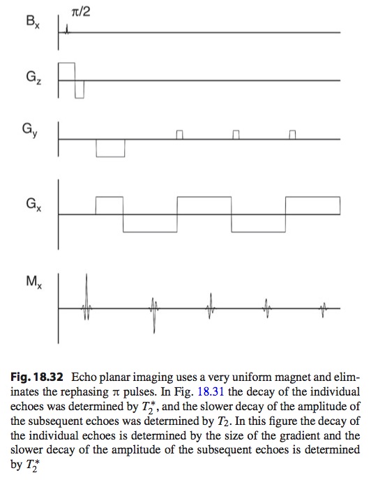
9.10.2  Power Frequency (50-60 Hz) Fields
9.10.2.1 Fields in Homes are Weak

Much weaker fields in homes are produced by power lines, house wiring, and electrical appliances. Barnes (1995) found average electric fields in air next to the body of about 7 V m−1, with peak values of 200 V m−1. (We will find that since the body is a conductor, the fields within the body are much less.) Average residential magnetic fields are about 0.1 μT, with peaks up to four times as large. Within the body they are about the same. Tenforde (1995) reviews both power-line and radio-frequency field intensities.
Electromagnetic Fields: Biological Interactions and Mechanisms, edited by Martin Blank, superimposed on Intermediate Physics for Medicine and Biology.
Electromagnetic Fields:
Biological Interactions and Mechanisms
,
edited by Martin Blank.
The reference is to
Tenforde TS (1995) Spectrum and intensity of environmental electromagnetic fields from natural and man-made sources. In: Blank M (ed.) Electromagnetic Fields: Biological Interactions and Mechanisms. American Chemical Society, Washington, DC, pp 13–35.
An obituary for Tenforde published online by the Health Physics Society begins
Thomas S. Tenforde died on 6 September 2019 in Berkeley, California, at the age of 78. He was born in Middletown, Ohio, on 15 December 1940. He received his BA degree from Harvard University with a major in physics. He earned a PhD in biophysics at the University of California at Berkeley. He was a senior scientist at the Lawrence Berkeley National Laboratory from 1969 to 1988. He moved to Richland, Washington, where he was a fellow at Battelle's Pacific Northwest National Laboratory from 1988 to 2002. His special areas of research included health effects of nonionizing radiation and the role of radionuclides for medical applications. He received the dArsonval Medal from the Bioelectromagnetics Society in 2001. He also received awards from the US Department of Energy in 2000 and the Federal Laboratory Consortium in 2001 for leading the development of 90Y as a therapeutic medical isotope that is used worldwide for the treatment of cancer and other medical disorders...
A photograph of Jacques-Arsène d'Arsonval (1851-1940).
Jacques-Arsène dArsonval
(1851-1940).
Photo from Wikipedia.
The d’Arsonval Medal is the most prestigious award bestowed by the Bioelectromagnetics Society. It’s named after French physician and physicist Jacques-Arsène d’Arsonval, a pioneer in electrophysiology. (Two scientists mentioned in last week's postEleanor Adair and Ken Foster—also were awarded the d'Arsonval Medal.) Tenforde’s award announcement states
Thomas S. Tenforde, senior chief scientist in the environmental technology division, Pacific Northwest National Laboratory, Richland, Wash., will receive the eighth dArsonval Award, presented by the Bioelectromagnetics Society to recognize “extraordinary accomplishment within the discipline of bioelectromagnetics.” This award recognizes Tenforde’s extensive research on dosimetry and biophysical interaction of static and low-frequency electric and magnetic fields with living systems….
Tenforde’s strong interest in bioelectromagnetics began with the use of static electric fields for single-cell micro-electrophoresis during his doctoral thesis work. In the 1970s and 1980s at Lawrence Berkeley National Laboratory he conducted a broad range of biological studies on static and ELF [extremely low frequency] magnetic fields. Tom and his colleagues at the Donner Laboratory at the University of California developed what soon became the foremost program investigating the biological effects of strong static magnetic fields. These studies looked at the cardiovascular system, the nervous system, thermoregulation, circadian rhythmicity, lipid bilayer membrane permeability, and animal behavior.
This work initially began because of concerns about human exposure to strong magnetic fields near thermonuclear fusion reactors, magneto-hydrodynamic power systems, and high-energy physics facilities such as cyclotrons and bubble chambers. Tom and his colleagues played a key role in the evaluation of potential risks to patients and workers from MRI facilities…

As manager of PNNL’s Hanford Radioisotopes Program, Tenforde supervised work which produced the medical isotope yttrium-90, which is now being used worldwide to treat cancer…
Tenforde provides yet another example of a scientist who made the transition from physics to biology. His career indicates how working at this interface can lead to new insights and great accomplishments. His interest in how magnetic fields affect animals overlaps with my work on the magnetic field of a nerve axon, magnetic stimulation and magnetoacoustic imaging. I think he would have enjoyed Chapter 8 of Intermediate Physics for Medicine and Biology, about biomagnetism.

I will let Tenforde have the last word. In an article based on his speech accepting the d’Arsonval Medal (Bioelectromagnetics, Volume 24, Pages 3-11, 2003), he wrote
A recurring theme of my work during the past 25 years has been the beneficial uses of magnetism in advancing our scientific knowledge of living systems, and hence I have chosen the title, “The Wonders of Magnetism.”

Friday, October 26, 2018

Earl Bakken (1924-2018)

Earl Bakken (1924-2018)
Earl Bakken. From the Bakken Museum, via wikipedia.
Earl Bakken, cofounder of the medical device company Medtronic, died Sunday at the age of 94. In 1957 he developed the first external, battery-operated artificial pacemaker. Russ Hobbie and I don’t mention Bakken by name in Intermediate Physics for Medicine and Biology, but we do discuss pacemakers.
Cardiac pacemakers are a useful treatment for certain heart diseases. The most frequent are an abnormally slow pulse rate (bradycardia) associated with symptoms such as dizziness, fainting (syncope), or heart failure. These may arise from a problem with the SA node (sick sinus syndrome) or with the conduction system (heart block). One of the first uses of pacemakers was to treat complete or third degree heart block. The SA node and the atria fire at a normal rate but the wave front cannot pass through the conduction system. The AV node or some other part of the conduction system then begins firing and driving the ventricles at its own, pathologically slower rate…. A pacemaker stimulating the ventricles can be used to restore a normal ventricular rate.
In the 1950s, famed cardiac surgeon Dr. Walt Lillehei was at the University of Minnesota, where he met Bakken. Kirk Jeffrey tells the story of their collaboration in Machines In Our Hearts.
Machines In Our Hearts, by Kirk Jeffrey
The myocardial pacing wire was the first electrical device ever to be implanted in the human body and left there for a period of time. Surgeons at Minnesota were now able to pace children for days or weeks after heart surgery. By October 1957, they had used the technique with 18 patients. But Lillehei now grew uneasy about the Grass stimulator because it was bulky and plugged into the electrical system. The surgeon wanted to get his heart patients out of bed and moving around, but the stimulator had to accompany them on a wheeled cart. The electrical cord was a further nuisance. ‘‘Many of these [patients] were kids. They wanted to wander around and get active. Well, they were active. They couldn’t go any further than the cord. We had to string wires down the hall. . . . And then, if they needed an X ray or something that couldn’t be done in the room, you couldn’t get on the elevator so you had to string them down the stairwells. It seemed that almost everything you wanted was on a different floor. We needed something battery-operated.’’

From Machines In Our Hearts.
The plug-in stimulator was more than an inconvenience, for by introducing the myocardial pacing wire, Lillehei and his associates had connected the hearts of their surgical patients to the 110-volt electrical system of the hospital. Everyone in the program knew that an electrical surge might send patients into ventricular fibrillation or that a power outage could leave them without pacemaker support. On October 31, 1957, an equipment failure at a large Twin Cities power plant caused an outage lasting nearly three hours in Minneapolis. The University hospital had auxiliary power in its surgical suites and recovery area, but not in patients’ rooms. None of his heart patients died—but Lillehei viewed the event as a warning. Lillehei…turned to Earl Bakken…a young engineer who owned a small medical electronics business called Medtronic and repaired and serviced equipment for the Department of Surgery.

Bakken…realized that he could simply build a stimulator that used transistors and small batteries. ‘‘It was kind of an interesting point in history,’’ he recalled—‘‘a joining of several technologies.’’ In constructing the external pulse generator, Bakken borrowed a circuit design for a metronome that he had noticed a few months earlier in an electronics magazine for hobbyists. It included two transistors. Invented a decade earlier, the transistor was just beginning to spread into general use in the mid-1950s. Hardly anyone had explored its applications in medical devices. Bakken used a nine-volt battery, housed the assemblage in an aluminum circuit box, and provided an on-off switch and control knobs for stimulus rate and amplitude.

At the electronics repair shop that he had founded with his brother-in-law in 1949, Bakken had customized many instruments for researchers at the University of Minnesota Medical School…When Bakken delivered the battery-powered external pulse generator to Walt Lillehei in January 1958, it seemed to the inventor another special order, nothing more. The pulse generator was hardly an aesthetic triumph, but it was small enough to hold in the hand and severed all connection between the patient’s heart and the hospital power system. Bakken’s business had no animal-testing facility, so he assumed that the surgeons would test the device by pacing laboratory dogs. They did ‘‘a few dogs,’’ then Lillehei put the pacemaker into clinical use. When Bakken next visited the university, he was surprised to find that his crude prototype was managing the heartbeat of a child recovering from open-heart surgery.
The Bakken Museum. From wikipedia.
Russ Hobbie is retired from the University of Minnesota, and still lives in the area, so he is particularly familiar with Earl Bakken. He served on the board of the Bakken Museum, which is devoted to bioelectricity (my kind of museum). Russ say he ‘‘was impressed by Bakken's vision, energy, and desire to help people. The Bakken Museum has an extensive outreach program which does a lot of good things.’’ They recently posted a statement honoring their founder.

Medtronic is one of the largest medical device companies. I had a job interview there years ago, but I didn’t get the position. I came away impressed by the company, and wish I had bought stock.

Bakken is a member of the dwindling greatest generation; he was an airborne radar maintenance instructor during World War II. He had a long, full life, and we will miss him.

Below are a couple of videos about Earl Bakken. Enjoy!


Friday, February 9, 2018

Suki Roth (2002-2018)

Intermediate Physics for Medicine and Biology: Suki Roth (2002-2018)
Suki Roth (2002-2018)
Suki Roth (2002-2018).
Regular readers of this blog are familiar with my dog Suki, who I’ve mentioned in more than a dozen posts. Suki passed away this week. She was a wonderful dog and I miss her dearly.

Suki and I used to take long walks when I would listen to audio books, such as The Immortal Life of Henrietta Lacks, Musicophilia, Destiny of the Republic, Galileo’s Daughter, and First American: The Life and Times of Benjamin Franklin. This list just scratches the surface. On my Goodreads account, I have a category called “listened-to-while-dog-walking” that includes 84 books, all of which Suki and I enjoyed together. 

Me holding Suki in the forest in Michigan among the fall color.In my post about the Physics of Phoxhounds, I mentioned that a photo of Suki and me (right) was included in Barb Oakley’s book A Mind for Numbers: How to Excel at Math and Science (Even if You Flunked Algebra). Recently I learned that Barb’s book has sold over 250,000 copies, making Suki something of a celebrity.

Suki Roth next to the textbook Intermediate Physics for Medicine and Biology.Suki helped me explain concepts from Intermediate Physics for Medicine and Biology, such as age-related hearing loss and the biomechanics of fleas. Few people knew that she had this secret career in biomedical education!

Thanks to Dr. Kelly Totin, and before her Dr. Ann Callahan, and all the folks at Rochester Veterinary Hospital for taking such good care of Suki. In particular I appreciate Dr. Totin’s help during Suki’s last, difficult days. As she said near the end, her focus was on the quality of Suki’s time left rather than the quantity; an important life lesson for us all.

I’ll close with a quote from one of my favorite authors, James Herriot. In his story “The Card Over The Bed,” the dying Miss Stubbs asks Herriot, a Yorkshire vet, if she will see her pets in heaven. She was worried because she had heard claims that animals have no soul. Herriot responded “If having a soul means being able to feel love and loyalty and gratitude, then animals are better off than a lot of humans. You’ve nothing to worry about there.”

Suki Roth resting in her bed.
Suki resting.

Suki Roth with her nephew Auggie, a foxhound.
Suki with her nephew Auggie.

Suki Roth with all five editions of Intermediate Physics for Medicine and Biology.
Suki with all five editions of IPMB.

Suki Roth (right), her niece Smokie Roth (the Greyhound, center), and her nephew Auggie Roth (the Foxhound, left), about to get treats from my wife Shirley.
Suki (right), her niece Smokie (the Greyhound, center),
and her nephew Auggie (the Foxhound, left),
about to get treats from my wife Shirley.

Suki and me, 15 years ago.
Suki Roth as a puppy.
Young Suki

Friday, October 13, 2017

John Clark, Biomedical Engineer (1936-2017)

The first page of an article by Clark and Plonsey about the extracellular potential of a nerve axon.
John W. Clark passed away on August 6, in Houston, Texas. He was a professor of Engineering at Rice University for 49 years.

When I was a graduate student at Vanderbilt University in the 1980s, I was influenced by the papers of Robert Plonsey and his graduate student Clark. They calculated the extracellular electrical potential outside a nerve axon from the transmembrane action potential by expressing the transmembrane potential in terms of its Fourier transform, and then using Bessel functions to calculate the Fourier transform of the extracellular potential. Russ Hobbie and I outline this technique in Problem 30 of Chapter 7 in Intermediate Physics for Medicine and Biology. James Woolsey, my PhD advisor John Wikswo, and I used a similar method—inspired by Clark and Plonsey’s work—to calculate the magnetic field of a nerve axon (see Problem 16 of Chapter 8 in IPMB). Moreover, my first work on the bidomain model of the heart was analyzing cylindrical strands of cardiac tissue using methods that were an extension of Clark and Plonsey’s work. If I were to list the articles that had the biggest impact on my own work, near the top of that list would be Clark and Plonsey’s 1968 paper in the Biophysical Journal (Volume 8, Pages 842-864).

Clark graduated from Case Western Reserve University at about the time this Biophysical Journal  paper was published, and joined the faculty at Rice. Rarely do you see a professor’s career span half a century at one institution. He was a Life Fellow of the Institute of Electrical and Electronics Engineers (IEEE) “for contributions to modeling in electrophysiology, and cardiopulmonary systems.” He played a role in establishing the field of biomedical engineering, and served as President of the IEEE Engineering in Medicine and Biology Society.

To learn more about Clark and his contributions, see obituaries here, here and here.

Friday, September 29, 2017

James Mattiello, Medical Physicist (1958-2017)

An article about James Mattiello that appeared in the spring 1984 issue of the Oakland University Magazine.
James Mattiello passed away on March 19, 2017, at the age of 59, in Utica, Michigan. Jim was a friend of mine from when we both worked at the National Institutes of Health, where he contributed to the development of a magnetic resonance imaging technique called Diffusion Tensor Imaging. He was the first graduate of the Oakland University Medical Physics PhD Program, which I now direct. When I was at NIH, I had never heard of Oakland University until Jim mentioned it as his alma mater. Little did I know that I would have a 20-year career at OU, teaching and doing research.

Jim performed his PhD research with Prof. Fred Hetzel, and graduated with his PhD in 1987. His dissertation described an in vivo experimental investigation on the interaction between photodynamic therapy and hyperthermia. A copy of his dissertation sits in our Physics Department office, and I often show it to prospective students because it is the thickest dissertation on the shelf, over 480 pages. Hetzel, Norm Tepley, Michael Chopp, and Abe Liboff formed the dissertation committee (I didn’t arrive at OU until ten years later). Three journal articles resulting from this work are:
Mattiello J, Hetzel FW (1986) Hematoporphyrin-derivative optical-fluorescence-detection instrument for localization of bladder and bronchous carcinoma in situ. Review of Scientific Instruments 57:2339–2342.
Mattiello J, Hetzel F, Vandenheede L (1987) Intratumor temperature measurements during photodynamic theorapy. Photochemistry and Photobiology 46:873–879.
Mattiello J, Evelhoch JL, Brown E, Schaap AP, Hetzel FW (1990) Effect of photodynamic therapy on RIF-1 tumor metabolism and blood flow examined by 31P and 2H NMR spectroscopy. NMR in Biomedicine 3:64–70.
A news article about Jim’s research appeared in the Spring 1984 issue of The Oakland University Magazine (above right).

After graduation, Jim obtained a fellowship to work at the intramural program of the National Institutes of Health in Bethesda, Maryland, where I first met him. Below I quote from an NIH oral history interview with Peter Basser, which describes how Basser, Denis LeBihan, and Jim developed Diffusion Tensor Imaging in the early 1990s.
Well actually this was an amazing story too, because there’s so many people involved and activities that had to be done in order to bring this from bench to bedside. So the first thing is Denis and I started corresponding, and Jim Mattiello then, who was working with Denis and who was also working in our program [Biomedical Engineering and Instrumentation Program], was a little frustrated with some of the projects he was working on and decided that he wanted to start working with us. So I was excited about that because Jim had a technical background in MRI, he had been working in the area for a few–maybe a year and a half at that point, and he would provide a lot of experimental help which I really couldn’t provide because my knowledge at that point of the NMRI [Nuclear Magnetic Resonance Imaging] hardware and sequences and things was almost nonexistent. And so we started doing diffusion experiments with water. The first thing that we – in pork loin – the first thing that we started doing was – Denis got us some magnetic time down at the NMRI center and we started to – since we had this mathematical framework that related the signal that we measured to the diffusion tensor the first thing that you want to do is show that the diffusion tensor in water is an isotropic tensor, which means that if you look at the diffusion process along any direction that it appears the same and that has a characteristic – a special form when you write it as a tensor and it’s something that if you can’t do that you can’t look at other materials that are more complex.
I can remember the morning when Peter came in to NIH carrying a pork loin from a local grocery store. I asked him why he brought a chunk of raw meat to work, and he told me that he and Jim were going to use it that day in their first DTI experiment on muscle. Later in the oral history interview, Basser describes this experiment.
We wrote our first abstract describing it [Anisotropic Diffusion Tensor Imaging] at the ISMRM [International Society for Magnetic Resonance in Medicine Conference] I think which we presented in Berlin in 1992, we looked at a sample of pork loin and we showed that we first measured the diffusion tensor for a large region of that pork loin specimen, and then we actually physically rotated that – Jim Mattiello actually physically rotated the pork loin specimen in the magnet. We repeated the experiments, calculated the tensor and we were able to show that the directions that we calculated for the pork loin muscles followed the direction of the rotation that he had applied physically on that sample, so that we were measuring something intrinsic to the tissue. These principle directions that we were able to extract from the diffusion tensor were fundamental to the tissue architecture and were independent of the coordinate system that we made the measurement in, which was really, I think, a very important demonstration then.
Jim is a coauthor on two classic papers about DTI that are widely cited in the medical literature.
Basser PJ, Mattiello J, LeBihan D (1994) MR Diffusion Tensor Spectroscopy and Imaging. Biophysical Journal 66:259–267. (4495 citations in Google Scholar as of 9-23-2017)

Basser PJ, Mattiello J, LeBihan D (1994) Estimation of the Effective Self-Diffusion Tensor From the NMR Spin Echo. Journal of Magnetic Resonance B 103:247–254. (3261 citations)
I know many scientists who have had long and successful careers, but few of them can claim they contributed to a paper with over 4000 citations, a significant achievement (that averages to one citation every other day for over two decades). My most cited article, published about the same time, has only 500 citations, and I consider myself to be a successful scientist. Jim was also the lead author on two related papers.
Mattiello J, Basser PJ, LeBihan D (1994) Analytical Expressions for the B Matrix in NMR Diffusion Imaging and Spectroscopy. Journal of Magnetic Resonance A, 108:131–141. (224 citations)

Mattiello J, Basser PJ, LeBihan D (1997) The B Matrix in Diffusion Tensor Echo-Planar Imaging. Magnetic Resonance in Medicine 37:292–300. (227 citations)
In addition, Jim is listed as an inventor on a key patent for DTI.
Basser PJ, Mattiello JH, LeBihan D. Method and System for Measuring the Diffusion Tensor and for Diffusion Tensor Imaging. US Patent 5,539,310.
Russ Hobbie and I cite the 1994 Biophysical Journal paper and the 1994 Journal of Magnetic Resonance A paper in Intermediate Physics for Medicine and Biology. Our Figure 18.40 is based in part on the pulse sequence he helped developed for DTI. Nowadays Diffusion Tensor Imaging is used to make beautiful maps of fiber tracts in the brain.

Jim spent the later part of his career teaching physics at St. Clair County Community College in Port Huron, Michigan. I last saw him when he returned to Oakland University in 2002 to give a physics colloquium about DTI.

James Mattiello’s contributions to magnetic resonance imaging, and specifically to diffusion tensor imaging, have had a lasting impact on the field of medical physics. He will be missed.

Friday, February 17, 2017

Sir Peter Mansfield (1933-2017)

MRI pioneer Peter Mansfield died last week. Russ Hobbie and I mention Mansfield in Chapter 18 of Intermediate Physics for Medicine and Biology
Many more techniques are available for imaging with magnetic resonance than for x-ray computed tomography. They are described by Brown et al. (1994), by Cho et al. (1993), by Vlaardingerbroek and den Boer (2004), and by Liang and Lauterbur (2000). One of these authors, Paul C. Lauterbur, shared with Sir Peter Mansfield the 2003 Nobel Prize in physiology or medicine for the invention of magnetic resonance imaging.
Mansfield made many contributions to the development of MRI, including the invention of echo-planar imaging. Russ and I write
Echo-planar imaging (EPI) eliminates the π pulses [normally used to rotate the spins in the x-y plane to form a spin echo]. It requires a magnet with a very uniform magnetic field, so that T2 [the transverse relaxation time, that is determined in part by dephasing of the spins in the x-y plane] (in the absence of a gradient) is only slightly greater than T2* [the experimentally observed transverse relaxation time]. The gradient fields are larger, and the gradient pulse durations shorter, than in conventional imaging. The goal is to complete all the k-space [all the points kx-ky in the spatial frequency domain] measurements in a time comparable to T2*. In EPI the echoes are not created using π pulses. Instead, they are created by dephasing the spins at different positions along the x axis using a Gx gradient, and then reversing that gradient to rephrase the spins, as shown in Fig. 18.32.
A magnetic resonance imaging pulse sequence for echo planar imaging, from Intermediate Physics for Medicine and Biology.
A MRI pulse sequence for echo planar imaging,
from Intermediate Physics for Medicine and Biology.

Mansfield tells about his first presentation on echo-planar imaging in his autobiography, The Long Road to Stockholm.
It was during the course of 1976 that Raymond Andrew convened a meeting in Nottingham of interested people in imaging…Most attendees brought us up to date with their images and gave us short talks on the goals that they were pursuing. Although my group had made considerable headway in a whole range of topics, I chose to speak about an entirely new imaging method that I had worked out theoretically but for which I had really no experimental results. The technique was called echo planar imaging (EPI), a condensation of planar imaging using spin echoes. I spoke for something like half an hour, talking in great detail, and at the end of the talk the audience seemed to be left in stunned silence. There were no questions, there was no discussion at all, and it was almost as though I had never spoken. In fact I had given a detailed talk about how one could produce very rapid images in a typically one shot process lasting, conservatively, for something like 40 or 50 milliseconds.
You can learn more about Mansfield in obituaries in the New York Times, in The Scientist, and from the BBC. Also, the Nobel Prize website has much information including a biography and his Nobel Prize address. Below, watch and listen to Mansfield talk about MRI.


Friday, October 14, 2016

John David Jackson (1925-2016)

Classical Electrodynamics, 3rd Ed, by John David Jackson, superimposed on Intermediate Physics for Medicine and Biology.
Classical Electrodynamics, 3rd Ed,
by John David Jackson.
John David Jackson died on May 20 of this year. I am familiar with Jackson mainly through his book Classical Electrodynamics. Russ Hobbie and I cite Jackson in Chapter 14 of Intermediate Physics for Medicine and Biology.
The classical analog of Compton scattering is Thomson scattering of an electromagnetic wave by a free electron. The electron experiences the electric field E of an incident plane electromagnetic wave and therefore has an acceleration −eE/m. Accelerated charges radiate electromagnetic waves, and the energy radiated in different directions can be calculated, giving Eqs. 15.17 and 15.19. (See, for example, Jackson 1999, Chap. 14.) In the classical limit of low photon energies and momenta, the energy of the recoil electron is negligible.
Classical Electrodynamics, 2nd Ed, by John David Jackson, superimposed on Intermediate Physics for Medicine and Biology.
Classical Electrodynamics, 2nd Ed,
by John David Jackson.
Classical Electrodynamics is usually known simply as “Jackson.” It is one of the top graduate textbooks in electricity and magnetism. When I was a graduate student at Vanderbilt University, I took an electricity and magnetism class based on the second edition of Jackson (the edition with the red cover). My copy of the 2nd edition is so worn that I have its spine held together by tape. Here at Oakland University I have taught from Jackson’s third edition (the blue cover). I remember my shock when I discovered Jackson had adopted SI units in the 3rd edition. He writes in the preface
My tardy adoption of the universally accepted SI system is a recognition that almost all undergraduate physics texts, as well as engineering books at all levels, employ SI units throughout. For many years Ed Purcell and I had a pact to support each other in the use of Gaussian units. Now I have betrayed him!
Classical Electrodynamics, by John David Jackson, editions 2 and 3, with Intermdiate Physics for Medicine and Biology.
Classical Electrodynamics,
by John David Jackson.
Jackson has been my primary reference when I need to solve problems in electricity and magnetism. For instance, I consider my calculation of the magnetic field of a single axon to be little more than a classic “Jackson problem.” Jackson is famous for solving complicated electricity and magnetism problems using the tools of mathematical physics. In Chapter 2 he uses the method of images to calculate the the force between a point charge q and a nearby conducting sphere having the same charge q distributed over its surface. When the distance between the charge and the sphere is large compared to the sphere radius, the repelling force is given by Coulombs law. When the distance is small, however, the charge induces a surface charge of opposite sign on the sphere near it, resulting in an attractive force. Later in Chapter 2, Jackson uses Fourier analysis to calculate the potential inside a two-dimension slot having a voltage V on the bottom surface and grounded on the sides. He finds a series solution, which I think I could have done myself, but then he springs an amazing trick with complex variables in order to sum the series and get an entirely nonintuitive analytical solution involving an inverse tangent of a sine divided by a hyperbolic sine. How lovely.

My favorite is Chapter 3, where Jackson solves Laplace’s equation in spherical and cylindrical coordinate systems. Nerve axons and strands of cardiac muscle are generally cylindrical, so I am a big user of his cylindrical solution based on Bessel functions and Fourier series. Many of my early papers were variations on the theme of solving Laplace’s equation in cylindrical coordinates. In Chapter 5, Jackson analyzes a spherical shell of ferromagnetic material, which is an excellent model for a magnetic shield used in biomagnetic studies.

I have spent most of my career applying what I learned in Jackson to problems in medicine and biology.

Friday, November 27, 2015

Steven Vogel (1940-2015)

Life in Moving Fluids,  by Steven Vogel, superimposed on Intermediate Physics for Medicine and Biology.
Life in Moving Fluids,
by Steven Vogel.
Steven Vogel died on Tuesday. He was the author of several excellent books about the interface between physics and biology. Two that Russ Hobbie and I cite in the first chapter of Intermediate Physics for Medicine and Biology are Vital Circuits (1992) and Life in Moving Fluids (1994), which is one of the books featured in the IPMB Ideal Bookshelf. I posted two blog entries about Vogel’s book Glimpses of Creatures in Their Physical Worlds, here and here. I quote him extensively in a blog entry about the Law of Laplace, in a blog entry about Murray’s law, and in a blog entry about the Reynolds number. His other books I have enjoyed include Life’s Devices, Cats’ Paws and Catapults, and Prime Mover. Reading The Life of a Leaf remains on my to-do list.

I learned the sad news of Vogel’s death from Raghuveer Parthasarathy’s blog The Eighteenth Elephant. There is little I can add to his eloquent tribute. I attended the same conference that Parthasarathy writes about, which is where I met Vogel. He was a delightful and fascinating man. You can listen to him talk about writing scientific papers here, and read his obituary here.

 Steven Vogel talking about writing scientific papers.

I leave you with Vogel’s own words, the first two paragraphs of the Preface from the second edition of Life in Moving Fluids. I don’t own the first edition, but I will try to hunt down for you the “first punning sentence” of the first edition Preface that Vogel refers to. I always love a good pun.
About a dozen years ago, calling up a degree of hubris I now find quite inexplicable, I wrote a book about the interface between biology and fluid dynamics. I had never deliberately written a book, and I had never taken a proper course in fluids. But I had learned through teaching—both something about the subject and something about the dearth of material that might provide a useful avenue of approach for biologist and engineer. Each seemed dazzled and dismayed by the complexity of the other’s domain. The book happened in a hurry, in a kind of race against the impending end of a sabbatical semester, and in a kind of mad fit of passion driven by simple realization (and astonishment) that it was actually happening.
The reception of Life in Moving Fluids turned out to surpass my most self-indulgent fantasies—it reached the people I hoped to reach, from ecologist and marine biologist to physical and applied scientists of various persuasions, and it seems to have played a catalytic or instigational role in quite a few instances. Quite clearly the book has been the most important thing of a professional sort that I’ve ever done: certainly that’s true if measured by the frequency with which the first punning sentence of its preface is flung back at me (That my writing has been more important than my research in furthering my area of science suggests that doing hands-on science, which I enjoy, is really just a personal indulgence—quite a curious state of affairs!)
Note added a few hours after the post: Russ has the first edition. He says the first line of the preface is “Fluid flow is not currently in the mainstream of biology, but it has its place.”

Friday, June 5, 2015

Robert Plonsey (1924-2015)

Bioelectricity: A Quantitative Approach, by Plonsey and Barr, superimposed on Intermediate Physics for Medicine and Biology.
Bioelectricity: A Quantitative Approach,
by Plonsey and Barr.
The eminent biomedical engineer Robert Plonsey died on March 14. Readers of the 5th edition of Intermediate Physics for Medicine and Biology will be familiar with Plonsey, as Russ Hobbie and I cite nine of his publications. I read Plonsey’s classic textbook Bioelectric Phenomena (1969) in graduate school, and I have taught from his book Bioelectricity: A Quantitative Approach with Roger Barr. I discussed previously in this blog his book Bioelectromagnetism with Jaakko Malmivuo.

Plonsey had an enormous impact on my research when I was in graduate school. For example, in 1968 John Clark and Plonsey calculated the intracellular and extracellular potentials produced by a propagating action potential along a nerve axon (“The Extracellular Potential Field of a Single Active Nerve Fiber in a Volume Conductor,” Biophysical Journal, Volume 8, Pages 842−864). Russ and I outline this calculation--which uses Bessel functions and Fourier transforms--in IPMB’s Homework Problem 30 of Chapter 6. In one of my first papers, Jim Woosley, my PhD advisor John Wikswo, and I extended Clark and Plonsey’s calculation to predict the axon’s magnetic field (Woosley, Roth, and Wikswo, 1985, “The Magnetic Field of a Single Axon: A Volume Conductor Model,” Mathematical Bioscience, Volume 76, Pages 1−36). I have described Clark and Plonsey’s groundbreaking work before in this blog.

I associate Plonsey most closely with the development of the bidomain model of cardiac tissue. The 1980s was an exciting time to be doing cardiac electrophysiology, and Duke University, where Plonsey worked, was the hub of this activity. Wikswo, Nestor Sepulveda, and I, all at Vanderbilt University, had to run fast to compete with the Duke juggernaut that included Plonsey, Barr, Ray Ideker, Theo Pilkington, and Madison Spach, as well as a triumvirate of then up-and-coming researchers from my generation: Natalia Trayanova, Wanda Krassowska, and Craig Henriquez. To get a glimpse of these times (to me, the “good old days”), read Henriquez’s “A Brief History of Tissue Models for Cardiac Electrophysiology” (IEEE Transaction on Biomedical Engineering, Volume 61, Pages 1457−1465) published last year.

My first work on the bidomain model was to extend Clark and Plonsey’s calculation of the potential along a nerve axon to an analogous calculation along a cylindrical strand of cardiac tissue, such as a papillary muscle (Roth and Wikswo, 1986, “A Bidomain Model for Extracellular Potential and Magnetic Field of Cardiac Tissue,” IEEE Transaction on Biomedical Engineering, Volume 33, Pages 467−469). I remember what an honor it was for me when Plonsey and Barr cited our paper (and mentioned John and me by name!) in their 1987 article “Interstitial Potentials and Their Change with Depth into Cardiac Tissue” (Biophysical Journal, Volume 51, Pages 547−555). That was heady stuff for a nobody graduate student who could count his citations on his ten fingers.

One day Wikswo returned from a conference and told us about a talk he heard, by either Plonsey or Barr (I don’t recall which), describing the action current distribution produced by a outwardly propagating wave front in a sheet of cardiac tissue (Plonsey and Barr, 1984, “Current Flow Patterns in Two-Dimensional Anisotropic Bisyncytia with Normal and Extreme Conductivities,” Biophysical Journal, Volume 45, Pages 557−571). Wikswo realized immediately that their calculations implied the wave front would have a distinctive magnetic signature, which he and Nestor Sepulveda reported in 1987 (“Electric and Magnetic Fields From Two-Dimensional Anisotropic Bisyncytia,” Biophysical Journal, Volume 51, Pages 557−568).

In another paper, Barr and Plonsey derived a numerical method to solve the bidomain equations including the nonlinear ion channel kinetics (Barr and Plonsey, 1984, “Propagation of Excitation in Idealized Anisotropic Two-Dimensional Tissue,” Biophysical Journal, Volume 45, Pages 1191−1202). This paper was the inspiration for my own numerical algorithm (Roth, 1991, “Action Potential Propagation in a Thick Strand of Cardiac Muscle,” Circulation Research, Volume 68, Pages 162−173). In my paper, I cited several of Plonsey’s articles, including one by Plonsey, Henriquez, and Trayanova about an “Extracellular (Volume Conductor) Effect on Adjoining Cardiac Muscle Electrophysiology” (1988, Medical and Biological Engineering and Computing, Volume 26, Pages 126−129), which shared the conclusion I reached that an adjacent bath can dramatically affect action potential propagation in cardiac tissue. Indeed, Henriquez (Plonsey’s graduate student) and Plonsey were following a similar line of research, resulting in two papers partially anticipating mine (Henriquez and Plonsey, 1990, “Simulation of Propagation Along a Cylindrical Bundle of Cardiac Tissue—I: Mathematical Formulation,” IEEE Transactions on Biomedical Engineering, Volume 37, Pages 850−860; and Henriquez and Plonsey, 1990, “Simulation of Propagation Along a Cylindrical Bundle of Cardiac Tissue—II: Results of Simulation,” IEEE Transactions on Biomedical Engineering, Volume 37, Pages 861−875.)

In parallel with this research, Ideker was analyzing how defibrillation shocks affected cardiac tissue, and in 1986 Plonsey and Barr published two papers presenting their saw tooth model (“Effect of Microscopic and Macroscopic Discontinuities on the Response of Cardiac Tissue to Defibrillating (Stimulating) Currents,” Medical and Biological Engineering and Computing, Volume 24, Pages 130−136; “Inclusion of Junction Elements in a Linear Cardiac Model Through Secondary Sources: Application to Defibrillation,” Volume 24, Pages 127−144). (It’s interesting how many of Plonsey’s papers were published as pairs.) I suspect that if in 1989 Sepulveda, Wikswo and I had not published our article about unipolar stimulation of cardiac tissue (“Current Injection into a Two-Dimensional Anisotropic Bidomain,” Biophysical Journal, Volume 55, Pages 987−999), one of the Duke researchers—perhaps Plonsey himself—would have soon performed the calculation. (To learn more about the Sepulveda et al paper, read my May 2009 blog entry.)

In January 1991 I visited Duke and gave a talk in the Emerging Cardiovascular Technologies Seminar Series, where I had the good fortune to meet with Plonsey. Somewhere I have a videotape of that talk; I suppose I should get it converted to a digital format. When I was working at the National Institutes of Health in the mid 1990s, Plonsey was a member of an external committee that assessed my work, as a sort of tenure review. I will always be grateful for the positive feedback I received, although it was to no avail because budget cuts and a hiring freeze led to my leaving NIH in 1995. Plonsey retired from Duke in 1996, and our paths didn’t cross again. He was a gracious gentleman who I will always have enormous respect for. Indeed, the first seven years of my professional life were spent traveling down a path parallel to and often intersecting his; to put it more aptly, I was dashing down a trail he had blazed.

Robert Plonsey was a World War Two veteran (we are losing them too fast these days), and a leader in establishing biomedical engineering as an academic discipline. You can read his obituary here and here.

I will miss him.

Friday, May 16, 2014

Paul Callaghan (1947-2012)

Principles of Nuclear Magnetic Resonance Microscopy, by Pual Callaghan, superimposed on Intermediate Physics for Medicine and Biology.
Principles of Nuclear
Magnetic Resonance Microscopy,
by Pual Callaghan.
Russ Hobbie and I are hard at work on the 5th edition of Intermediate Physics for Medicine and Biology, which has me browsing through many books—some new and some old classics—looking for appropriate texts to cite. The one I’m looking at now is Paul Callaghan’s Principles of Nuclear Magnetic Resonance Microscopy (Oxford University Press, 1991). Callaghan was the PhD mentor of my good friend and Oakland University colleague Yang Xia. You probably won’t be surprised to know that, like Callaghan, Xia is a MRI microscopy expert. He uses the technique to study the ultrastructure of cartilage at a resolution of tens of microns. Xia assigns Callaghan’s book when he teaches Oakland’s graduate MRI class.

Callaghan gives a brief history of MRI on the first page of his book.
Until the discovery of X-rays by Roentgen in 1895 our ability to view the spatial organization of matter depended on the use of visible light with our eyes being used as primary detectors. Unaided, the human eye is a remarkable instrument, capable of resolving separations of 0.1 mm on an object placed at the near point of vision and, with bifocal vision, obtaining a depth resolution of around 0.3 mm. However, because of the strong absorption and reflection of light by most solid materials, our vision is restricted to inspecting the appearance of surfaces. “X-ray vision” gave us the capacity, for the first time, to see inside intact biological, mineral, and synthetic materials and observe structural features.

The early X-ray photographs gave a planar representation of absorption arising from elements right across the object. In 1972 the first X-ray CT scanner was developed with reconstructive tomography being used to produce a two-dimensional absorption image from a thin axial layer.1 The mathematical methods used in such image reconstruction were originally employed in radio astronomy by Bracewell2 in 1956 and later developed for optical and X-ray applications by Cormack3 in 1963. A key element in the growth of tomographic techniques has been the availability of high speed digital computers. These machines have permitted not only the rapid computation of the image from primary data but have also made possible a wide variety of subsequent display and processing operations. The principles of reconstructive tomography have been applied widely in the use of other radiations. In 1973, Lauterbur4 reported the first reconstruction of a proton spin density map using nuclear magnetic resonance (NMR), and in the same year Mansfield and Grannell5 independently demonstrated the Fourier relationship between the spin density and the NMR signal acquired in the presence of a magnetic field gradient. Since that time the field has advanced rapidly to the point where magnetic resonance imaging (MRI) is now a routine, if expensive, complement to X-ray tomography in many major hospitals. Like X-ray tomography, conventional MRI has a spatial resolution coarser than that of the unaided human eye with volume elements of order (1 mm)3 or larger. Unlike X-ray CT however, where resolution is limited by the beam collimation, MRI can in principle achieve a resolution considerably finer than 0.1 mm and, where the resolved volume elements are smaller than (0.1 mm)3, this method of imaging may be termed microscopic.

1. Hounsfield, G. N. (1973). British Patent No. 1283915 (1972) and Br. J. Radiol. 46, 1016.

2. Bracewell, R. N. (1956). Austr. J. Phys. 9, 109–217.

3. Cormack, A. M. (1963). J. Appl. Phys. 34, 2722–7.

4. Lauterbur, P. C. (1973). Nature 242, 190.

5. Mansfield, P. and Grannell, P. K. (1973). J. Phys. C 6, L422.
Callaghan was an excellent teacher, and he prepared a series of videos about MRI. You can watch them for free here. They really are “must see” videos for people wanting to understand nuclear magnetic resonance. He was a professor at Massey University in Wellington, New Zealand. In 2011 he was named New Zealander of the Year, and you can hear him talk about scientific innovation in New Zealand here.

Callaghan died about two years ago. You can see his obituary here, here and here. Finally, here you can listen to an audio recording of Yang Xia speaking about his mentor at the Professor Sir Paul Callaghan Symposium in February 2013.

Video 1

Video2

Video3

Video 4

Video 5

Video 6

Video 7

Video 8

Video 9a

Video 9b

Video 10

Friday, August 9, 2013

Martha Chase (1927-2003)

Ten years ago yesterday, the American biologist Martha Chase passed away. Chase is famous for her participation in a fundamental genetics experiment. In collaboration with Alfred Hershey, she performed this experiment in 1952 at Cold Spring Harbor Laboratory (see last week's blog entry).  Their results supported the hypothesis that DNA is the biological molecule that carries genetic information. They showed that the DNA, not the protein, of the bacteriophage T2 (a virus that infects bacteria) entered E. coli upon infection.

The Eighth Day of Creation: The Makers of the Revolution in Biology, by Horance Freeland Judson, suuperimposed on Intermediate Physics for Medicine and Biology.
The Eighth Day of Creation:
The Makers of the Revolution in Biology,
by Horace Freeland Judson.
To describe this experiment, I quote from Horace Freeland Judson’s wonderful book The Eighth Day of Creation: The Makers of the Revolution in Biology.
Hershey and Chase decided to see if they could strip off the empty phage ghosts from the bacteria and find out what they were and where their contents had gone. DNA contains no sulphur; phage protein has no phosphorus. Accordingly, they began by growing phage in a bacterial culture with a radioactive isotope as the only phosphorus in the soup [P32], which was taken up in all the phosphate groups as the DNA of the phage progeny was assembled, or, in the parallel experiment, by growing phage whose coat protein was labelled with hot sulphur [S35]. They used the phage to infect fresh bacteria in broths that were not radioactive, and a few minutes after infection tried to separate the bacteria from the emptied phage coats. “We tried various grinding arrangements, with results that weren’t very encouraging,” Hershey wrote later. Then they made a technological breakthrough, in the best Delbruck fashion of homely improvisation. “When Margaret McDonald loaned us her blender the experiment promptly succeeded.”
This ordinary kitchen blender provided just the right shear forces to strip the empty bacteriophage coats off the bacteria. When tested, those bacteria infected by phages containing radioactive phosphorus were themselves radioactive, but those infected by phages containing radioactive sulphur were not. Thus, the DNA and not the protein is the genetic material responsible for infection. This was truly an elegant experiment. They key was the use of radioactive tracers. Russ Hobbie and I discuss nuclear physics and nuclear medicine in Chapter 17 of the 4th edition of Intermediate Physics for Medicine and Biology. We focus on medical applications of radioactive isotopes, but we should remember that these tracers also have played a crucial role in experiments in basic biology.

Hershey and Chase’s experiment, often called the Warring Blender experiment, is a classic studied in introductory biology classes. It was the high point of Chase’s career. She obtained her bachelor’s degree from the College of Wooster and was then hired by Hershey to work in his Cold Spring Harbor laboratory. She stayed at Cold Spring Harbor only three years, but in that time she and Hershey performed their famous experiment. In 1964 she obtained her PhD from the University of Southern California. Unfortunately, things did not go so well from Chase after that. Writer Milly Dawson tells the story.
In the late 1950s in California, she had met and married a fellow scientist, Richard Epstein, but they soon divorced… Chase suffered several other personal setbacks, including a job loss, in the late 1960s, a period that saw the end of her scientific career. Later, she experienced decades of dementia, with long-term but no short-term memory. [Waclow] Szybalski [a colleague at Cold Spring Harbor Laboratory in the 1950s] remembered his friend as “a remarkable but tragic person.”
A good description of the Hershey-Chase experiment can be found here. You can learn more about life of Martha Chase in obituaries here and here.  Szybalski’s reminiscences are recording in a Cold Spring Harbor oral history available here. Dawson’s tribute can be found here. And most importantly, the 1952 Hershey-Chase paper can be found here.