Showing posts sorted by relevance for query 150. Sort by date Show all posts

Friday, February 17, 2012

Measurement of Blood Pressure

Last week I was in the hospital with pneumonia. I’m fine now, thank you, but I was there three days, and last week’s blog entry was posted from my hospital bed (doesn’t everyone bring their laptop with them to the hospital?).

A hospital is a rich environment for a lover of physics applied to medicine. One thing that particularly caught my eye is their way of measuring blood pressure. I got interested when, after a cuff was inflated around my arm, instead of feeling the familiar slow steady release of pressure as the nurse listened to my arm (that’s the way they still do it at the blood drive), this cuff started gripping and ungripping my arm in a strange and almost belligerent way. I had several opportunities to observe the measurement of blood pressure, and I decided that it would be a good topic for this blog.

First, a bit about the basic physics and physiology. In Chapter 1 of the 4th edition of Intermediate Physics for Medicine and Biology, Russ Hobbie and I write
As the heart beats, the pressure in the blood leaving the heart rises and falls. The maximum pressure during the cardiac cycle is the systolic pressure. The minimum is the diastolic pressure. (A blood pressure reading is in the form systolic/diastolic, measured in torr.)
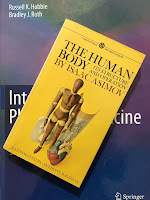
The Human Body, by Isaac Asimov, superimposed on Intermediate Physics for Medicine and Biology.
The Human Body,
by Isaac Asimov.
To explain how blood pressure is measured traditionally, I will turn to my hero Isaac Asimov’s book The Human Body (I quote from my 1963 paperback copy).
When blood is forced into the aorta, it exerts a pressure against the walls that is referred to as blood pressure. This pressure is measured by a device called a sphygmomanometer (sfig’ moh-ma-nom’ i-ter; “to measure the pressure of the pulse” [Greek]), an instrument which, next to the stethoscope, is surely the darling of the general practitioner. The sphygmomanometer consists of a flat rubber bag some 5 inches wide and 8 inches long. This is kept in a cloth bag that can be wrapped snugly about the upper arm, just over the elbow. The interior of the rubber bag is pumped up with air by means of a little rubber bulb fitted with a one-way valve. As the bag is pumped up, the pressure within it increases and that pressure is measured by a small mercury manometer to which the interior of the bag is connected by a second tube.

As the bag is pumped up, the arm is compressed until, at a certain point, the pressure of the bag against the arm equals the blood pressure. At that point, the main artery of the arm is pinched closed and the pulse in the lower arm (where the physician is listening with a stethoscope) ceases.

Now air is allowed to escape from the bag and, as it does so, the level of mercury in the manometer begins to fall and blood begins to make its way through the gradually opening artery. The person measuring the blood pressure can hear the first weak beats and the reading of the manometer at that point is the systolic pressure, for those first beats can be heard only during systole, when the blood pressure is highest. As the air continues to escape and the mercury level to fall, there comes a characteristic quality of the beat that indicates the diastolic pressure; the pressure when the heart is relaxed.
What I experienced in the hospital was different than Asimov’s explanation, and was more automated. I’m having a difficult time finding good technical literature about automated blood pressure monitors, but I’m going out on a limb here and guess how they work. In the hospital there was an inflatable cuff around my forearm, but there was no one listening with a stethoscope. That person is replaced by an optical device (similar to a pulse oximeter, see Section 14.6, Biological Applications of Infrared Scattering, in Intermediate Physics for Medicine and Biology) clipped to my finger, which presumably can detect flow. The cuff then inflates and flow is measured, the cuff changes the pressure to a new level and flow is measured again, etc. This all happens rapidly; each new cuff pressure level was maintained for at most one second, implying that only a single heart beat sufficed to make the flow measurement. The cuff and optical device are attached to a computer, and the computer made the decision about when to increase or decrease pressure, and what values to use. It seemed to be doing some sort of binary search, first going above and then below the level that allows flow. The algorithm slowed as the threshold level was approached, and I suspect that in such cases several heartbeats were required to accurately determine if flow occurred. The device also output the pulse rate and, if my memory serves me well, blood oxygenation level. Both were recorded after the cuff had completed its measurement of blood pressure.

I like this method. It does not depend on someone carefully listening for delicate blood flow noises (also known as Korotkoff sounds). In fact, while the blood pressure was being measured, the nurse was usually checking my IV line or doing some other task; the method is truly automated. One time, I did a little experiment and fidgeted with the finger clip during the measurement. The nurse got a fright when she saw my blood pressure up around 220/150. But a quick repeat measurement (during which I behaved myself) revealed that my blood pressure was actually normal (about 110/70). I suspect that the use of the binary search and pulse oximeter provides a more accurate measurement than does the traditional method, although I have no evidence to support that opinion. Automated blood pressure recording is an excellent example of how physics and engineering can contribute to medicine and biology.


Friday, September 9, 2022

An Immense World

“Earth teems with sights and textures, sounds and vibrations, smells and tastes, electric and magnetic fields. But every animal can only tap into a small fraction of reality’s fullness. Each is enclosed within its own unique sensory bubble, perceiving but a tiny sliver of our immense world.”
An Immense World, by Ed Yong, superimposed on Intermediate Physics for Medicine and Biology.
An Immense World,
by Ed Yong.
Those three sentences sum up Ed Yong’s new book An Immense World: How Animal Senses Reveal the Hidden Realms Around Us. Yong is a science writer for The Atlantic who won a Pulitzer Prize for his reporting about the COVID-19 pandemic. I’ve mentioned Yong in this blog before when quoting advice from his chapter in the book Science Blogging: “you have to have something worth writing about, and you have to write it well.” In An Immense World, Yong does both.

An Immense World sometimes overlaps with Intermediate Physics for Medicine and Biology. For example, both books discuss vision. Yong points out the human eye has better visual acuity than most other animals. He writes “we assume that if we can see it, they [other animals] can, and that if it’s eye-catching to us, it’s grabbing their attention… That’s not the case.” Throughout his book, Yong returns to this idea of how sensory perception differs among animals, and how misleading it can be for us to interpret animal perceptions from our own point of view.

Like IPMB, An Immense World examines color vision. Yong speculates about what a bee would think of the color red, if bees could think like humans.
Imagine what a bee might say. They are trichromats, with opsins that are most sensitive to green, blue, and ultraviolet. If bees were scientists, they might marvel at the color we know as red, which they cannot see and which they might call “ultrayellow” [I would have thought “infrayellow”]. They might assert at first that other creatures can’t see ultrayellow, and then later wonder why so many do. They might ask if it is special. They might photograph roses through ultrayellow cameras and rhapsodize about how different they look. They might wonder whether the large bipedal animals that see this color exchange secret messages through their flushed cheeks. They might eventually realize that it is just another color, special mainly in its absence from their vision.
Both An Immense World and IPMB also analyze hearing. Yong says
Human hearing typically bottoms out at around 20 Hz. Below those frequencies, sounds are known as infrasound, and they’re mostly inaudible to us unless they’re very loud. Infrasounds can travel over incredibly long distances, especially in water. Knowing that fin whales also produce infrasound, [scientist Roger] Payne calculated, to his shock, that their calls could conceivably travel for 13,000 miles. No ocean is that wide.…

Like infrasound, the term ultrasound… refers to sound waves with frequencies higher than 20 kHz, which marks the upper limit of the average human ear. It seems special—ultra, even—because we can’t hear it. But the vast majority of mammals actually hear very well into that range, and it’s likely that the ancestors of our group did, too. Even our closest relatives, chimpanzees, can hear close to 30 kHz. A dog can hear 45 kHz; a cat, 85 kHz; a mouse, 100 kHz; and a bottlenose dolphin, 150 kHz. For all of these creatures, ultrasound is just sound.
In IPMB, Russ Hobbie and I introduce the decibel scale for measuring sound intensity, or how loud a sound is. Yong uses this concept when discussing bats.
The sonar call of the big brown bat can leave its mouth at 138 decibels—roughly as loud as a siren or jet engine. Even the so-called whispering bats, which are meant to be quiet, will emit 110-decibel shrieks, comparable to chainsaws and leaf blowers. These are among the loudest sounds of any land animal, and it’s a huge mercy that they’re too high-pitched for us to hear.

Yong examines senses that Russ and I never consider, such as smell, taste, surface vibrations, contact, and flow. He wonders about the relative value of nociception [a reflex action to avoid a noxious stimulus] and the sensation of pain [a subjective feeling created by the brain].

The evolutionary benefit of nociception is abundantly clear. It’s an alarm system that allows animals to detect things that might harm or kill them, and take steps to protect themselves. But the origin of pain, on top of that, is less obvious. What is the adaptive value of suffering?

On the continuum ranging from life’s unity to diversity, Yong excels at celebrating the diverse, while Russ and I focus on how physics reveals unifying principles. I’m sometimes frustrated that Yong doesn’t delve into the physics of these topics more, but I am in awe of how he highlights so many strange and wonderful animals. There’s a saying that “nothing in biology makes sense except in light of evolution.” That’s true for An Immense World, which is a survey of how the evolution of sensory perception shapes they way animals interact, mate, hunt their prey, and avoid their predators.

Two chapters of An Immense World I found especially interesting were about sensing electric and magnetic fields. When discussing the black ghost knifefish’s ability to sense electric fields, Yong writes

Just as sighted people create images of the world from patterns of light shining onto their retinas, an electric fish creates electric images of its surroundings from patterns of voltage dancing across its skin. Conductors shine brightly upon it. Insulators cast electric shadows.
Then he notes that
Fish use electric fields not just to sense their environment but also to communicate. They court mates, claim territory, and settle fights with electric signals in the same way other animals might use colors or songs.
Even bees can detect electric fields. For instance, the 100 V/m electric field that exists at the earth’s surface can be sensed by bees.
Although flowers are negatively charged, they grow into the positively charged air. Their very presence greatly strengthens the electric fields around them, and this effect is especially pronounced at points and edges, like leaf tips, petal rims, stigmas, and anthers. Based on its shape and size, every flower is surrounded by its own distinctive electric field. As [scientist Daniel] Robert pondered these fields, “suddenly the question came: Do bees know about this?” he recalls. “And the answer was yes.”
The chapter on sensing magnetic fields is different from the others, because we don’t yet know how animals sense these fields.
Magnetoreception research has been polluted by fierce rivalries and confusing errors, and the sense itself is famously difficult both to study and to comprehend. There are open questions about all the senses, but at least with vision, smell, or even electroreception, researchers know roughly how they work and which sense organs are involved. Neither is true for magnetoreception. It remains the sense that we know least about, even though its existence was confirmed decades ago.

Yong lists three possible mechanisms for magnetoreception: 1) magnetite, 2) electromagnetic induction, and 3) magnetic effects on radical pairs. Russ and I discuss the first two in IPMB. I get the impression that the third is Yong’s favorite, but I remain skeptical. In my book Are Electromagnetic Fields Making Me Ill? I say that “they jury is still out” on the radical pair hypothesis.

If you want to read a beautifully written book that explores how much of the physics in Intermediate Physics for Medicine and Biology can be used by species throughout the animal kingdom to sense their environment, I recommend An Immense World. You’ll love it.

 Umwelt: The hidden sensory world of animals. By Ed Yong.

https://www.youtube.com/watch?v=Pzsjw-i6PNc


 Ed Yong on An Immense World

https://www.youtube.com/watch?v=bQS0Ioch05E

Friday, December 28, 2018

The Pitfalls of Using Handbooks and Formulae

A photo of three books: (left) Structures: Or Why Things Don't Fall Down, (center) Intermediate Physics for Medicine and Biology, and (right) The New Science of Strong Materials: Or Why You Don’t Fall Through the Floor.
Structures: Or Why Things Don't Fall Down, by J. E. Gordon.
Last week I discussed James Gordon’s book Structures: Or Why Things Don’t Fall Down. The book contains several appendices. The first appendix is ostensibly about using handbooks and formulas to make structural calculations.
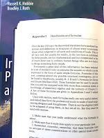
Over the last 150 years the theoretical elasticians have analysed the stresses and deflections of structures of almost every conceivable shape when subjected to all sorts and conditions of loads…Fortunately a great deal of this information has been reduced to a set of standard cases or examples the answers to which can be expressed in the form of quite simple formulae.
Then, to my surprise, Gordon changes tack and warns about pitfalls when using these formulas. His counsel, however, applies to all calculations, not just mechanical ones. In fact, his advice is invaluable for any young scientist or engineer. Below, I quote parts of this appendix. Read carefully, and whenever you encounter a word specific to mechanics substitute a general one, or one related to your own field.
[Formulae] must be used with caution.
A photo of Appendix 1 from Structures: Or Why Things Don't Fall Down, superimposed on the cover of Intermediate Physics for Medicine and Biology.
Appendix 1 of Structures.
  1. Make sure that you really understand what the formula is about.
  2. Make sure that it really does apply to your particular case.
  3. Remember, remember, remember, that these formulae take no account of stress concentrations or other special local conditions.
After this, plug the appropriate loads and dimensions into the formula—making sure that the units are consistent and that the noughts are right. [I’m not sure what “noughts” are, but I think the Englishman Gordon is saying to make sure the decimal point is in the right place.] Then do a little elementary arithmetic and out will drop a figure representing a stress or a deflection.

Now look at this figure with a nasty suspicious eye and think if it looks and feels right. In any case you had better check your arithmetic; are you sure that you haven’t dropped a two?...

If the structure you propose to have made is an important one, the next thing to do, and a very right and proper thing, is to worry about it like blazes. When I was concerned with the introduction of plastic components into aircraft I used to lie awake night after night worrying about them, and I attribute the fact that none of these components ever gave trouble almost entirely to the beneficent effects of worry. It is confidence that causes accidents and worry which prevents them. So go over your sums not once or twice but again and again and again.
Appendix 1 in J. E. Gordon's book Structures: Or Why Things Don't Fall Down has an important lesson for students studying from Intermediate Physics for Medicine and Biology.
Structures: Or Why Things Don't Fall Down.
This is the attitude I try to instill in my students when teaching from Intermediate Physics for Medicine and Biology. I implore them to think before they calculate, and then think again to judge if their answer makes sense. Students sometimes submit an answer to a homework problem (almost always given to five or six significant figures) that is absurd because they didn't look at their answer with a “nasty suspicious eye.” I insist they "remember, remember, remember" the assumptions and limitations of a mathematical model and its resulting formulas. Maybe Gordon goes a little overboard with his “night after night” of lost sleep, but at least he cares enough about his calculation to wonder “again and again and again” if it is correct. A little worry is indeed a “right and proper thing.”

Who would of expected such wisdom tucked away in an appendix about handbooks and formulae?

Friday, December 11, 2020

Selig Hecht (1892-1947)

A photo of Selig Hecht
Selig Hecht,
History of the Marine Biological Laboratory,
http://hpsrepository.asu.edu/handle/10776/3269.
In Chapter 14 of Intermediate Physics for Medicine and Biology, Russ Hobbie and I analyze the “classic experiment on scotopic vision” by Hecht, Shlaer, and Pirenne. George Wald wrote an obituary about Selig Hecht in 1948 (Journal of General Physiology, Volume 32, Pages 1–16). He writes that Hecht was
Intensely interested in the relation of light quanta (photons) to vision. Reexamining earlier measurements of the minimum threshold for human rod vision, he and his colleagues confirmed that vision requires only fifty to 150 photons. When all allowances had been made for surface reflections, the absorption of light by ocular tissues, and the absorption by rhodopsin (which alone is an effective stimulant), it emerged that the minimum visual sensation corresponds to the absorption in the rods of, at most, five to fourteen photons. An entirely independent statistical analysis suggested that an absolute threshold involves about five to seven photons. Both procedures, then, confirmed the estimation of the minimum visual stimulus at five to fourteen photons. Since the test field in which these measurements were performed contained about 500 rods, it was difficult to escape the conclusion that one rod is stimulated by a single photon.
Wald also describes the coauthor on the study, Shlaer.
Among Hecht’s first students was Simon Shlaer, who became Hecht’s assistant in his first year at Columbia and continued as his associate for twenty years thereafter. A man infinitely patient with things and impatient with people, Shlaer gave Hecht his entire devotion. He was a master of instrumentation, and though he also had a keen grasp of theory, he devoted himself by choice to the development of new technical devices. Hecht and Shlaer built a succession of precise instruments for visual measurement, among them an adaptometer and an anomaloscope that have since gone into general use. The entire laboratory came to rely on Shlaer’s ingenuity and skill. “I am like a man who has lost his right arm,” remarked Hecht on leaving Columbia—and Shlaer—in 1947, “and his right leg.”

In his Columbia laboratory, Hecht instituted investigations of human dark adaptation, brightness discrimination, visual acuity, the visual response to flickered light, the mechanism of the visual threshold, and normal and anomalous color vision. His lab also made important contributions regarding the biochemistry of visual pigments, the relation of night blindness to vitamin A deficiency in humans, the spectral sensitivities of man and other animals, and the light reactions of plants—phototropism, photosynthesis, and chlorophyll formation.
Hecht and Shlaer both contributed to the war effort during the Second World War.
Throughout the late years of World War II, Hecht devoted his energies and the resources of his laboratory to military problems. He and Shlaer developed a special adaptometer for night-vision testing that was adopted as standard equipment by several Allied military services. Hecht also directed a number of visual projects for the Army and Navy and was consultant and advisor on many others. He was a member of the National Research Council Committee on Visual Problems and of the executive board of the Army-Navy Office of Scientific Research and Development Vision Committee.

Explaining the Atom, by Selig Hecht, superimposed on Intermediate Physics for Medicine and Biology.
Explaining the Atom,
by Selig Hecht.
Hecht straddled the fields of physics and physiology, and was comfortable with both math and medicine. He entered college studying mathematics. After World War II ended, he wrote the book Explaining the Atom, which Wald described as “a lay approach to atomic theory and its recent developments that the New York Times (in a September 20, 1947, editorial) called ‘by far the best so far written for the multitude.’”

An obituary in Nature by Maurice Henri Pirenne concludes

The death of Prof. Selig Hecht in New York on September 18, 1947, at the age of fifty-five, deprives the physiology of vision of one of its most outstanding workers. Hecht was born in Austria and was brought to the United States as a child. He studied and worked in the United States, in England, Germany and Italy. After a broad biological training, he devoted his life to the study of the mechanisms of vision, considered as a branch of general physiology. He became professor of biophysics at Columbia University and made his laboratory an international centre of visual research.

Friday, June 4, 2021

The Bidomain Model of Cardiac Tissue: Predictions and Experimental Verification

“The Bidomain Model of Cardiac Tissue: Predictions and Experimental Verification” superimopsed on Intermediate Physics for Medicine and Biology.
“The Bidomain Model of Cardiac Tissue:
Predictions and Experimental Verification”

In the early 1990s, I was asked to write a chapter for a book titled Neural Engineering. My chapter had nothing to do with nerves, but instead was about cardiac tissue analyzed with the bidomain model. (You can learn more about the bidomain model in Chapter 7 of Intermediate Physics for Medicine and Biology.) 

“The Bidomain Model of Cardiac Tissue: Predictions and Experimental Verification” was submitted to the editors in January, 1993. Alas, the book was never published. However, I still have a copy of the chapter, and you can download it here. Now—after nearly thirty years—it’s obsolete, but provides a glimpse into the pressing issues of that time.

I was a impudent young buck back in those days. Three times in the chapter I recast the arguments of other scientists (my competitors) as syllogisms. Then, I asserted that their premise was false, so their conclusion was invalid (I'm sure this endeared me to them). All three syllogisms dealt with whether or not cardiac tissue could be treated as a continuous tissue, as opposed to a discrete collection of cells.

The Spach Experiment

The first example had to do with the claim by Madison Spach that the rate of rise of the cardiac action potential, and time constant of the action potential foot, varied with direction.

Continuous cable theory predicts that the time course of the action potential does not depend on differences in axial resistance with direction.

The rate of rise of the cardiac wave front is observed experimentally to depend on the direction of propagation.

Therefore, cardiac tissue does not behave like a continuous tissue.
I then argued that their first premise is incorrect. In one-dimensional cable theory, the time course of the action potential doesn’t depend on axial resistance, as Spach claimed. But in a three-dimensional slab of tissue superfused by a bath, the time course of the action potential depends on the direction of propagation. Therefore, I contended, their conclusion didn’t hold; their experiment did not prove that cardiac tissue isn’t continuous. To this day the issue is unresolved.

Defibrillation

A second example considered the question of defibrillation. When a large shock is applied to the heart, can its response be predicted using a continuous model, or are discrete effects essential for describing the behavior?
An applied current depolarizes or hyperpolarizes the membrane only in a small region near the ends of a continuous fiber.

For successful defibrillation, a large fraction of the heart must be influenced by the stimulus.

Therefore, defibrillation cannot be explained by a continuous model.
I argued that the problem is again with the first premise, which is true for tissue having “equal anisotropy ratios” (the same ratio of conductivity parallel and perpendicular to the fibers, in both the intracellular and extracellular spaces), but is not true for “unequal anisotropy ratios.” (Homework Problem 50 in Chapter 7 of IPMB examines unequal anisotropy ratios in more detail). If the premise is false, the conclusion is not proven. This issue is not definitively resolved even today, although the sophisticated simulations of realistically shaped hearts with their curving fiber geometry, performed by Natalia Trayanova and others, suggest that I was right.

Reentry Induction

The final example deals with the induction of reentry by successive stimulation through a point electrode. As usual, I condensed the existing dogma to a syllogism.
In a continuous tissue, the anisotropy can be removed by a coordinate transformation, so reentry caused by successive stimulation through a single point electrode cannot occur, since there is no mechanism to break the directional symmetry.

Reentry has been produced experimentally by successive stimulation through a single point electrode.

Therefore, cardiac tissue is not continuous.

Once again, that pesky first premise is the problem. In tissue with equal anisotropy ratios you can remove anisotropy by a coordinate transformation, so reentry is impossible. However, if the tissue has unequal anisotropy ratios the symmetry is broken, and reentry is possible. Therefore, you can’t conclude that the observed induction of reentry by successive stimulation through a point electrode implies the tissue is discrete.


I always liked this book chapter, in part because of the syllogisms, in part because of its emphasis on predictions and experiments, but mainly because it provides a devastating counterargument to claims that cardiac tissue acts discretely. Although it was never published, I did send preprints around to some of my friends, and the chapter took on a life of its own. This unpublished manuscript has been cited 13 times!

Trayanova N, Pilkington T (1992) “The use of spectral methods in bidomain studies,” Critical Reviews in Biomedical Engineering, Volume 20, Pages 255–277.

Winfree AT (1993) “How does ventricular tachycardia turn into fibrillation?” In: Borgreffe M, Breithardt G, Shenasa M (eds), Cardiac Mapping, Mt. Kisco NY, Futura, Chapter 41, Pages 655–680.

Henriquez CS (1993) “Simulating the electrical behavior of cardiac tissue using thebidomain model,” Critical Reviews of Biomedical Engineering, Volume 21, Pages 1–77.

Wikswo JP (1994) “The complexities of cardiac cables: Virtual electrode effects,” Biophysical Journal, Volume 66, Pages 551–553.

Winfree AT (1994) “Puzzles about excitable media and sudden death,” Lecture Notes in Biomathematics, Volume 100, Pages 139–150.

Roth BJ (1994) “Mechanisms for electrical stimulation of excitable tissue,” Critical Reviews in Biomedical Engineering, Volume 22, Pages 253–305.

Roth BJ (1995) “A mathematical model of make and break electrical stimulation ofcardiac tissue by a unipolar anode or cathode,” IEEE Transactions on Biomedical Engineering, Volume 42, Pages 1174–1184.

Wikswo JP Jr, Lin S-F, Abbas RA (1995) “Virtual electrodes in cardiac tissue: A common mechanism for anodal and cathodal stimulation,” Biophysical Journal, Volume 69, Pages 2195–2210.

Roth BJ, Wikswo JP Jr (1996) “The effect of externally applied electrical fields on myocardial tissue,” Proceedings of the IEEE, Volume 84, Pages 379–391.

Goode PV, Nagle HT (1996) “On-line control of propagating cardiac wavefronts,” The 18th Annual International Conference of the IEEE Engineering in Medicine and Biology Society, Amsterdam.

Winfree AT (1997) “Rotors, fibrillation, and dimensionality,” In: Holden AV, Panfilov AV (eds): Computational Biology of the Heart, Chichester, Wiley, Pages 101–135.

Winfree AT (1997) “Heart muscle as a reaction-diffusion medium: The roles of electric potential diffusion, activation front curvature, and anisotropy,” International Journal of Bifurcation and Chaos, Volume 7, Pages 487–526.

Winfree AT (1998) “A spatial scale factor for electrophysiological models of myocardium,” Progress in Biophysics and Molecular Biology, Volume 69, Pages 185–203.
I’ll end with the closing paragraph of the chapter.
The bidomain model ignores the discrete nature of cardiac cells, representing the tissue as a continuum instead. Experimental evidence is often cited to support the hypothesis that the discrete nature of the cells plays a key role in cardiac electrophysiology. In each case, the bidomain model offers an alternative explanation for the phenomena. It seems wise at this point to reconsider the evidence that indicates the significance of discrete effects in healthy cardiac tissue. The continuous bidomain model explains the data, recorded by Spach and his colleagues, showing different rates of rise during propagation parallel and perpendicular to the fibers, anodal stimulation, arrhythmia development by successive stimulation from a point source, and possibly defibrillation. Of course, these alternative explanations do not imply that discrete effects are not responsible for these phenomena, but only that two possible mechanisms exist rather than one. Experiments must be found that differentiate unambiguously between alternative models. In addition, discrete junctional resistance must be incorporated into the bidomain model. Only when such experiments are performed and the models are further developed will we be able to say with any certainty that cardiac tissue can be described as a continuum.