Friday, December 31, 2021

Adrianus Kalmijn (1933–2021)

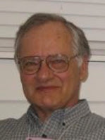
Adrianus Kalmijn, a biophysicist known for his studies of electroreception in sharks, died December 7, 2021, at the age of 88.

In Chapter 9 of Intermediate Physics for Medicine and Biology, Russ Hobbie and I discuss Kalmijn’s work.

Kalmijn et al. discovered that the ocean flounder generates a current dipole of 3 × 10⁻⁷ A m. Sea water with resistivity of 0.23 Ω m gives an electric field of 2 × 10⁻⁵ V m−1 at a distance 10 cm in front of the flounder. They were able to show in a beautiful series of behavioral experiments that dogfish (a small shark) could detect the electric field 0.4 m from a current dipole of 4 × 10⁻⁷ A m, corresponding to an electric field of 5 × 10⁻⁷ V m−1. The fish would bite at the electrodes, ignoring a nearby odor source. A field of 10−4 V m−1 would elicit the startle response. A field ⅒ as large caused a physiologic response. 
The first page of Kalmijn, A. J. (1977) The electric and magnetic sense of sharks, skates, and rays. Oceanus 20:45–52, superimposed on Intermediate Physics for Medicine and Biology.
Kalmijn, A. J. (1977) The electric and
magnetic sense of sharks, skates,
and rays. Oceanus 20:45–52.

 My favorite paper by Kalmijn is

Kalmijn, A. J. (1977) The electric and magnetic sense of sharks, skates, and rays. Oceanus 20:45–52.
Below is an excerpt.
During the summer of 1976, we learned from longline fishing off Cape Cod that the smooth dogfish Mustelus regularly frequents the shallow, inshore waters of Vineyard Sound on its nightly feeding excursions. This predatory shark is a warm-season visitor, arriving at Woods Hole in May and leaving for the South again in late October or shortly thereafter. It is an active bottom hunter, preying on small fish as well as crustaceans and other invertebrate animals. The females reach an average length of 115 centimeters; the males are slightly smaller. The smooth dogfish is truly live-bearing; the new-born measure 29 to 37 centimeters.

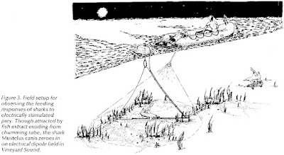
To observe the sharks’ feeding behavior, we worked from an inflatable rubber raft (Zodiac Mark II) free of any metal under the waterline. On station in 2.5 to 3.0-meter-deep water over a sand patch devoid of seaweed, we attracted the sharks by squeezing liquified herring through a long Tygon tube that ran from the raft to the bottom of the sea. The Tygon chumming tube was attached to a polypropylene line, suspended from a Styrofoam float and stretched over the ocean floor between two polyvinyl pipes anchored in low-profile cinder blocks (Figure 3). Starting after dark, we illuminated the area with a 100-watt, battery-operated underwater light. To break the water surface, we used a glass-bottom viewing box secured behind the stern of the raft. 
Figure 3 from Kalmijn, A. J. (1977) Oceanus 20:45–52.
With permission from the Woods Hole Oceanographic Institution.
Two pairs of agar-filled, salt-bridge electrodes were tied to the polypropylene line and positioned on the sand, one on either side of the odor source and 30 centimeters from it. Mekka underwater plugs with stainless steel pins and integral cables connected the thin, 30 to 90-centimeter-long Silastic salt-bridge tubes to the electrical equipment set up in the rubber raft. The use of a constant-current source virtually eliminated the adverse effects of polarization at the stainless steel/seawater interfaces. From the raft, we could conveniently vary the strength of the field and select the pair of electrodes to be energized, the other pair functioning as the control. The applied direct-current dipole moments ranged from 1 to 8 microamperes × 5 centimeters (dipole current × distance between electrodes), roughly corresponding to the bioelectric fields of small prey at a seawater resistivity of 20.0 to 20.5 ohm·centimeters and a temperature of 19 to 22 degrees Celsius.
After entering the area, the smooth dogfish began frantically searching over the sand, apparently trying to locate the odor source. Both young and mature sharks were observed, sometimes alone, sometimes in groups of two to five. Neither the raft nor the underwater light appeared to bother them. Most interestingly, when nearing the odor source, the animals did not bite at the opening of the chumming tube but from distances up to 25 centimeters turned sharply to the current-electrodes, viciously attacking the electrically simulated prey. After snapping the line with their teeth right at the position of the electrodes, the sharks usually attempted to rip them apart—and one night they succeeded. When the current was switched to the other pair of electrodes, the animals let go, circled around for awhile, and attacked again, but at the electrodes on the other side of the odor source. At the lower current levels, the sharks kept responding, though from increasingly shorter distances.

These observations convincingly demonstrate that odor-motivated sharks are capable of detecting and taking prey by the exclusive use of their electric sense, not only under well-controlled laboratory conditions, but also in their electrically more noisy, ocean habitat.
You can find an obituary of Kalmijn at the Scripps Institution of Oceanography website. It states
Family members remember Kalmijn as a renaissance man and a maverick. His work was his passion. He set a very high standard of integrity in his work and sought truth, accuracy, and scientific insight.

As a scientist, you can’t ask for more than that.

Friday, December 24, 2021

Michael Faraday and the Royal Institution Christmas Lectures

Tonight is Christmas Eve. 

At this time of the year, I think of the great English physicist Michael Faraday giving the Royal Institution Christmas Lectures. Below is a famous illustration of Faraday delivering one of his talks.

Professor Faraday lecturing at the Royal Institution, December 27, 1855, from a painting by Alexander Blaikley, commemorating the Attendance of HRH the Prince of Wales and HRH Prince Alfred, at the Juvenile Course of Lectures, 1855–1856.
https://commons.wikimedia.org/wiki/File:Faraday_Michael_Christmas_lecture.jpg

Here’s a photo of the statue of Michael Faraday at the Royal Institution in London.

Faraday was very closely associated with the Royal Institution. He was first appointed as laboratory assistant there in 1813, became director of the laboratory in 1825, and Fullerian Professor of Chemistry there from 1833 to 1867. It was there too that he conducted his electricity experiments; as superintendent of the institution, he also lived in a flat there with his wife Sarah, until the couple were given a house near Hampton Court in 1858. Photograph, caption, and commentary by Jacqueline Banerjee
https://victorianweb.org/sculpture/foley/4.html

I’m fond of Michael Faraday because he discovered electromagnetic induction. Induction is the process that underlies transcranial magnetic stimulation of the brain, a technique that I worked on in the 1990s at the National Institutes of Health. In Intermediate Physics for Medicine and Biology, Russ Hobbie and I write

In 1831 Michael Faraday discovered that a changing magnetic field causes an electric current to flow in a circuit. It does not matter whether the magnetic field is from a permanent magnet moving with respect to the circuit or from the changing current in another circuit. The results of many experiments can be summarized in the Faraday induction law: Eds = – d/dtBdS.
I usually associate the Royal Institution with Victorian scientists like Michael Faraday, John Tyndall, and James Dewar. But the Royal Institution—nowadays referred to as the Ri—is alive and well! I love its vision:

A world where everyone is inspired to think more deeply about science and its place in our lives.

The Ri continues to host the Christmas Lectures every year. This year, the lecture is—of course—about Covid-19. Its title is “Going Viral: How Covid Changed Science Forever.” England’s Deputy Chief Medical Officer, Jonathan Van-Tam, will be joined by top UK scientists to examine the science of viruses. Typically these lectures contain many demonstrations and are especially aimed at a young audience, but I like them too. Eventually, the Ri will post these lectures online for all to see. I can’t wait.

I learned on the Ri website that 2021 is the 200th anniversary of Faraday's invention of the electric motor.

The Royal Institution will begin a year-long series of activities from September, to mark the 200th anniversary of Michael Faraday’s development of the world’s first electric motor, the science charity announced today.

Activities will begin on Friday 3 September – 200 years to the day since Faraday’s world-shaping breakthrough – with an opportunity to name one of 200 seats in the very same theatre in which Faraday lectured on many occasions to an audience of Ri Members and the general public. The ‘200 seats for 200 years’ fundraising campaign is designed to help secure the future of the Ri, after it’s income was severely impacted by the Covid-19 pandemic.
The Royal Institution also has a lot of great videos. Here are some I enjoyed, which are related to Intermediate Physics for Medicine and Biology

Merry Christmas.

How do Medicine and Physics Overlap?

Celebrating Crystallography - An Animated Adventure

How Does Convection Work? - Christmas Lectures with George Porter

Michael Faraday's Electric Frogs

Rutherford, Radioactivity and the Future of Physics - with the Cosmic Shambles Network

Friday, December 17, 2021

Russell Hobbie (1934–2021)

Russell Hobbie
Russell Hobbie
I am heartbroken to have to tell you that Russ Hobbie, my coauthor and friend, passed away this week, after a long battle with Parkinson’s disease. Russ was the sole author on the first three editions of Intermediate Physics for Medicine and Biology, and was the senior author with me on the fourth and fifth editions.

Below are excerpts from Russ’s preface to the first edition, describing how he came to write IPMB.
Between 1971 and 1973 I audited all of the courses medical students take in their first two years at the University of Minnesota. I was amazed at the amount of physics I found in those courses and how little of it is discussed in the general physics course.

I found a great discrepancy between the physics in some papers in the biological research literature and what I knew to be the level of understanding of most biology majors who had taken a year of physics. It was clear that an intermediate level physics course would help these students. It would provide the physics they needed and would relate it directly to the biological problems where it is useful. Making the connection with biology is something that we tend to leave for the student. When we do that, I think we overestimate the ability of most students to see the application and work out the details. Seeing few applications is also a powerful motivation for mastering difficult material.

This book is the result of my having taught such a course since 1973. It is intended to serve as a text for an intermediate course taught in a physics department and taken by a variety of majors… 

Here is a list from Google Scholar of some of his publications.

First ten listings in the Google Scholar entry for Russ Hobbie.

His most highly cited publication, by far, was IPMB (all editions are combined into one citation score). His most highly cited research article was a 2010 paper that he coauthored with his daughter Sarah. Some of the figures in that paper found their way, with slight modifications, into the 5th edition of IPMB: exponential decay assuming constant error bars (Fig. 2.6 in IPMB) or a constant percentage error bars (Fig. 2.7). He has articles going back to 1960, and the most recent edition of IPMB was in 2015, implying an amazing 55 year publication history. His earliest papers in nuclear physics were published during his time at the Harvard cyclotron laboratory

You can learn more about Russ by reading the transcript of his 1994 interview as part of an oral history project at the University of Minnesota. Some excerpts:

I grew up as a college brat. My parents both taught at Skidmore College in upstate New York. I was born in 1934. One of my earliest recollections is at age three or four falling in the college fishpond and being fished out by some of the Skidmore students. My father taught physics there...
[My high school had] a standard college preparatory course except that they had machine shop and mechanical drawing. I don’t know how I decided it, but I decided that I wanted to go to college at MIT [Massachusetts Institute of Technology], which I did and I thoroughly enjoyed that experience. I liked Cambridge so much that I wanted to stay in Cambridge. Everybody told me that I ought to change schools for graduate school; so, I went up the river to Harvard as a graduate student...
[At Harvard] my TA [teaching assistant] assignment was to work with Ed[ward Mills] Purcell, who is a Nobel Prize winning physicist, a very wonderful and humble person, redesigning the junior electricity and magnetism lab. That was a very great experience...

I became an RA [research assistant] at the Harvard cyclotron and drifted into doing my thesis in experimental nuclear physics on the cyclotron. I took my final Ph.D. oral exam on April Fool's Day in 1960, which means that I had done my Ph.D. in slightly under four years because I graduated from college in 1956...

I had met my wife [Cynthia], a public health nurse, in Boston. She had grown up in Iowa, had gone to the University of Iowa, and had then come out to work for the Visiting Nurses’ Association. She had some interest in moving closer to her home and I had seen this about Minnesota...
I got out here [an interview at the University of Minnesota] and discovered that I was actually being interviewed by Morris Blair to be a post-doc on the old Van de Graaff generator. I spent the day with him. About two o’clock that afternoon, he offered me a job and I accepted. All of this is a little bit different from the way things are done now...

I was attracted by the fact that it appeared to me at that time that at the University of Minnesota, research was important. It was a research university. Having grown up seeing a small college, I thought that that was a bit stifling and I didn’t want that. I thought that one could combine teaching and a research career here...
I also saw coming up the fact that nuclear physics was going to change and that the experiments were going to have to be done at national laboratories because individual universities could not afford to keep these things going; so, I made a conscious decision about that time—I was now an associate professor—that I wanted not to continue in nuclear physics. The department was fairly supportive of that and I became the director of undergraduate studies in physics for a few years...

I’d come out here [Minnesota] and happened, at some point along here, to be invited to the neighbors for a dinner party at which I met a pathologist named Richard Riess [spelled Reece], who was a pathologist at, then, St. Barnabas Hospital, a principal in Lufkin Medical Laboratories, and who was interested in using computers for interpreting lab test results...
So, Riess was making things that he called diagnotes, which were just lists of what could cause an elevated uric acid, or a low calcium, or a high calcium, and so on. At that dinner party, he started asking, "Was there any way that one could computerize this?" Having just put in this online computer at the Tandem Lab, I started using it to try to do some pattern matching. This, then, led to, for several years, my working with Riess as a collaborator and Lufkin Medical Labs having a research contract with the University of Minnesota that supported a couple of students over the years. We did a lot of work on developing automatic interpretation of the clinical laboratory results and published several papers in this area. I can remember still being the director of undergraduate studies in Physics and Mort[on] Hamennesh, who was the department head, coming in one day to tell me that they were promoting me to full professor based on the work that I had done at the Tandem in nuclear physics and the online computer...
[Reece] got me to thinking that it might be interesting to put some [medical] examples in the pre-med physics course; so, I wrote Al Sullivan who was the assistant dean of the Medical School asking if it was possible to snoop around over there. Al asked me to have lunch with him one day—it was in October—and said, "What you really ought to do is to attend Medical School." I said, "I can’t. I’m director of undergraduate studies in Physics. I’m teaching a full load, which is a course each quarter. There’s just no time to do that." He said, "You could just audit things and skip the labs." So, for two years, I did that....

Probably around 1972 or 1973, I started teaching that course, developing it as I went. That turned into a book [Intermediate Physics/or Medicine and Biology] that was published by Wiley in 1978 with a second edition about 1988. I’m trying, without much success, to do a third edition right now [Russ was ultimately successful]....

Included in the interview was a story about Russ’s daughter Ann.

It is now time to do the second edition of my book. I have the solutions manual and the new problems, I have already put into the computer. The new problems are there, but the old ones aren’t; so, Wiley agreed to hire my youngest daughter, Anne [Ann], to type all of the old solutions manual into the computer. She thought she had a summer job. She was done in two and half weeks.

If you still want to know more about Russ, check out the December 2006 issue of The Biological Physicist, the newsletter of the Division of Biological Physics in the American Physical Society. Russ and I (mostly Russ) answered questions about IPMB. Here is what I said about becoming Russ’s coauthor.

THE BIOLOGICAL PHYSICIST (to Brad Roth): Tell us a little about how you first became acquainted with Hobbie’s text, and how you see it has having influenced the field. 

Brad Roth: I used the first edition of Hobbie’s book for a class taught by John Wikswo when I was a graduate student at Vanderbilt University. This was a very crucial time in my education, when I was changing from a physics undergraduate student to a biological physics graduate student. The book had a huge impact on me and my career.

When visitors come by my office at OaklandUniversity, they sit politely and listen to me describe my research. Then I mention “Oh, by the way, I am also going to be second author on the 4th edition of Hobbie’s book Intermediate Physics in Medicine and Biology.” At this point, their eyes usually light up and they say, almost with disbelief, “Really? I know that book.”

At the end of the preface to the first edition of IPMB, Russ wrote

Every list of acknowledgments seems to close with thanks to a long-suffering family. I never knew what those words really meant, nor how deep the indebtedness, until I wrote this book.

 Below is the obituary prepared by his family.

Russell Klyver Hobbie, 87, died peacefully at home surrounded with love on December 16. Russ was born on November 3, 1934. His parents were Eulin Klyver and John Remington Hobbie. He grew up in Saratoga Springs, NY and Springfield, MA. He graduated from MIT with a BS in physics, and earned a PhD in physics from Harvard University. In 1960, he and his wife Cynthia moved with their young family to Minneapolis, where he joined the faculty at the University of Minnesota. Over his 38-year career, he was a wonderful professor and stalwart advocate for students. 
After auditing two years of medical school, Russ changed his specialty from nuclear physics to biophysics. He developed a new medical physics course which led to the first edition of his text book, Intermediate Physics for Medicine and Biology. For 12 years, Russ served as Associate Dean of Student Affairs in the Institute of Technology. After retirement, despite having been diagnosed with Parkinson’s disease, Russ completed the fourth and fifth edition of the book. 
Russell had many interests, among them canoeing and fishing in the BWCA, the Quetico and the Canadian Arctic. He enjoyed working on genealogy, and developed his skills as a woodworker, making beautiful furniture for his children and grandchildren. He and Cynthia took many trips in the US and abroad and loved spending time at the family cabin on Burntside Lake. They enjoyed theater, music, and visual arts in the Twin Cities. 
Russ is survived by his wife of 64 years, and his children, Lynn (Kevin Little), Erik (Pam Gahr), Sarah (Jacques Finlay) and Ann (Jeff Benjamin), and by his six grandchildren, Henry Benjamin, Grace Little, William Benjamin, Owen Finlay, Phoebe Finlay, and Rosie Hobbie. His sister Jane Bacon, two nieces and several grand nieces and nephews also survive him. 
Russ was a most wonderful and kind person and we will miss him very much. There will be a Memorial Service over Zoom from First Congregational Church in SE Minneapolis on Sunday January 2 at 2PM. We invite people to join us on-line. In lieu of flowers, Memorials may be directed to Save the Boundary Waters or Friends of the Boundary Waters.

Russ, I will miss you. Your work left a legacy that will influence how physics is taught to medical and biology students for years to come.

Russ Hobbie demonstratg the computer program MacDose. Part 1.
https://www.youtube.com/watch?v=eZGfHBYMuHg

Russ Hobbie demonstrating the computer program MacDose. Part 2.
https://www.youtube.com/watch?v=Rwp8eeKFycQ

Russ Hobbie demonstrating the computer program MacDose. Part 3.

Biology’s Built-In Faraday Cages

Klee (2014) "Biology's Built-In Faraday Cage," American Journal of Physics, 82:451–459, superimposed on the cover of Intermediate Physics for Medicine and Biology.
Klee, M. (2014)
Amer
. J. Phys. 82:451-459.
In Chapter 9 of Intermediate Physics for Medicine and Biology, Russ Hobbie and I discuss how well an external electric field penetrates the human body. We examine a sinusoidally varying field, but another problem is an field that turns on suddenly and then remains constant (a step function). This case is analyzed by Maurice Klee in his American Journal of Physics article “Biology’s Built-In Faraday Cages” (Volume 82, Pages 451-459, 2014). Klee obtained a PhD in Biomedical Engineering from Case Western Reserve University, under the direction of Robert Plonsey, and a law degree from George Washington University. His abstract states
Biological fluids are water-based, ionic conductors. As such, they have both high relative dielectric constants and substantial conductivities, meaning they are lossy dielectrics. These fluids contain charged molecules (free charges), whose movements play roles in essentially all cellular processes from metabolism to communication with other cells. Using the problem of a point source in air above a biological fluid of semi-infinite extent, the bound charges in the fluid are shown to perform the function of a fast-acting Faraday cage, which protects the interior of the fluid from external electric fields. Free charges replace bound charges in accordance with the fluid’s relaxation time, thereby providing a smooth transition between the initial protection provided by the bound charges and the steady state protection provided by the free charges. The electric fields within the biological fluid are thus small for all times just as they would be inside a classical Faraday cage.
The most interesting part of this article is the interplay between bound and free charges at the tissue surface. Klee assumes that bound charge, arising primarily from the rotation of the polarized water molecules, responds to the external electric field instantaneously, while free charge responds with a delay. The rapid bound charge shields the tissue immediately, but incompletely. Over time the free charge replaces the bound charge, eventually providing complete shielding of the tissue from the external electric field.

Klee addresses two questions: how completely does the bound charge shield the tissue, and how fast does the free charge replace the bound charge? (1) He shows that the initial bound charge is less than the final free charge by a factor of (κ – 1)/(κ + 1), where κ is the dielectric constant. Klee uses the value of 80 for κ, and finds that the bound charge provides 97.5% of the shielding that the free charge ultimately contributes. Russ and I point out that the dielectric constant of tissue can be much greater than water, and suggest a value of one million might be more appropriate. (2) The time constant for free charge to replace bound charge is κεo/σ, where εo is the permittivity of free space and σ is the conductivity. If you use σ = 0.5 S/m and κ = 80, the time constant is 1.4 nanoseconds. If you use κ = 1,000,000, the time required for complete shielding by the free charge is much longer (about 2 microseconds) but the initial shielding from the bound charge is 99.9998% complete.

Klee concludes that
Through a combination of bound and free charges, a biological fluid surrounded by a non-conductor of low relative dielectric constant does not develop large internal electric fields as a result of charges located outside the fluid. The fluid is thus its own robust Faraday cage, thereby ensuring that biological molecules within the fluid do not experience large electric fields due to outside sources.

Friday, December 10, 2021

Physical Models of Living Systems, Second Edition

Physical Models of Living Systems, 2nd Edition, by Philip Nelson, superimposed on Intermediate Physics for Medicine and Biology.
Physical Models of Living Systems, 2nd Edition,
by Philip Nelson.
In a 2015 blog post, I discussed Philip Nelson’s then-new book Physical Models of Living Systems. I wrote that “It’s an excellent book, well written and beautifully illustrated.” Recently, Nelson published a second edition of Physical Models of Living Systems. All the nice things I wrote about the first edition remain true in the second, but now there are four new chapters to increase your fun. In this post, I’ll focus on the new chapters.

Chapter 6: Random Walks on an Energy Landscape

I like how Nelson organizes each chapter around a biological question and a physical idea.
Biological question: How can pulling two things apart strengthen their bond?

Physical idea: Bond breaking is a first passage process, controlled by the lowest activation barrier, and that barrier can increase upon moderate loading.

The chapter describes slip bonds and catch bonds. A slip bond is the normal case when the bond’s strength decreases as you pull on it, and a catch bond is the unusual case when its strength increases as you pull. Wikipedia compares a catch bond to one of those Chinese finger traps.

Photograph by Carol Spears on Wikipedia. 
https://commons.wikimedia.org/wiki/File:Finger_trap_toys.jpg
 
Nelson explains catch bonds using random walk simulations; first a free random walk, then one with an applied force, next one in a harmonic oscillator potential, and finally one with a oscillator potential plus a barrier, where if you reach the top of the barrier the bond breaks. The “strength” of the bond then becomes the walking time before reaching the barrier (a “first passage process”). By manipulating the potential shape, he finds clutch bond behavior. He then relates these simple simulations (which the reader can easily perform on their own computer) to T cell activation and leukocyte rolling. In each chapter, he sums up the analysis with a section he calls “The Big Picture.” For this chapter, he writes

Our physical model… was absurdly simple, but it nevertheless contained a lot of buried treasure: the basic facts about free Brownian motion, drift under constant force, equilibration in a trapping potential field, the Boltzmann distribution in equilibrium, the Arrhenius rule for escape in quasiequilibrium, and the entire surprising phenomenon of catch bonding. The key step was to understand bond breaking as a first passage problem.

Chapter 8: Single Particle Reconstruction in Cryo-electron Microscopy

Biological question: How can we combine many noisy images of a viral protein to get one clean image?

Physical idea: We must first align the images, but our best estimate of the required alignment is actually a probability distribution.
In this chapter, Nelson examines how to take noisy electron microscope images of an object that are each rotated or shifted relative to each other, and align them to get a clear picture. He warns us “You can’t win by averaging noisy signals unless you know the proper alignment.” What biological example does he look at? The coronavirus spike protein! Apparently the procedure described in this chapter played a big role in the development of the covid-19 vaccine. The story makes me want to seek out the scientists who developed this method and give them a big hug. 

Chapter 14: Demographic Variation in Epidemic Spread

Biological question: Why do some outbreaks of a communicable illness spread explosively, whereas others, in similar communities, fizzle after the first few cases?

Physical idea: A tiny subpopulation of superspreader individuals can have a huge effect on the course of an epidemic.
This chapter starts with the SIR model of an epidemic (S = susceptible, I = infected, and R = recovered) that I’ve discussed before in this blog. Nelson tweaked it to examine what happens just as the epidemic begins if you have a handful of superspreaders. Once again, the model is applied to understanding covid. In the big picture Nelson writes
We have found that because outbreaks always begin with just one or a few infective individuals, the discrete, stochastic character of transmission has a large effect on outbreak dynamics. Thus, a community that is lucky to get only a mild outbreak in the first instance must not become complacent, imagining themselves to be somehow protected: Always some outbreaks fizzle, but any such instance is just as likely to be followed by a severe outbreak on a later introduction as in any other community. 
There are many ways to improve the realism of the SIR model, but we focused on just one: the well documented fact that some illnesses have superspreader individuals. The implications are profound. Although Figure 14.5a is frightening, such time courses can be replaced by the milder ones in Figure 14.3 by promptly identifying and quarantining just a few percent of the infected population. For example, backward contact tracing seeks to identify contacts of each sick individual who may have been the source of that person’s infection. When multiple backward trails point to the same person, that person may be a superspreader.

Chapter 15: Bet-Hedging Via Stochastic, Excitable Dynamics

Biological question: How can a pathogen hide from the immune system?

Physical idea: Positive feedback with small copy numbers can lead to a stochastic toggle that transiently changes state after a long, random delay.
I like this chapter because it makes good use of phase portrait plots. The pathogen behaves almost like a nerve, which can either sit at rest or fire an action potential, with the all-or-none response relying on a positive feedback loop. 

What bet is being hedged? If you’re in a situation where normally one type of behavior is favored, but on rare occasions the environment changes and an unusual behavior may be needed to save the species, then sometimes organisms will keep most individuals in the normal state but will have a few random individuals in the unusual state just in case.


The second edition of Physical Models of Living Systems still has all the good stuff from the first edition: lovely color figures (including some by David Goodsell), lots of homework problems, comparisons to real data, and a winning combination of words, pictures, equations, and computer code. Add in the four new chapters—and a kindle price under ten dollars!—and you have a masterpiece.

My favorite part of the second edition: Like in the first edition, Nelson cites Intermediate Physics in Medicine and Biology. And, he remembers to update the citation to IPMB's 5th edition!

Friday, December 3, 2021

Resource Letter BP-1: Biological Physics

The December, 2021 issue of the American Journal of Physics contains Resource Letter BP-1: Biological Physics (Volume 89, Pages 1071–1078), by Raghuveer Parthasarathy. A Resource Letter is a guide to the literature, websites, and other teaching aids about a particular topic. Parthasarathy’s abstract states
This Resource Letter provides an overview of the literature in biological physics, a vast, active, and expanding field that links the phenomena of the living world to the tools and perspectives of physics. While no survey of this area could be complete, this list and commentary are intended to help provide an entry point for upper-level undergraduates, graduate students, researchers new to biophysics, or workers in subfields of biophysics who wish to expand their horizons. Topics covered include subcellular structure and function, cell-scale mechanics and organization, collective behaviors and embryogenesis, genetic networks, and ecological dynamics.
I particularly like the opening paragraph of his resource letter, which reflects what Russ Hobbie and I have tried to convey in Intermediate Physics for Medicine and Biology.
Life is full of variety, vigor, and clever solutions to daunting challenges. Physics provides deep, elegant insights into how nature works and tools with which to gain ever-greater insights. In biological physics, we find the merger of physics and biology. The resulting combination of diversity and depth, together with a wealth of practical applications, contributes to the vitality and size of the field.

The Resource Letter cites many books that I have discussed in my blog: Physical Biology of the Cell, Biophysics: Searching for Principles, From Photon to Neuron, Molecular Biology of the Cell, Cell Biology by the Numbers, Physical Models of Living Systems, Sync, The Machinery of Life, The Eighth Day of Creation, Random Walks in Biology, Life in Moving Fluids, and On Being the Right Size. It also includes many items I have not written about, such as: Biological Physics/Physical Biology Virtual Seminars, BioRxiv, and The Way of the Cell: Molecules, Organisms, and the Order of Life. I’ve always wondered what exactly “systems biology” is, so I should read A First Course in Systems Biology. The Vital Question: Energy, Evolution, and the Origins of Complex Life sounds fascinating.

I am looking forward to Parthasarathy’s new book So Simple a Beginning: How Four Physical Principles Shape Our Living World, due out next year (I see there is even an audiobook version, so I can listen to it on my phone while dog walking!). For some reason, he didn’t cite his blog in the Resource Letter, so I will cite it here: the Eighteenth Elephant. You can find out what the name of his blog means by reading the charming story told here. Another item lacking in the Resource Letter are Parthasarathy’s wonderful watercolor paintings, which grace each post in his blog. You can see a few of them in the video below.

What is the best thing about Resource Letter BP-1: Biological Physics? It cites both Intermediate Physics for Medicine and Biology and this blog. As far as I know, this is the only citation my blog has ever received. Thanks Raghu! 

So Simple a Beginning: How Four Physical Principles Shape Our Living World

https://www.youtube.com/watch?v=fxnqq9Dv18o


BPPB Virtual Seminar, Raghuveer Parthasarathy, Seeing Gut Microbes Swim, Stick, and Survive.

(At the start of the video, you can see me in the zoom meeting, second row on the left.)

https://www.youtube.com/watch?v=g2gYKvMXh-s

Friday, November 26, 2021

When Death Becomes Life

When Death Becomes Life, by Joshua Mezrich, superimposed on Intermediate Physics for Medicine and Biology.
When Death Becomes Life,
by Joshua Mezrich.
In last week’s post, I told you about a book I didn’t like. This week, I’ll tell you about one I liked. About a year ago, Russ Hobbie suggested I read When Death Becomes Life: Notes From a Transplant Surgeon, by Joshua Mezrich. Mezrich is a transplant surgeon at the Wisconsin School of Medicine and Public Health. The book starts
The following book is neither a memoir nor a complete history of transplantation. I am not old enough to write a memoir, and a few excellent complete histories of transplantation exist already (and are listed in the bibliography). My goal is not to provide a chronological depiction of my coming-of-age as a surgeon, but rather, to use my experiences and those of my patients to give context for the story of the modern pioneers who made transplantation a reality.
Russ and I discuss transplants briefly in Chapter 5 of Intermediate Physics for Medicine and Biology, when describing the artificial kidney.
The artificial kidney provides an example of the use of the transport equations to solve an engineering problem. The problem has been extensively considered by chemical engineers, and we will give only a simple description here… The reader should also be aware that this “high-technology” solution to the problem of chronic renal disease is not entirely satisfactory… The alternative treatment, a transplant, has its own problems, related primarily to the immunosuppressive therapy.
Mezrich describes the kidney in this way.
The kidney is an exquisite organ. I like to tell my residents that “the dumbest kidney is smarter than the smartest doctor.” In a healthy person with a working organ, blood flows into the kidney and goes through an ingenious system of glomeruli—that is, circular tufts of thin blood vessels surrounding the tubules of the kidney. Across the kidney’s membranes and structures, toxins, wastes, and electrolytes are filtered out into the tubules to be secreted as urine. Kidneys are also involved in controlling blood pressure and stimulating the production of red blood cells. It’s amazing how a working kidney seems to know exactly what to do with fluids and reabsorption, whereas we doctors have so much trouble regulating fluid in patients, no matter how many labs and vitals we check.
After Mezrich told of the challenges he faced in his first kidney transplant, he wrote
Since then, I have done hundreds of kidney transplants, and I promise much more smoothly than that first one. To this day, though, I experience the same feeling of amazement when the organ pinks up and urine squirts out. To this day, I still can’t believe it works—and not just for a few days or a few months. With a little luck, the little beans I successfully transplant into patients should keep pumping out urine for years.

Mezrich tells the story of Willem Kolff’s invention of the dialysis machine (the artificial kidney) in Nazi-occupied Holland. There’s a lot of physics in dialysis, but even more in Jack Gibbon’s development of the first heart-lung machine. Mezrich also reviews the discovery of the immunosuppressive drug cyclosporine (he calls it the penicillin of transplantation), which made long-term kidney, liver, pancreas, and heart transplants possible.

By juxtaposing the history of transplantation with his own career as a transplant surgeon, Mezrich makes clear both the historical development and the special challenges of his field. Anyone applying physics or engineering to medicine would benefit from his unique insights. His look at the human side of medicine contrasts with the more technical information found in Intermediate Physics for Medicine and Biology.

Somehow, Thanksgiving seems like the appropriate time to write about When Death Becomes Life. Certainly, we all owe a great debt to the doctors, nurses, support staff, researchers, organ donors, and their families for this lifesaving surgery. I urge you to sign up to be an organ donor at https://www.organdonor.gov/sign-up

I’ll give Mezrich the final word.

By illustrating what it took for me to practice transplantation, and by painting a picture, with the stories of my patients, of how the discipline has touched so many, I hope to highlight the incredible gift transplantation is to all involved, from the doctors to the recipients to those of us lucky enough to be the stewards of the organs. I also will show the true courage of the pioneers in transplant, those who had the courage to fail but also the courage to succeed.

How Death Becomes Life, by Joshua Mezrich. Talks at Google. 

https://www.youtube.com/watch?v=oA-EZ2Tsv1I

Friday, November 19, 2021

The Body Electric

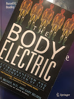
The Body Electric, superimposed on the cover of Intermediate Physics for Medicine and Biology
The Body Electric,
By Robert Becker and Gary Selden
Go to www.amazon.com and look up the best selling book in the category “biophysics.” You’ll often find #1 is The Body Electric: Electromagnetism and the Foundation of Life, by Robert Becker and Gary Selden. (Selden helped with the writing, but the book tells Becker’s story.) The purpose of today’s post is to explain why this book is awful.

1. Let’s begin with Becker’s views on nerve conduction (page 86).
“According to the theory, an impulse should travel with equal ease in either direction along the nerve fiber. If the nerve is stimulated in the middle, an impulse should travel in both directions to opposite ends. Instead, impulses travel only in one direction; in experiments they can be made to travel ‘upstream,’ but only with great difficulty. This may not seem like such a big deal, but it is very significant. Something seems to polarize the nerve.”

I stimulated many nerves as a graduate student, back in the days when I did experiments. Action potentials propagate just fine in either direction. I had no difficulty making one travel upstream.


2. Becker didn’t understand why nerves, which fire all-or-none action potentials, can produce smooth, coordinated muscle movements (page 87).

“In addition, impulses always have the same magnitude and speed. This may not seem like such a big thing either, but think about it. It means the nerve can carry only one message, like the digital computer’s 1 or 0… The motor activities we take for granted—getting out of a chair and walking across the room, picking up a cup and drinking coffee, and so on—require integration of all the muscles and sensory organs working smoothly together to produce coordinated movements that we don’t even have to think about. No one has ever explained how the simple code of impulses can do all that.”
A muscle contains many motor units. Each motor unit is controlled by a single motor neuron. If you want a muscle to contract with a small force, you activate one motor unit. If you want a muscle to contract more forcefully, you activate many motor units. Motor unit recruitment, plus changes in nerve firing rate, explains the smooth operation of muscles. 


3. Becker didn’t believe that nerves worked using ionic currents, meaning the movement of ions like sodium, potassium, and chloride dissolved in the salt water that makes up our tissues (page 92).

“At that earlier time, there had been only two known modes of current conduction, ionic and metallic. Metallic conduction can be visualized as a cloud of electrons moving along the surface of metal, usually a wire. It can be automatically excluded from living creatures because no one has ever found any wires in them. Ionic current is conducted in solutions by the movement of ions—atoms or molecules charged by having more or fewer than the number of electrons needed to balance their protons’ positive charges. Since ions are much bigger than electrons, they move more laboriously through the conducting medium, and ionic currents die out after short distances. They work fine across the thin membrane of the nerve fiber, but it would be impossible to sustain an ionic current down the length of even the shortest nerve.”

Regular readers of this blog will recall my post about Baker, Hodgkin and Shaw’s experiment in which they squeezed the axoplasm out of a squid nerve axon and replaced it with salt water. The nerve worked just fine. 

Some individual molecules work by transfer of electrons (for instance, the electron transport chain in mitochondria), but currents flowing through tissue are produced by ions. Ionic currents don’t “die out” after short distances.
 

4. Instead of ionic conduction, Becker believed that nerves conducted electricity by semiconduction (page 94).

“I postulated a primitive, analog-coded information system that was closely related to the nerves but not necessarily located in the nerve fibers themselves. I theorized that this system used semiconducting direct currents and that, either alone or in concert with the nerve impulse system, it regulated growth, healing, and perhaps other basic processes.”
Later, he performed measurements of the Hall effect (a voltage induced by current flowing in a magnetic field) and wrote (page 102):
“The experiment demonstrated unequivocally that there was a real electric current flowing along the salamander’s foreleg, and it virtually proved that the current was semiconducting. In fact, the half-dozen tests I’d performed supported every point of my hypothesis.”
Scientists have made semiconductors based on biological ideas: organic semiconductors and semiconductors based on synthetic biology. But there’s no evidence that semiconductors play a role in our normal physiology. Our bodies are basically all salt water. Ionic conduction is the way currents flow through our tissue.
 

5. Becker declared he had discovered the mechanism of acupuncture (page 234).

“The acupuncture meridians, I suggested, were electrical conductors that carried an injury message to the brain, which responded by sending back the appropriate level of direct current to stimulate healing in the troubled area…. If the lines and points [corresponding to acupuncture meridians] really were conductors and amplifiers, the skin above them would show specific electrical differences compared to the surrounding skin.”
Acupuncture is based on pseudoscience; No anatomical structures such as “meridians” exist, and the vital force “qi” has never been observed. Listen to Harriet Hall describe acupuncture. Read what Edzard Ernst says.

6. Becker asserted that static magnetic fields could act as an anesthetic (page 238).

“A strong enough magnetic field oriented at right angles to a current magnetically ‘clamped it’, stopping the flow [of current]. By placing frogs and salamanders between the poles of an electromagnet so that the back-to-front current in their heads was perpendicular to the magnetic lines of force, we could anesthetize the animals just as well as we could with chemicals.”
Such neurological effects are not caused by static magnetic fields. Patients have undergone magnetic resonance imaging in static magnetic fields far larger than what Becker used, and no one has been anesthetized, regardless of the orientation of their head. Using magnets for pain has been discredited.

7. Becker thought that the cells forming the myelin sheaths surrounding myelinated nerve axons carried their own electric current that could have biological effects (page 239).

Electron microscope work has shown that the cytoplasm of all Schwann cells is linked together through holes in the adjacent membranes, forming a syncytium that could provide the uninterrupted pathway needed by the current.”
The Schwann cells make up the myelin sheath. Myelin consists of layers of fat with little cytoplasm between the layers. Its purpose is to insulate a nerve between openings called nodes of Ranvier. There is no evidence myelin carries significant current, but even if it did carry current along a nerve through the myelin, it would be interrupted ever millimeter or so by a node. 
 

8. Becker believed that magnetoencephalography confirmed his claim that DC current existed in the brain (page 241)

“The MEG research so far seems to be establishing that every electrical evoked potential is accompanied by a magnetic evoked potential. This would mean that the evoked potentials and the EEG of which they’re a part reflect true electrical activity, not some artifact of nerve impulses being discharged in unison, as was earlier theorized. Some of the MEG’s components could come from such additive nerve impulses, but other aspects of it clearly indicate direct currents in the brain.”
DC currents in the brain are uncommon, and primarily associated with brain injury or migraines. Researchers in biomagnetism interpret their results as arising from additive nerve impulses, discharged in unison.

9. Becker promoted the idea that extrasensory perception was a result of DC or extremely low frequency (ELF) electromagnetic fields (page 267).

“At this time the DC perineural system [myelin sheaths around nerves] and its electromagnetic fields provide the only theory of parapsychology that’s amenable to direct experiment. And it yields hypotheses for almost all such phenomena except precognition. Telepathy may be transmission and reception via a biologically programmed channel of ELF vibrations in the perineural system’s electromagnetic field.”
What can I say? I don’t believe in extrasensory perception.
 

10. Becker suggested electromagnetic effects could explain psychokinesis, such as spoon-bending by pure thought (page 269).

“Once we admit the idea of this kind of influence, then the same kind of willed action of biofields on the electromagnetic structure of inanimate matter becomes a possibility. This encompasses all forms of psychokinesis, from metal-bending experiments in which trickery has been excluded to more rigidly controlled tests with interferometers, strain gauges, and random number generators.”
I don’t believe in psychokinesis either.  Neither did James Randi, who died just a year ago.


11. Becker claimed that weak magnetic fields could affect cognitive ability in humans (page 276).

“We exposed volunteers to magnetic fields placed so the lines of force passed through the brain from ear to ear, cutting across the brainstem-frontal current. The fields were 5 to 11 gauss [0.0005 to 0.0011 tesla], not much compared with the 3,000 gauss needed to put a salamander to sleep, but ten to twenty times earth’s background and well above the level of most magnetic storms. We measured their influence on a standard test of reaction time—having subjects press a button as fast as possible in response to a red light. Steady fields produced no effect, but when we modulated the field with a slow pulse of a cycle every five seconds (one of the delta-wave frequencies we’d observed in salamander brains during a change from one level of consciousness to another), people’s reaction slowed down.”
Many reviews of the biological effects of magnetic fields conclude there are no such effects.
 

12. Becker championed the idea that 60-Hz, power line magnetic fields could cause cancer. But he went even further, saying the such “electropollution” could threaten human existence (page 327).

“Everyone worries about nuclear weapons as the most serious threat to our survival. Their danger is indeed immediate and overwhelming. In the long run, however, I believe the ultimate weapon is manipulation of our electromagnetic environment, because it’s imperceptibility subtle and strikes at the core of life itself. We’re dealing here with the most important scientific discovery ever—the nature of life. Even if we survive the chemical and atomic threats to our existence, there’s a strong possibility that increasing electropollution could set in motion irreversible changes leading to our extinction before we’re even aware of them.” 

The “electropollution” Becker speaks of is weak electric and magnetic fields, such as produced by power lines. Power line magnetic fields are safe, and earlier claims that they are not have been shown to be false (see my previous post). “Electropollution” is closer to an imaginary threat than an existential one.


What do I make of all this? Becker’s book is full of nonsense. Moreover, I know little about some of the topics in the book, such as regeneration, bone growth, and injury currents. There could well be more mistakes than just those I’ve caught.

Becker died almost twelve years ago. Am I beating a dead horse? No. According to Google Scholar, The Body Electric has been cited more than 1000 times in the scientific literature (twice as many times as Intermediate Physics for Medicine and Biology), including over 25 times in 2021 already. It’s cited by the supporters of the worst kind of alternative medicine foolishness. The 5G opponents quote him. The power lines and cancer folks quote him. The magnets for pain promoters quote him.

You might wonder: am I upset just because The Body Electric gets more sales and citations than IPMB? Well, maybe that’s part of it. But I believe debunking Becker’s book is a public service. People need to learn real science.

My favorite story in The Body Electric is the time a bigwig physiologist visited Becker’s lab, and told him outright that his results were “artifact, all artifact” (page 106). Thereafter, Becker and his colleagues referred to this fellow derisively as “Artifact Man” and held him up as a symbol for dogmatism. I love Artifact Man.

Chapter 1 sums up Becker’s view of medicine with a defense of “faith healing, magic healing, psychic healing, and spontaneous healing” (page 25). He goes on to say (page 29) 
“The more I consider the origins of medicine, the more I’m convinced that all true physicians seek the same thing. The gulf between folk therapy and our own stainless-steel version is illusory. Western medicine springs from the same roots and, in the final analysis, acts through the same little-understood forces as its country cousins. Our doctors ignore this kinship at their—and worse, their patients’—peril. All worthwhile medical research and every medicine man’s intuition is part of the same quest for knowledge of the same elusive healing energy.”
No, No, No. The origins of medicine should be science. The gulf between folk therapy and modern medicine is wide and must get wider. Our doctors ignore science at their—and worse, their patients’—peril.

Okay, I’m done now. I realize this post is more of a rant than is usual for me. Sorry about that, but there’s something about The Body Electric that really gets my goat.

Friday, November 12, 2021

Bidomain Modeling of Electrical and Mechanical Properties of Cardiac Tissue

This week Biophysics Reviews published my article “Bidomain Modeling of Electrical and Mechanical Properties of Cardiac Tissue” (Volume 2, Article Number 041301, 2021). The introduction states
This review discusses the bidomain model, a mathematical description of cardiac tissue. Most of the review covers the electrical bidomain model, used to study pacing and defibrillation of the heart. For a book-length analysis of this topic, consult the recently published second edition of Cardiac Bioelectric Therapy. In particular, one chapter in that book complements this review: it contains a table listing many bidomain predictions and their experimental confirmation, includes many original figures from earlier publications, and cites additional references. Near the end, the review covers the mechanical bidomain model, which describes mechanotransduction and the resulting growth and remodeling of cardiac tissue.

The review has several aims: to (1) introduce the bidomain model to younger investigators who are bringing new technologies from outside biophysics into cardiac physiology; (2) examine the interaction of theory and experiment in biological physics; (3) emphasize intuitive understanding by focusing on simple models and qualitative explanations of mechanisms; and (4) highlight unresolved controversies and open questions. The overall goal is to enable technologists entering the field to more effectively contribute to some of the pressing scientific questions facing physiologists.

My manuscript traveled a long and winding road. The initial version was a personal account of my career as I worked on the bidomain model (Russ Hobbie and I discuss the bidomain concept in Chapter 7 of Intermediate Physics for Medicine and Biology), and was organized around ten papers I published between 1986 and 2010, with an emphasis on the 1990s. My first draft (and all subsequent ones) benefited from thoughtful comments by my former graduate student, Dilmini Wijesinghe. After I fixed all the problems Dilmini found, I sent the initial version to the editor. He responded that the journal board wanted a more traditional, authoritative review article. That was fine, so I transformed the paper from a memoir into a review, and submitted it officially to the journal. Then the reviewers had a couple rounds of helpful comments, leading to more revisions. Next, there were changes in the page proofs to fulfill all the journal editorial rules. At last, it was published.

The final version is unlike the initial one. I changed the perspective from first person to third; added figures; increased the number of references by almost 50%; and deleted all the reminiscences, colorful anecdotes, and old war stories. 

I hope you enjoy the peer-reviewed, published article. If you want to read the original version (the one with the war stories), you can find it here.  

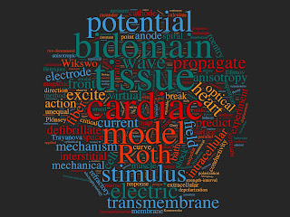
I made a word cloud based on the article. The giant “Roth” is embarrassing, but otherwise it provides a nice summary of what the paper is about.

Word Cloud of "Bidomain Modeling of Electrical and Mechanical Properties of Cardiac Tissue."

Biophysics Reviews is a new journal, edited by my old friend Kit Parker. Long-time readers of this blog may remember Parker as the guy who said “our job is to find stupid and get rid of it.” Listen to him describe his goals as Editor-in-Chief.

Kit Parker, Editor-in-Chief of Biophysics Reviews, introduces the journal.

https://www.youtube.com/watch?v=2V1fpskjJtM

Friday, November 5, 2021

Electroreception

Suppose you’re reading Homework Problem 4 in Chapter 8 of Intermediate Physics for Medicine and Biology, and you run across the phrase “If a shark can detect an electric field strength of 0.5 μV m−1…”. What’s your first reaction? Probably you suspect a typo (it isn’t). An electric field with a strength of 0.5 μV m−1 is tiny. By comparison, you need a field of about 10 V m−1 to stimulate a neuron in the brain. How can a shark detect a field of only 0.0000005 V m−1? The answer makes for an interesting story.

Some of the first studies of electroreception—the ability of some animals, such as sharks, to sense weak electric fields—were performed by a biophysicist at Woods Hole Oceanographic Institute named Adrianus Kalmijn. He observed dogfish sharks while sitting in an inflatable rubber raft in the ten-foot deep water of the Atlantic Ocean near Martha’s Vineyard. Kalmijn attracted the sharks using liquified herring placed on the ocean floor. On either side of the herring was a pair of electrodes that could be used to pass current. The dogfish were initially attracted by the smell of the herring, and “began frantically searching over the sand, apparently trying to locate the odor source” (Kalmijn, 1977). But when current was turned on, the dogfish stopped searching for the herring and “viciously attacked” the electrodes! Using experiments like these, Kalmijn was able to characterize how sharks respond to electric fields. 


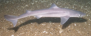
Spiny dogfish (Squalus acanthias).
Spiny dogfish (Squalus acanthias) at the Josephine Marie shipwreck, Stellwagen National Marine Laboratory. From Wikipedia.


Sharks detect weak electric fields using sensory organs called the ampullae of Lorenzini. The ampullae consist of highly conducting jelly-filled tubes about 30 cm long (a little more than a foot). The shark detects the voltage across the length of the tube, and then places that entire voltage difference across a single cell membrane. An electric field of 0.5 μV m−1 multiplied by a distance of 0.3 m gives you a voltage of 0.15 μV. There’s an extra factor of three arising from the distortion of the field by the shark, so you end up with a transmembrane voltage of about half a microvolt.

A membrane voltage of 0.5 μV is minuscule. The typical resting membrane voltage of a cell is approximately 70 mV, so half a microvolt is less than ten parts per million. How can such a small voltage change be detected? To answer this question, William Pickard, an engineer at Washington University in St. Louis, assumed that this membrane voltage does not cause a neuron to fire (it’s far too weak for that), but instead modulates its spontaneous firing rate. The neuron normally operates in a regime where this rate is very sensitive to the membrane voltage, which has the effect of magnifying a small change in voltage into a large change in rate (Pickard, 1988).

Many ampullae of Lorenzini influence a single neuron. Their summation has the effect of averaging out any background noise. The size of thermal voltage fluctuations across a neuron’s membrane were estimated by Yale physicist Robert Adair to be about 1 μV (Adair, 1991), which is twice as large as the membrane voltage produced by the smallest electric field a shark can detect. Integrating the signal over hundreds of ampullae suppresses these fluctuations, allowing the system to pick a signal out of the thermal background. This sensory mechanism has been honed by evolution to be about as sensitive as it can be without detecting the constant roar of random noise. 

To learn more about electroreception, see Section 9.9 of Intermediate Physics for Medicine and Biology.

  1. Kalmijn, A. J. (1977) “The electric and magnetic sense of sharks, skates, and rays.” Oceanus Volume 20, Pages 45-52.
  2. Pickard, W. F. (1988) “A model for the acute electrosensitivity of cartilaginous fishes.” IEEE Transactions on Biomedical Engineering Volume 35, Pages 243-249. 
  3. Adair, R. K. (1991) “Constraints on biological effects of weak extremely low-frequency electromagnetic fields.” Physical Review A Volume 43, Pages 1039-1048. 

The ampullae of Lorenzini. https://www.youtube.com/watch?v=9S8a5hSc22s