Friday, December 25, 2020

The Mathematical Approach to Physiological Problems

In Chapter 2 of Intermediate Physics for Medicine and Biology, Russ Hobbie and I analyze decay with multiple half-lives, and practice fitting data to exponentials. Near the end of this discussion, we write (really, Russ wrote these words, since they go back to his solo first edition of IPMB)
Estimating the parameters [governing exponential decay] for the longest-lived term may be difficult because of the potentially large error bars associated with the data for small values of y. For a discussion of this problem, see Riggs (1970, pp. 146–163).
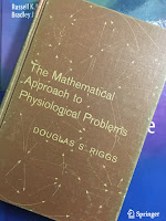
The Mathematical Approach of Physiological Problems, by Douglas S. Riggs, superimposed on Intermediate Physics for Medicine and Biology.
The Mathematical Approach
to Physiological Problems
,
by Douglas S. Riggs.
The citation is to the book The Mathematical Approach to Physiological Problems, by Douglas Riggs. I  wanted to take a look, so I went to the Oakland University library and checked out the 1963 first edition.

It’s a gold mine. I particularly like the beginning of the preface. Riggs starts with what seems like an odd digression about hiking in the mountains, but then in the second paragraph he skillfully brings us back to math.

Before settling down in the village of Shepreth, in Cambridgeshire, England, to start working in earnest upon this book, I had the unusual pleasure of taking my family on a month-long youth hosteling trip through Northern England and the Scottish Highlands. For the most part, we bicycled, but occasionally we would make an excursion on foot up the steep hillsides and along the rocky ridges where no bicycle could go. We soon learned that the British discriminate carefully between “hill walking” and “mountain climbing.” To be a mountain climber, you must coil 100 feet of nylon rope around you slantwise from shoulder to waist, and have a few pitons dangling somewhere about. You are then entitled to adopt an ever-so-faintly condescending attitude toward any hill-walkers whom you may encounter along the trail, even if you meet them where the trail is practically level. Hill-walkers, on the other hand, remain hill-walkers even when the “walk” turns into a hands-and-knees job up a 40° slope of talus which is barely anchored to the mountain by a few wisps of grass and a clump or two of scraggly heather.

Mathematically speaking, this is a hill-walking book. It is necessarily so, since I myself have never learned the ropes of higher mathematics. But I do believe that the amount of wandering I have done on the lower slopes, the number of sorry hours I have spent lost in a mathematical fog, and the miles I have stumbled down false trails have made me a kind of backwoods expert on mathematical pitfalls, and have given me some practical knowledge of how to plan a safe mathematical ascent of the more accessible physiological hills.
One theme I stress in this blog is the value of simple models. Riggs agrees.
Precisely because living systems are so very complex, one can never expect to achieve anything like a complete mathematical description of their behavior. Before the mathematical analysis itself is begun, it is therefore invariably necessary to reduce the complexity of the real system by making various simplifying assumptions about how it behaves. In effect, these assumptions allow us to replace the actual biological system by an imaginary model system which is simple enough to be described mathematically. The results of our mathematical analysis will then be rigorously applicable to the model. But they will be applicable to the original biological system only to the extent that our underlying assumptions are reasonable. Hence, the ultimate value of our mathematical labors will be determined in large part by our choice of simplifying assumptions.
He then advocates for an approach that I call “think before you calculation.”
An investigator who publishes an erroneous equation has no place to hide! It is therefore prudent to check each calculation, each algebraic manipulation, and each transcription from a table of figures before going on to the next step. Above all, whenever you are engaged in mathematical work you should keep asking yourself over and over and over again, “Does this make sense?” and “Is this of the correct magnitude?
Riggs’s introduction ends with some wise advice.
All too frequently, students are willing to accept on faith whatever mathematical formulations they encounter in their reading. And why not? After all, mathematics is the exact science, and presumably an author would not express his theories or his conclusions mathematically without due regard for mathematical rigor and precision. It is only by bitter experience that we learn never to trust a published mathematical statement or equation, particularly in a biological publication, unless we ourselves have checked it to see whether or not it makes sense… Misprints are common. Copying errors are common. Blunders are common. Editors rarely have the time or training to check mathematical derivations. The author may be ignorant of mathematical laws, or he may use ambiguous notation. His basic premises may be fallacious even though he uses impressive mathematical expressions to formulate his conclusions… Caveat lector! Let the reader beware!
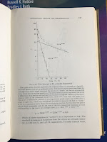
What about the problem of fitting multiple exponentials to data, which is why Russ and I cite Riggs in the first place? After analyzing several specific examples, Riggs concludes
Page 157 of The Mathematical Approach to Physiological Problems, showing a fit to a double exponential, superimposed on Intermediate Physics for Medicine and Biology.
Page 157 of The Mathematical
Approach to Physiological Problems
,
showing a fit to a double exponential.
These examples warn us not to take too seriously any particular set of coefficients and rate constants which we may get by plotting data on semi-logarithmic paper and ferreting out the exponential terms in the fashion described above. The need for such a warning is all too evident from the preposterously elaborate exponential equations which are sometimes published. The technique of “peeling off” successive terms is so deceptively easy! Fit a straight line, subtract, plot the differences, fit another straight line, subtract, plot the differences. How solid and impressive the resulting sum of exponentials looks! And how remarkably well the curve agrees with the observations. Surely the investigator can be pardoned a certain self-satisfaction for having so clearly identified the individual components which were contributing to the overall change. Yet, the examples discussed above show how groundless his satisfaction may be. It is undoubtedly true that the particular sum of exponentials which he happened to pick, plot, and publish fits the points with gratifying accuracy. But so also may other equations of the same general form but with quite different parameters. It is not great trick to have found one such equation. Even with a single exponential declining from a known value at time zero toward an unknown constant asymptote there are two parameters—the rate constant and the asymptote—to be fitted to the data. The effect of a considerable change in either may be largely offset by making a compensatory change in the other… Add a second exponential term with two more parameters to be estimated from the data, and the number and variety of “closely fitting” equations becomes truly bewildering. Worst of all, there are no simple statistical measures of the precision with which any of the parameters have been estimated. These considerations do not destroy the value of fitting an exponential equation to experimental data when it is suggested by some underlying theory or when it provides a convenient empirical way of summarizing a group of observations mathematically… But they make very clear the danger of using an empirical exponential equation to predict what may happen beyond the period actually covered by the observations. It is equally clear that we must be exceedingly skeptical when attempts are made to match the individual terms of an empirical exponential equation with supposedly corresponding processes or regions of the body.
Riggs’s book examines many of the same topics that appear in the first half of IPMB, such as exponential growth, diffusion, and feedback. He has a wonderful chapter suggesting questions you should ask when checking the validity of an equation. Is it dimensionally correct? How does it behave when variables approach zero or infinity? Does it give reasonable answers after numerical substitution?

I’m impressed that Riggs, a professor and head of a Department of Pharmacology at SUNY Buffalo, could write so insightfully about mathematics. I give the book two thumbs up.
Friday, December 18, 2020

Life at Low Reynolds Number

The first page of "Life at Low Reynolds Number," by Edward Purcell, superimposed on Intermediate Physics for Medicine and Biology.
“Life at Low Reynolds Number,”
by Edward Purcell.

In Chapter 1 of Intermediate Physics for Medicine and Biology, Russ Hobbie and I cite Edward Purcell’s wonderful article “Life at Low Reynolds Number” (American Journal of Physics, Volume 45, Pages 3–11, 1977). This paper is a transcript of a talk Purcell gave to honor physicist Victor Weisskopf. The transcript captures the casual tone of the talk, and the hand-drawn figures are charming. Below I quote excerpts from the article, including my own versions of a couple of the drawings. Notice how his words emphasize insight. As Purcell says, “some essential hand-waving could not be reproduced.” Enjoy!

I’m going to talk about a world which, as physicists, we almost never think about. The physicist hears about viscosity in high school when he’s repeating Millikan’s oil drop experiment and never hears about it again, as least not in what I teach. And Reynolds’s number, of course, is something for the engineers. And the low Reynolds number regime most engineers aren’t even interested in… But I want to take you into the world of very low Reynolds number—a world which is inhabited by the overwhelming majority of the organisms in this room. This world is quite different from the one that we have developed our intuitions in…

Based on Figure 1 from
“Life at Low Reynolds Number.”

In Fig. 1, you see an object which is moving through a fluid with velocity v. It has dimension a,… η and ρ are the viscosity and density of the fluid. The ratio of the inertial forces to the viscous forces, as Osborne Reynolds pointed out slightly less than a hundred years ago, is given by avρ/η or av/ν, where ν is called the kinematic viscosity. It’s easier to remember its dimensions; for water, ν = 10−2 cm2/sec. The ratio is called the Reynolds number and when that number is small the viscous forces dominate… Now consider things that move through a liquid... The Reynolds number for a man swimming in water might be 104, if we put in reasonable dimensions. For a goldfish or a tiny guppy it might get down to 102. For the animals that we’re going to be talking about, as we’ll see in a moment, it’s about 10−4 or 10−5. For these animals inertia is totally irrelevant. We know F = ma, but they could scarcely care less. I’ll show you a picture of the real animals in a bit but we are going to be talking about objects which are the order of a micron in size… In water where the kinematic viscosity is 10−2 cm2/sec these things move around with a typical speed of 10 μm/sec. If I have to push that animal to move it, and suddenly I stop pushing, how far will it coast before it slows down? The answer is, about 0.1 Å. And it takes it about 0.6 μsec to slow down. I think this makes it clear what low Reynolds number means. Inertia plays no role whatsoever. If you are at very low Reynolds number, what you are doing at the moment is entirely determined by the forces that are exerted on you at that moment, and by nothing in the past…

Based on Figure 18 from
“Life at Low Reynolds Number.”

Diffusion is important because of [a] very peculiar feature of the world at low Reynolds number, and that is, stirring isn’t any good… At low Reynolds number you can’t shake off your environment. If you move, you take it along; it only gradually falls behind. We can use elementary physics to look at this in a very simple way. The time for transporting anything a distance by stirring is about divided by the stirring speed v. Whereas, for transport by diffusion, it’s 2 divided by D, the diffusion constant. The ratio of those two times is a measure of the effectiveness of stirring versus that of diffusion for any given distance and diffusion constant. I’m sure this ratio has someone’s name but I don’t know the literature and I don’t know whose number that’s called. Call it S for stirring number. It’s just v/D. You’ll notice by the way that the Reynolds number was v/ν. ν is the kinematic viscosity in cm2/sec, and D is the diffusion constant in cm2/sec, for whatever it is that we are interested in following—let’s say a nutrient molecule in water. Now, in water the diffusion constant is pretty much the same for every reasonably sized molecule, something like 10−5 cm2/sec. In the size domain that we’re interested in, of micron distances, we find that the stirring number S is 10−2, for the velocities that we are talking about (Fig. 18). In other words, this bug can’t do anything by stirring its local surroundings. It might as well wait for things to diffuse, either in or out. The transport of wastes away from the animal or food to the animal is entirely controlled locally by diffusion. You can thrash around a lot, but the fellow who just sits there quietly waiting for stuff to diffuse will collect just as much.


The Physics of Life: Life at Low Reynolds Number
https://www.youtube.com/watch?v=gZk2bMaqs1E


 Edward Purcell
https://www.youtube.com/watch?v=0uATCx7WMMs

Friday, December 11, 2020

Selig Hecht (1892-1947)

A photo of Selig Hecht
Selig Hecht,
History of the Marine Biological Laboratory,
http://hpsrepository.asu.edu/handle/10776/3269.
In Chapter 14 of Intermediate Physics for Medicine and Biology, Russ Hobbie and I analyze the “classic experiment on scotopic vision” by Hecht, Shlaer, and Pirenne. George Wald wrote an obituary about Selig Hecht in 1948 (Journal of General Physiology, Volume 32, Pages 1–16). He writes that Hecht was
Intensely interested in the relation of light quanta (photons) to vision. Reexamining earlier measurements of the minimum threshold for human rod vision, he and his colleagues confirmed that vision requires only fifty to 150 photons. When all allowances had been made for surface reflections, the absorption of light by ocular tissues, and the absorption by rhodopsin (which alone is an effective stimulant), it emerged that the minimum visual sensation corresponds to the absorption in the rods of, at most, five to fourteen photons. An entirely independent statistical analysis suggested that an absolute threshold involves about five to seven photons. Both procedures, then, confirmed the estimation of the minimum visual stimulus at five to fourteen photons. Since the test field in which these measurements were performed contained about 500 rods, it was difficult to escape the conclusion that one rod is stimulated by a single photon.
Wald also describes the coauthor on the study, Shlaer.
Among Hecht’s first students was Simon Shlaer, who became Hecht’s assistant in his first year at Columbia and continued as his associate for twenty years thereafter. A man infinitely patient with things and impatient with people, Shlaer gave Hecht his entire devotion. He was a master of instrumentation, and though he also had a keen grasp of theory, he devoted himself by choice to the development of new technical devices. Hecht and Shlaer built a succession of precise instruments for visual measurement, among them an adaptometer and an anomaloscope that have since gone into general use. The entire laboratory came to rely on Shlaer’s ingenuity and skill. “I am like a man who has lost his right arm,” remarked Hecht on leaving Columbia—and Shlaer—in 1947, “and his right leg.”

In his Columbia laboratory, Hecht instituted investigations of human dark adaptation, brightness discrimination, visual acuity, the visual response to flickered light, the mechanism of the visual threshold, and normal and anomalous color vision. His lab also made important contributions regarding the biochemistry of visual pigments, the relation of night blindness to vitamin A deficiency in humans, the spectral sensitivities of man and other animals, and the light reactions of plants—phototropism, photosynthesis, and chlorophyll formation.
Hecht and Shlaer both contributed to the war effort during the Second World War.
Throughout the late years of World War II, Hecht devoted his energies and the resources of his laboratory to military problems. He and Shlaer developed a special adaptometer for night-vision testing that was adopted as standard equipment by several Allied military services. Hecht also directed a number of visual projects for the Army and Navy and was consultant and advisor on many others. He was a member of the National Research Council Committee on Visual Problems and of the executive board of the Army-Navy Office of Scientific Research and Development Vision Committee.

Explaining the Atom, by Selig Hecht, superimposed on Intermediate Physics for Medicine and Biology.
Explaining the Atom,
by Selig Hecht.
Hecht straddled the fields of physics and physiology, and was comfortable with both math and medicine. He entered college studying mathematics. After World War II ended, he wrote the book Explaining the Atom, which Wald described as “a lay approach to atomic theory and its recent developments that the New York Times (in a September 20, 1947, editorial) called ‘by far the best so far written for the multitude.’”

An obituary in Nature by Maurice Henri Pirenne concludes

The death of Prof. Selig Hecht in New York on September 18, 1947, at the age of fifty-five, deprives the physiology of vision of one of its most outstanding workers. Hecht was born in Austria and was brought to the United States as a child. He studied and worked in the United States, in England, Germany and Italy. After a broad biological training, he devoted his life to the study of the mechanisms of vision, considered as a branch of general physiology. He became professor of biophysics at Columbia University and made his laboratory an international centre of visual research.

Friday, December 4, 2020

Role of Virtual Electrodes in Arrhythmogenesis: Pinwheel Experiment Revisited

The Journal of Cardiovascular Electrophysiology, with a figure from Lindblom et al. on the cover, superimposed on Intermediate Physics for Medicine and Biology.
The Journal of Cardiovascular Electrophysiology,
with a figure from Lindblom et al. on the cover.

Twenty years ago, I published an article with Natalia Trayanova and her student Annette Lindblom about initiating an arrhythmia in cardiac muscle (“Role of Virtual Electrodes in Arrhythmogenesis: Pinwheel Experiment Revisited,” Journal of Cardiovascular Electrophysiology, Volume 11, Pages 274-285, 2000). We performed computer simulations based on the bidomain model, which Russ Hobbie and I discuss in Section 7.9 of Intermediate Physics for Medicine and Biology. A key feature of a bidomain is anisotropy: the electrical conductivity varies with direction relative to the axis of the myocardial fibers.

Our results are summarized in the figure below (Fig. 14 of our article). An initial stimulus (S1) launched a planar wavefront through the tissue, either parallel to (longitudinal, L) or perpendicular to (transverse, T) the fibers (horizontal). As the tissue recovered from the first wave front, we applied a second stimulus (S2) to a point cathodal electrode (C), inducing a complicated pattern of depolarization under the cathode and two regions of hyperpolarization (virtual anodes) adjacent to the cathode along the fiber axis (see my previous blog post for more about how cardiac tissue responds to a point stimulus). In some simulations, we reversed the polarity of S2 so the electrode was an anode (A). This pair of stimuli (S1-S2) underlies the “pinwheel experiment” that has been studied by many investigators, but never before using the anisotropic bidomain model. 

Fig. 14 from Lindblom et al. (2000).

We found a variety of behaviors, depending on the direction of the S1 wave front, the polarity of the S2 stimulus, and the time between S1 and S2, known as the coupling interval (CI). In some cases, we induced a figure-of-eight reentrant circuit: an arrhythmia consisting of two spiral waves, one rotating clockwise and the other counterclockwise. In other cases, we induced quatrefoil reentry: an arrhythmia consisting of four spiral waves (see my previous post for more about the difference between these two behaviors).

I began working on these calculations in the winter of 1999, shortly after I arrived at Oakland University as an Assistant Professor. The photograph below is of a page from my research notebook on March 5 showing initial results, including my first observation of quatrefoil reentry in the pinwheel experiment (look for “Quatrefoil!”).

The March 5, 1999 entry from my research notebook,
showing my first observation of quatrefoil reentry
induced during the pinwheel experiment.

A few weeks later I got a call from my friend Natalia (see my previous post about an earlier collaboration with her). She was organizing a session for the IEEE Engineering in Medicine and Biology Society conference, to be held in Atlanta that October, and asked me to give a talk. We got to chatting and she started to describe simulations she and Lindblom were doing. They were the same calculations I was analyzing! I told her about my results, and we decided to collaborate on the project, which ultimately led to our Journal of Cardiovascular Electrophysiology paper.

Our article was full of beautiful color figures showing the different types of arrhythmias. Below is a photo of two pages of the article. Those familiar with my previous publications will notice that the color scheme representing the transmembrane potential is different than what I usually used. Lindblom and Trayanova had their own color scale, and we decided to adopt it rather than mine. One of the figures was featured on the cover of the March, 2000 issue the journal. Lindblom made some lovely movies to go along with these figures, but they’re now lost in antiquity. I later discovered that a simple cellular automata model could reproduce many of these results (see my previous post for details).

Two pages from Lindblom et al. (2000),
showing some of the color figures.

The editor asked Art Winfree to write an editorial to go along with our article (see my previous post about Winfree). I especially like his closing remarks.

This is clearly a landmark event in cardiac electrophysiology at the end of our century. It is sure to have major implications for clinical electrophysiologic work and for defibrillator design.
In retrospect, he was overly optimistic; the paper was an incremental contribution, not a landmark event of the 20th century. But I appreciated his kind words.

Friday, November 27, 2020

Defibrillation Mechanisms: The Parable of the Blind Men and the Elephant

“Defibrillation Mechanisms:
The Parable of the Blind Men
and the Elephant,”
by Ideker, Chattipakorn, and Gray.

I’ve read many scientific papers, but only one began with an eight-stanza poem about an elephant. Twenty years ago, Ray Ideker, Nipon Chattipakorn, and Rick Gray published “Defibrillation Mechanisms: The Parable of the Blind Men and the Elephant” in the Journal of Cardiovascular Electrophysiology (Volume 11, Pages 1008-1013, 2000). The opening poem by John Godfrey Saxe is reproduced below.

The purpose of the article was to review the different hypotheses that explain defibrillation of the heart. Russ Hobbie and I discuss defibrillation in Chapter 7 of Intermediate Physics for Medicine and Biology.

Ventricular fibrillation occurs when the ventricles contain many interacting reentrant wavefronts that propagate chaotically… During fibrillation the ventricles no longer contract properly, blood is no longer pumped through the body, and the patient dies in a few minutes. Implantable defibrillators are similar to pacemakers, but are slightly larger. An implanted defibrillator continually measures the [electrocardiogram]. When a signal indicating fibrillation is sensed, it delivers a much stronger shock that can eliminate the reentrant wavefronts and restore normal heart rhythm.
Ideker et al. discuss several possible mechanisms that explain how an electrical shock terminates fibrillation. This is a difficult problem, and I’ve spent much of my career trying to figure it out (I guess I’m one of the blind men).
It is possible that most of the electrical and optical mapping studies and the associated hypotheses about the mechanism of interaction of electrical stimuli with myocardium are all valid. It may be that shocks of low strength do not halt the activation fronts of fibrillation; and shocks of higher strength, depending on the circumstances, cause polarization critical points, field-recovery critical points, and/or action potential prolongation; whereas still stronger shocks slightly below the defibrillation threshold cause activation that appears focal on the epicardium either by intramural reentry, by reentry involving the Purkinje fibers, or by true focal activity, perhaps caused by delayed or early afterdepolarization… If so, then just as in the parable of the blind men and elephant, most of the reported studies and proposed defibrillation mechanisms all may be partially correct, yet all may be partially wrong because they are incomplete.
Defibrillation is a fine example of how a knowledge of physics can help solve a critical problem in medicine. Apparently a knowledge of poetry helps too.

Friday, November 20, 2020

The Virtual Museum of Medical Physics

How would you like to visit a museum dedicated solely to medical physics? Well, with COVID-19 raging, we shouldn’t visit any museums in person. But how about visiting a virtual museum? The History Committee of the American Association of Physicists in Medicine has recently opened a Virtual Museum of Medical Physics.

The Virtual Museum was launched to celebrate the 125th anniversary of the discovery of x-rays on November 8, 1895 by Wilhelm Roentgen. Existing exhibits include those about Roentgen, Fluoroscopy, Mammography, and External Beam Radiotherapy. Exhibits under construction include Computed Tomography, Ultrasonic Imaging, Magnetic Resonance Imaging, and Nuclear Medicine. Once it’s done, the Virtual Museum will be a wonderful adjunct to Intermediate Physics for Medicine and Biology. If you want to contribute to developing an exhibit, contact the Virtual Museum.

Asimov's Biographical Encyclopedia of Science & Technology, superimposed on Intermediate Physics for Medicine and Biology.
Asimov’s Biographical Encyclopedia
of Science & Technology
.
And now, the story of how Roentgen discovered x-rays, as told in Asimov’s Biographical Encyclopedia of Science & Technology.
ROENTGEN, Wilhelm Konrad 
German physicist 
Born: Lennep, Rhenish Prussia, March 27, 1845 
Died: Munich, Bavaria, February 10, 1923

...The great moment that lifted Roentgen out of mere competence and made him immortal came in the autumn of 1895 when he was head of the department of physics at the University of Wurzburg in Bavaria. He was working on cathode rays and repeating some of the experiments of Lenard and Crookes. He was particularly interested in the luminescence these rays set up in certain chemicals.

In order to observe the faint luminescence, he darkened the room and enclosed the cathode ray tube in thin black cardboard. On November 5, 1895, he set the enclosed cathode ray tube into action and a flash of light that did not come from the tube caught his eye. He looked up and quite a distance from the tube he noted that a sheet of paper coated with barium platinocyanide was glowing. It was one of the luminescent substances, but it was luminescing now even though the cathode rays, blocked off by cardboard, could not possibly be reaching it.

He turned off the tube; the coated paper darkened. He turned it on again; it glowed. He walked into the next room with the coated paper, closed the door, and pulled down the blinds. The paper continued to glow while the tube was in operation.

It seemed to Roentgen that some sort of radiation was emerging from the cathode-ray tube, a radiation that was highly penetrating and yet invisible to the eye. By experiment he found the radiation could pass through considerable thicknesses of paper and even through thin layers of metal. Since he had no idea of the nature of the radiation, he called it X rays, X being the usual mathematical symbol for the unknown. For a time, there was a tendency to call them Roentgen rays, but the inability of the non-Teutonic tongue to wrap itself about the German œ diphthong militated against that. The unit of X-ray dosage is, however, officially called the roentgen.

Friday, November 13, 2020

The SIR Model of Epidemics

In Chapter 10 of Intermediate Physics for Medicine and Biology, Russ Hobbie and I discuss models described by nonlinear differential equations. We provide several examples in the text and homework problems, but one topic we never address is epidemics.

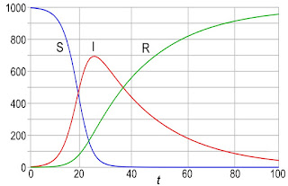
The archetype mathematical description of an epidemic is the SIR model. A population is divided into three categories, corresponding to three dynamic variables:

    S: the number of susceptible people

    I: the number of infected people

    R: the number of recovered people.

Three differential equations govern the number of people in each category.

    dS/dt = - (β/N) I S

    dI/dt = (β/N) I Sγ I

    dR/dt = γ I

where N is the total population, and β and γ are constants. Rather than analyze these equations myself, I’ll let you do it in a new homework problem.
Section 10.8

Problem 36 ½. The SIR model describes the dynamics of an epidemic. 
(a) Add the three differential equations and determine how the total number of people (S + I + R) changes with time. Does this model include people who die from the disease? 
(b) Write the equation governing the number of infected people as dI/dt = γI (r0 – 1). Find an expression for r0. Initially, when S = N, what does r0 reduce to? This value of r0 is known as the basic reproduction number. If r0 is less than what value will the number of infected people decay, preventing an epidemic?
(c) Suppose r0 is greater than one, so the number of infected people grows and the epidemic spreads. How low must the ratio S/N become for I to begin decreasing? Once this value of S/N is reached, the population is said to have herd immunity and the epidemic decays away.
Results from a numerical simulation of the SIR model.
Results from a numerical simulation of the SIR model, using S(0) = 997, I(0) = 3, R(0) = 0, β = 0.4, and γ = 0.04. By Klaus-Dieter Keller, CC0, https://commons.wikimedia.org/w/index.php?curid=77633956

The SIR model provides insight into the COVID-19 pandemic. It’s a simple model, and many researchers have modified it to be more realistic. Yet, there is value in a toy model like SIR. It lets you to gain intuition about a dynamical system without being overwhelmed by complexity. I always encourage students to first master a toy model, and only then add additional detail.

Friday, November 6, 2020

International Day of Medical Physics

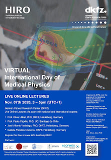
Poster for the International Day of Medical Physics.
Poster for the
International Day of
Medical Physics.

Tomorrow is the International Day of Medical Physics! This year’s theme is the “Medical Physicist as a Health Professional.”

The second half of Intermediate Physics for Medicine and Biology focuses on medical physics topics, such as ultrasound, radiation therapy, tomography, nuclear medicine, and magnetic resonance imaging. These concepts are central to the work of medical physicists in our hospitals. The COVID-19 pandemic reminds us of how important health care professionals are. They are truly essential workers.

Nine years ago the International Organization for Medical Physics established this annual celebration of medical physics. The IOMP represents tens of thousands of medical physicists worldwide. It’s mission is to advance medical physics practice by “disseminating scientific and technical information, fostering the educational and professional development of medical physicists, and promoting the highest quality medical services for patients.” Below is a message from the President of the IOMP, Madan Rehani.

A message from Madan Rehani, President of the International Organization for Medical Physics.
https://www.youtube.com/watch?v=yFlOi7k8IjA

German Cancer Research Center will host a series of live online lectures celebrating the International Day of Medical Physics

How can you celebrate this special day? Tomorrow the German Cancer Research Center will host a series of live online lectures about medical physics aimed at a general audience. They will take place 3–5 PM their time, which would be 9–11 AM my time (Eastern Standard Time in the United States). You have to register to get the zoom link, but it’s free.

November 7 was chosen for the International Day of Medical Physics because it’s the birthday of Marie Curie. Below are excerpts from the Asimov’s Biographical Encyclopedia of Science & Technology entry about Curie. Enjoy!


Asimov's Biographical Encyclopedia of Science & Technology, by Isaac Asimov.
Asimov's Biographical
Encyclopedia of
Science & Technology.

CURIE, Marie Sklodowska (kyoo-ree’) 

Polish-French chemist 

Born: Warsaw, Poland, November 7, 1867 

Died: Haute Savoie, France, July 4, 1934

Asimov begins by discussing Curie’s education.

Marie was unable to obtain any education past the high school level in repressed Poland. An older brother and sister had left for Paris in search of education and Marie worked to help meet their expenses and to save money for her own trip there, meanwhile teaching herself as best she could out of books. In 1891 her earnings had accumulated to the minimum necessary, and off she went to Paris where she entered the Sorbonne. She lived with the greatest frugality during this period (fainting with hunger in the classroom at one time), but when she graduated, it was at the top of the class….
He then describes the scientific discoveries that underlie Curie’s research
The discovery of X rays by Roentgen and of uranium radiations by A. H. Becquerel galvanized Marie Curie into activity. It was she who named the process whereby uranium gave off rays “radioactivity.” She studied the radiations given off by uranium and her reports coincided with those of Ernest Rutherford and Becquerel in showing that there were three different kinds of rays, alpha, beta, and gamma….
Later, he explains the Nobel Prize winning research Marie Curie performed with her husband Pierre.
At the physics school where the Curies worked there was an old wooden shed with a leaky roof, no floor, and very inadequate heat. The two obtained permission to work there and for four years (during which Marie Curie lost fifteen pounds) they carefully purified and repurified the tons of [uranium] ore into smaller and smaller samples of more and more intensely radioactive material… Marie’s burning determination kept the husband-and-wife team going in the face of mountainous difficulties. By 1902 they had succeeded in preparing a tenth of a gram of radium after several thousand crystallizations. Eventually, eight tons of pitchblende gave them a full gram of the [radium] salt
Asimov ends with Curie’s final years.
Her last decades were spent in the supervision of the Paris Institute of Radium. She had made no attempt to patent any part of the extraction process of radium and it remained in the glamorous forefront of the news for nearly a generation, thanks to its ability to stave off the inroads of cancer under the proper circumstances. But in the end Marie died of leukemia (a form of cancer of the leukocyte-forming cells of the body) caused by overexposure to radioactive radiation.

Marie Curie - Scientist. https://www.youtube.com/watch?v=ZEV4KJBJvEg

Friday, October 30, 2020

Fundamental Limits of Spatial Resolution in PET

In Chapter 17 of Intermediate Physics for Medicine and Biology, Russ Hobbie and I discuss positron emission tomography.

17.10 Positron Emission Tomography

If a positron emitter is used as the radionuclide, the positron comes to rest and annihilates an electron, emitting two annihilation photons back to back. In positron emission tomography (PET) these are detected in coincidence.
Anyone who has looked at PET images will be struck by their low spatial resolution. They provide valuable functional information, but little anatomical detail. Why?
The first page of “Fundamental Limits of Spatial Resolution in PET,” by William Moses, superimposed on Intermediate Physics for Medicine and Biology.
“Fundamental Limits of
Spatial Resolution in PET,”
by William Moses.

In a 2011 article in Nuclear Instruments and Methods in Physics Research A (“Fundamental Limits of Spatial Resolution in PET,” Volume 648, Supplement 1, Pages S236–S240), William Moses analyses what factors contribute to PET spatial resolution.
Abstract: The fundamental limits of spatial resolution in positron emission tomography (PET) have been understood for many years. The physical size of the detector element usually plays the dominant role in determining resolution, but the combined contributions from acollinearity, positron range, penetration into the detector ring, and decoding errors in the detector modules often combine to be of similar size. In addition, the sampling geometry and statistical noise further degrade the effective resolution. This paper quantitatively describes these effects, discusses potential methods for reducing the magnitude of these effects, and computes the ultimately achievable spatial resolution for clinical and pre-clinical PET cameras.

Detector size

The most obvious limitation of spatial resolution comes from the detector size. Usually detection occurs in a scintillator crystal that converts a gamma ray to visible light, which is detected by a photomultiplier. The width of the scintillator limits the spatial solution of the image. A typical detector size is about 4 mm.

Positron range

A positron is emitted with an energy of about a million electronvolts. It then travels through tissue until it slows enough to capture an electron and and give off two 0.511 MeV photons. The range of the positron sets a limit to the spatial resolution. Different isotopes emit positrons with different energies. One of the most widely used isotopes for functional studies is 18F, which has a range of about half a millimeter. Most other common isotopes used in PET have longer ranges.

Acollinearity

If a positron and electron are at rest when they annihilate, they emit two 0.511 MeV photons. To conserve momentum, these photons must travel in opposite directions. If, however, the positron-electron pair has some kinetic energy when they annihilate, the photons are not emitted exactly in opposite directions. Usually they deviate from 180° by up to 0.25°. This translates into about one to two millimeters of blurring in typical detector rings.

Decoding

Decoding is complicated. Many PET devices have more scintillators than photomultipliers, so the photomultipliers take turns recording from different scintillators (a process called multiplexing). The PET scanner must then decode all this information, and this decoding process is not perfect. Moses estimates that decoding introduces an uncertainty of about a third of the detector width, or around a couple millimeters in spatial resolution.

Penetration

The 0.511MeV photons can penetrate into the ring of detectors used in a PET device, causing blurring. In the illustration below, if the source (green) contains an isotope that emits two photons, then for some angles those photons (red) are detected by a single detector, but for other angles (blue) they are detected by multiple detectors. 

An illustration based on Fig. 2 of
“Fundamental Limits of Spatial Resolution in PET,”
by William Moses, showing how penetration of a
photon into different detectors causes blurring.

Sampling Error

A detector ring is more sensitive to sources at some positions compared to others (see Moses’s Fig. 3 for an explanation). This effect tends to degrade by spatial resolution by about 25%.


If you add all these uncertainties in quadrature, you get a spatial resolution of about 6 mm. This is worse resolution that you would have for magnetic resonance imaging or computed tomography, which is why PET images look so blurry. They are often overlaid on an MRI (see Fig. 17.25 in IPMB).

If you decided to build a PET system with the best possible spatial resolution (regardless of complexity or cost), you could eliminate all of the sources of uncertainty except positron range and acollinearity, implying a spatial resolution of about 2 mm (worse for isotopes other than 18F). PET is never going to image small-scale anatomical detail.

Friday, October 23, 2020

Qualifying Exams

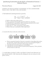
First page of the 2020 Physics Qualifying Exam.
First page of the 2020 Physics
Qualifying Exam.
I have discussed the Oakland University Medical Physics PhD Qualifying Exam previously in this blog. It’s a series of three written exams over math, physics, and biology, plus an oral exam.

I’ve collected the written qualifying exams over the last ten years (2011-2020) in a single file that you’re welcome to download. These exams are broad but not deep. They cover material at a level similar to, or somewhat higher than, Intermediate Physics for Medicine and Biology. Anyone working at the intersection of physics with biology should master these topics. The exams allow you to hone your problem solving skills. If you’re a student interested in applying physics to physiology, or math to medicine, but are stuck at home and not able to attend classes because of the covid-19 pandemic, you might want to try solving the 300 problems in this collection. (Slightly less than 300, because there wasn’t a math exam in 2014 and because we sometimes repeated questions from previous years.) If you want to try some of the exams from 2010 or earlier, you can find them at https://sites.google.com/view/bradroth/home/medical-physics-graduate-program/qualifying-exams?authuser=0.

Sorry, but I don’t have written solutions for these exams. You can always email me (roth@oakland.edu) if you’re stuck.

Enjoy!

Friday, October 16, 2020

Boron Neutron Capture Therapy

In Chapter 16 of Intermediate Physics for Medicine and Biology, Russ Hobbie and I discuss boron neutron capture therapy.

Boron neutron capture therapy (BNCT) is based on a nuclear reaction which occurs when the stable isotope 10B is irradiated with neutrons, leading to the nuclear reaction (in the notation of Chap. 17)
Both the alpha particle and lithium are heavily ionizing and travel only about one cell diameter. BNCT has been tried since the 1950s; success requires boron-containing drugs that accumulate in the tumor. The field has been reviewed by Barth (2003).
The first page of Barth, RF (2003) A Critical Assessment of Boron Neutron Capture Therapy: An Overview. Journal of Neuro-Oncology, Volume 62, Pages 1–5, superimposed on Intermediate Physics for Medicine and Biology.
Barth (2003)
J Neurooncol 62:1–5.
The citation is to an article by Rolf Barth of Ohio State University.
Barth, RF (2003) A Critical Assessment of Boron Neutron Capture Therapy: An Overview. Journal of Neuro-Oncology, Volume 62, Pages 1–5.
The abstract of this seventeen-year-old review states
Boron neutron capture therapy (BNCT) is based on the nuclear reaction that occurs when boron-10 is irradiated with neutrons of the appropriate energy to produce high-energy alpha particles and recoiling lithium-7 nuclei. BNCT has been used clinically to treat patients with high-grade gliomas, and a much smaller number with primary and metastatic melanoma. The purpose of this special issue of the Journal of Neuro-Oncology is to provide a critical and realistic assessment of various aspects of basic and clinical BNCT research in order to better understand its present status and future potential. Topics that are covered include neutron sources, tumor-targeted boron delivery agents, brain tumor models to assess therapeutic efficacy, computational dosimetry and treatment planning, results of clinical trails in the United States, Japan and Europe, pharmacokinetic studies of sodium borocaptate and boronophenylalanine (BPA), positron emission tomography imaging of BPA for treatment planning, and finally an overview of the challenges and problems that must be faced if BNCT is to become a useful treatment modality for brain tumors. Clinical studies have demonstrated the safety of BNCT. The next challenge is an unequivocal demonstration of therapeutic efficacy in one or more of the clinical trails that either are in progress or are planned over the next few years.
I was wondering what’s happening in this field lately, so I searched the Physics World website and found a fascinating and recent article by Tami Freeman.
Boron neutron capture therapy (BNCT), a technique that deposits highly targeted radiation into tumour cells, was first investigated as a cancer treatment back in the 1950s. But the field remains small, with only 1700 to 1800 patients treated to date worldwide. This may be about to change.

“The field of BNCT seems to be progressing rapidly at the moment,” said Stuart Green, director of medical physics at University Hospital Birmingham. “The big difference compared with five or ten years ago is that the commercial interest from a variety of companies is strong now and this is driving the field…”

Speaking at the Medical Physics & Engineering Conference (MPEC), Green updated on the status of BNCT programmes worldwide, noting that clinical experience is continually increasing. The US Food and Drug Administration has now approved two boron drugs for clinical use. But by far the majority of treatments, over 1150 to date, have taken place in Japan, initially using the Kyoto University Reactor in the early 2000s, and more recently using three Sumitomo accelerator systems in Kyoto, Fukushima and Osaka.

“Very importantly, earlier this year we had the first ever medical device approval for BNCT, for treatment in Japan of recurrent head-and-neck cancer,” said Green. “This is a significant marker for the entire field....”
“For the first time, there’s a substantial and sustained effort in the commercial sector to drive this field forward,” Green concluded. “We should keep an eye on BNCT over the next few years, there’s a lot happening, and hopefully our community can play a key role.”

Why the renewed interest in this technique? First, the original clinical applications of BNCT used neutrons from a nuclear reactor. Now accelerator-based neutron sources are available that can be installed in a hospital. Second, researchers are working hard on boron-containing drugs. Currently, boronophenylalanine and sodium borocaptate are the most common drugs used clinically. Improving the delivery of these drugs, or designing entirely new drugs, could increase the usefulness of BNCT. 

We live in exciting times.

Boron Neutron Capture Therapy Animation. 

https://www.youtube.com/watch?v=cQrbfBiQpfk

Friday, October 9, 2020

Generation of Unidirectionally Propagating Action Potentials Using a Monopolar Electrode Cuff

The first page from Ungar et al. (1986) “Generation of Unidirectionally Propagating Action Potentials Using a Monopolar Electrode Cuff,” Ann. Biomed. Engin., 14:437-450, superimposed on Intermediate Physics for Medicine and Biology.
Ungar et al. (1986) Ann.
Biomed. Engin.
, 14:437–450.
In Chapter 7 of Intermediate Physics for Medicine and Biology, Russ Hobbie and I discuss electrical stimulation of nerves. One of the end-of-the-chapter homework problems asks the students to
Design a stimulator that will result in one-way propagation… For an application of such [a device] during functional electrical stimulation, see Ungar et al. (1986).
The reference is
Ungar IJ, Mortimer JT, Sweeney JD (1986) “Generation of Unidirectionally Propagating Action Potentials Using a Monopolar Electrode Cuff,” Annals of Biomedical Engineering, Volume 14, Pages 437–450.
The abstract to their article states
Unidirectionally propagating action potentials, which can be used to implement transmission failure on peripheral nerve through “collision block,” have been generated electrically on cat myelinated peripheral nerve using a monopolar electrode cuff with the conductor positioned closest to the “arrest” end of the cuff. A single cathode located at least 5 mm from the arrest end resulted in unidirectional propagation with minimal current and charge injection. The range of stimulus current values that produced unidirectional propagation increased with increases in longitudinal asymmetry of cathode placement over the range of asymmetries tested (1.7:1 to 7:1). The stimulus current pulse that minimized charge injection was quasitrapezoidal in shape with a plateau pulse width of approximately 350 μsec and an exponential trailing phase having a fall time (90%–10%) of approximately 600 μsec. These parameters were found to be independent of cuff geometry. Arrest efficiency was not degraded using a cuff of sufficient internal diameter to prevent nerve compression in chronic implantation. The critical current density within the extracellular space of the electrode cuff required to produce conduction failure at the arrest end was estimated to be 0.47 ± 0.08 mA/mm2.
I’ll explain how to make a one-way stimulator using the illustration below, adapted from Ungar et al.’s Figure 1.
The design of a one-way neural stimulator.
Adapted from Fig. 1 of Ungar et al. (1986).

The nerve (blue) is threaded through a cylindrical insulating cuff (red), which resembles a short segment of a plastic drinking straw. The cathode (green) is inside the cuff; it stimulates the action potential. The anode (not shown) is far away. Current (purple curves) comes out of the cathode and enters the nerve axons, depolarizing them. Once it reaches the end of the cuff the current spreads out as it returns to the distance anode. Current there leaves the axons, hyperpolarizing them, and lowering their transmembrane potential. The locations where the axons are hyperpolarized are labeled the virtual anodes.

The key to making a one-way stimulator is to place the cathode off-center in the cuff. Most of the current leaves the cuff through the end nearest the cathode (right end), so the current density is stronger there (the purple current lines are crowded together). Only a small fraction of the current leaves the cuff through the end farther from the cathode (left end), so the virtual anode is weaker there.

Depolarization under a cathode excites an action potential, which then propagates outward in both directions (left and right). If, however, the stimulus strength is strong enough, the hyperpolarization at a virtual anode can block propagation. If the current has the correct strength, the stronger virtual anode on the right will block propagation, while the weaker virtual anode on the left won’t. In that case, an action potential will propagate to the left (the escape end of the stimulator) but will not propagate to the right (the arrest end).

Ira Ungar and his collaborators were able to test their stimulator for different cathodal current strengths. For a very weak stimulus, the cathode is below threshold and no action potential is excited. For a moderately weak stimulus, the cathode excites an action potential that then propagates to both the left and the right; both virtual anodes are too weak to block propagation. For a moderately strong stimulus, the right virtual anode is strong enough to block the action potential and you have one-way propagation. For a very strong stimulus, both ends block propagation, and no action potential leaves the stimulator.

Why construct a one-way stimulator? Suppose you have a nerve that’s constantly firing action potentials, causing unwanted muscle contraction (spasticity). You could stop the downstream propagation of those action potentials by electrically stimulating action potentials further downstream. The stimulated action potentials propagating upstream will collide with the original action potentials propagating downstream, annihilating them (colliding action potentials can’t pass through each other because they’re each followed by a region of refracotoriness). This works great unless your stimulator sends its own volley of action potentials propagating downstream to excite the muscle. To avoid this problem, you need a one-way stimulator, so you only excite action potentials propagating upstream to block those causing the trouble, but none propagating downstream.

The senior author on this article was J. Thomas Mortimer, now emeritus professor in the Neural Engineering Center at Case Western Reserve University. He earned his PhD from Case and then spent his entire career there. He has developed an online Applied Neural Control Toolkit to teach how nerves work. I heard Mortimer speak during one of the Neural Prosthesis Workshops at the National Institutes of Health; he was inspirational.

Mortimer and his team’s development of a one-way stimulator is a classic example of how physics and engineering can contribute to medicine and biology.

Friday, October 2, 2020

The Death and Life of the Great Lakes

Many readers of this blog live far from me—sometimes in countries on the other side of the world—and may not be interested in local topics germane to Michigan. Yet the Great Lakes are important to everyone; they hold 20% of the earth’s liquid freshwater. The challenges faced by the Great Lakes aren’t unique; they’re relevant to other watersheds worldwide.

For those who aren’t Michiganders, a drawing of the five Great Lakes looks like a map of Michigan

The Great Lakes
From Phizzy at Wikipedia.

Between its upper and lower peninsulas, Michigan borders four of the five lakes. I live half way between the southern tip of Lake Huron and the western edge of Lake Erie. I’ve visited all five lakes, and I’m concerned about their welfare.
The Death and Life of the Great Lakes,
by Dan Egan.
 
I recently read The Death and Life of the Great Lakes (2017), by Dan Egan. (This book was recommended to me by Congresswoman Elissa Slotkin, who’s now running for reelection in my home district). I’ve lived in Michigan for 22 years, but much of this story was new to me. In his introduction, Egan writes
Iconic disasters have a history of prompting government action. Three years after the Cuyahoga River fire of 1969, Congress passed the Clean Water Act. Two decades later, when the Exxon Valdez ran aground and dumped 10.8 million gallons of crude oil into Alaska’s Prince William Sound, images of cleanup crews using paper towels to cleanse tarred birds helped press Congress into doing something it should have done years earlier. It mandated double-hulled oil tankers.

But the disaster unfolding today on the Great Lakes didn’t ignite like a polluted river or gush like oil from a cracked hull, and so far there is no galvanizing image of this slow-motion catastrophe, though a few come to my mind. One is the bow of an overseas ship easing its way into the first navigation lock on the St. Lawrence Seaway, the Great Lakes’ “front door” to fresh waves of biological pollution. Another is a satellite photo of a green-as-paint toxic algae slick smothering as much as 2,000 square miles of Lake Erie.

Yet another is the grotesque mug of an Asian carp, a monster-sized carp imported to the United States in the 1960s and used in government experiments to gobble up excrement in Arkansas sewage lagoons. The fish, which can grow to 70 pounds and eat up to 20 percent of their weight in plankton per day, escaped into the Mississippi River basin decades ago and have been migrating north ever since. There are now mustering at the Great Lakes’ “back door”—the Chicago canal system that created a manmade connection between the previously isolated Great Lakes and the Mississippi basin, which covers about 40 percent of the continental United States. The only thing blocking the fish’s swim through downtown Chicago and into Lake Michigan is an electrical barrier in the canal—one that has a history of unexpected shutdowns.

The Chicago canal has also turned the Great Lakes’ ballast water problem [ships from the Atlantic Ocean having exotic species in their ballast water and then dumping it into the Great Lakes] into a national one, because there are dozens of invasive species poised to ride its waters out of the lakes and into the rivers and water bodies throughout the heart of the continent. Species like the spiny water flea, the threespine stickleback, the bloody red shrimp and the fishhook water flea. All organisms you probably haven’t heard of. Yet.

Few out West, after all, had ever heard of quagga mussels—until they tumbled down the Chicago canal and metastasized across the Mississippi basin and, eventually, into the arid West, likely as hitchhikers aboard recreational boats towed over the Rocky Mountains. The mussels have since unleashed havoc on hydroelectric dams, drinking water systems and irrigation networks in Utah, Nevada and California and the federal government estimates that if the mussels make their way into the Northwest’s Columbia River hydroelectric dam system they could do a half billion dollars of damage—per year.
Much of Egan’s book is about how invasive species interact with native animals. In Chapter 2 of Intermediate Physics for Medicine and Biology, Russ Hobbie and I discuss the predator-prey problem modeled by the Lotka-Volterra equations. It is a useful toy model for understanding the interplay of two species, but it doesn’t explain the workings of a large and complex ecosystem.

The Great Lakes (at least the four that border Michigan) were once isolated from the Atlantic Ocean by Niagara Falls, a barrier than even the most determined fish could not surmount. The opening of the Saint Lawrence Seaway allowed a series of nonnative animals to enter the lakes. First came the sickening sea lamprey that nearly caused the native lake trout to go extinct. Next was an explosion of alewife (a river herring). Scientists introduced coho and Chinook salmon to prey upon the alewives. In each case, the ramifications of a new species were difficult to predict. Then zebra mussels and the quagga muscles arrived, and finally the round goby. It’s a horror story, and I suspect an invasion of the Asian carp is inevitable.

Besides these exotic species, the Great Lakes (especially Lake Erie) suffer from algae blooms triggered by fertilizer runoff. Add in climate change, which is causing the water level in the lakes to fluctuate, and you have an unstable and unhealthy ecosystem. 

I’ll give Egan the last word.
If we can close these doors to future invasions [of invasive species], we may give the lakes, and the rest of the country, time to reach a new equilibrium, a balance between what is left of the natural inhabitants and all the newcomers... And if we do this, then we can focus on the major problems that still plague the lakes, which include the overapplication of farm fertilizer that is helping to trigger the massive toxic algae outbreaks, the impact a warming globe is having on the lake’s increasingly unstable water levels and the need to protect lake waters from outsiders seeking to drain them for their own profit.

Dan Egan on The Death and Life of the Great Lakes at the 2018 L.A. Times Festival of Books. https://www.youtube.com/watch?v=jtq_cN1kPkg