Showing posts with label my own research. Show all posts

Friday, July 18, 2025

Millikan and the Magnetic Field of a Single Axon

“The Magnetic Field of a Single Axon: A Comparison of Theory and Experiment” superimposed on Intermediate Physics for Medicine and Biology.
The Magnetic Field of a Single Axon:
A Comparison of Theory and Experiment.”

Forty years ago this month, I published one of my first scientific papers. “The Magnetic Field of a Single Axon: A Comparison of Theory and Experiment” appeared in the July, 1985 issue of the Biophysical Journal (Volume 48, Pages 93–109). I was a graduate student at Vanderbilt University at the time, and my coauthor was my PhD advisor John Wikswo. When discussing the paper below, I will write “I did this…” and “I thought that…” because I was the one in the lab doing the experiments, but of course it was really Wikswo and I together writing the paper and analyzing the results.

Selected Papers of Great American Physicists superimpsed on the cover of Intermediate Physics for Medicine and Biology.
Selected Papers of
Great American Physicists
.
In those days I planned to be an experimentalist (like Wikswo). About the time I was writing “The Magnetic Field of a Single Axon,” I read “On the Elementary Electrical Charge and The Avogadro Constant” by Robert Millikan (Physical Review, Volume 11, Pages 109–143, 1913). It had been reprinted in the book Selected Papers of Great American Physicists, published by the American Institute of Physics.

If you are reading this blog, you’re probably are familiar with Millikan’s oil drop experiment. He measured the speed of small droplets of oil suspended in air and placed in gravitational and electric fields, and was able to determine the charge of a single electron. I remember doing this experiment as a undergraduate physics major at the University of Kansas. I was particularly impressed by the way Millikan analyzed his experiment for possible systematic errors: He worried about deviations of the frictional force experienced by the drops from Stokes’ law and corrected for it; he analyzed the possible changes to the density of the oil in small drops; he checked that his 5300 volt battery was calibrated correctly and supplied a constant voltage; and he fussed about convection currents in the air influencing his results. He was especially concerned about his value of the viscosity of air, which he estimated was known to about one part in a thousand. Rooting out systematic errors is a hallmark of a good experimentalist. I wanted to be like Millikan, so I analyzed my magnetic field measurement for a variety of systematic errors.

The first type of error in my experiment was in the parameters used to calculate the magnetic field (so I could compare it to the measured field). I estimated that my largest source of error was in my measurement of the axon radius. This was done using a reticle in the dissecting microscope eyepiece. I only knew the radius to 10% accuracy, in part because I could see that it was not altogether uniform along the axon, and because I could not be sure the axon’s cross section was circular. It was my biggest source of error for calculating the magnitude of the magnetic field, because the field varied as the axon cross-sectional area, which is proportional to the radius squared.
Figure 1 from "The Magnetic Field of a Single Axon."
Figure 1 from "The Magnetic
Field of a Single Axon."

I measured the magnetic field by threading the axon through a wire-wound ferrite-core toroid (I’ve written about these toroid measurements before in this blog). I assumed the axon was at the center of the toroid, but this was not always the case. I performed calculations assuming the toroid averaged the magnetic field for an off-axis axon, and was able to set an upper limit on this error of about 2%. The magnetic field was not measured at a point but was averaged over the cross-sectional area of the ferrite core. More numerical analysis suggested that I could account for the core area to within about 1%. I was able to show that inductive effects from the toroid were utterly negligible. Finally, I assumed the high permeability ferrite did not affect the magnetic field distribution. This should be true if the axon is concentric with the toroid and aligned properly. I didn’t have a good way to estimate the size of this error.

Figure 2 from "The Magnetic Field of a Single Axon."
Figure 2 from "The Magnetic
Field of a Single Axon."
The toroid and axon were suspended in a saline bath (technically, Van Harreveld's solution), and this bath gave rise to other sources of error. I analyzed the magnetic field for different sized baths (the default assumption was an unbounded bath), and for when the bath had a planar insulating boundary. I could do the experiment of measuring the magnetic field as we raised and lowered the volume of fluid in the bath. The effect was negligible. I spent a lot of time worrying about the heterogeneity caused by the axon being embedded in a nerve bundle. I didn’t really know the conductivity of the surrounding nerve bundle, but for reasonable assumptions it didn’t seem to have much effect. Perhaps the biggest heterogeneity in our experiment was the “giant” (~1 mm inner radius, 2 mm outer radius, 1 mm thick) toroid, which was embedded in an insulated epoxy coating. This big chunk of epoxy certainly influenced the current density in the surrounding saline. I had to develop a new way of calculating the extracellular current entirely numerically to estimate this effect. The calculation was so complicated that Wikswo and I didn’t describe it in our paper, but instead cited another paper that we listed as “in preparation” but that in fact never was published. I concluded that the toroid was not a big effect for my nerve axon measurements, although it seemed to be more important when I later studied strands of cardiac tissue.

Figure 3 of "The Magnetic Field of a Single Axon."
Figure 3 of "The Magnetic
Field of a Single Axon."
Other miscellaneous potential sources of error include capacitive effects in the saline and an uncertainty in the action potential conduction velocity (measured using a second toroid). I determined the transmembrane potential by taking the difference between the intracellular potential (measured by a glass microelectrode, see more here) and a metal extracellular electrode. However, I could not position the two electrodes too accurately, and the extracellular potential varies considerably over small distances from the axon, so my resulting transmembrane potential certainly had a little bit of error. Measurement of the intracellular potential using the microelectrode was susceptible to capacitive coupling to the surrounding saline bath. I used a “frequency compensator” to supply “negative capacitance” and correct for this coupling, but I could not be sure the correction was accurate enough to avoid introducing any error. One of my goals was to calculate the magnetic field from the transmembrane potential, so any systematic errors in my voltage measurements were concerning. Finally, I worried about cell damage when I pushed the glass microelectrode into the axon. I could check this by putting a second glass microelectrode in nearby and I didn’t see any significant effect, but such things are difficult to be sure about.

All of this analysis of systematic errors, and more, went into our rather long Biophysical Journal paper. It remains one of my favorite publications. I hope Millikan would have been proud. If you want to learn more, see Chapter 8 about Biomagnetism in Intermediate Physics for Medicine and Biology

Forty years is a long time, but to this old man it seems like just yesterday.

Friday, March 14, 2025

The First Measurement of the Magnetocardiogram

Biomagnetism: The First Sixty Years, superimposed on Intermediate Physics for Medicine and Biology.
Biomagnetism: The First Sixty Years.
A couple years ago, I published a review article titled “Biomagnetism: The First Sixty Years. I wrote about that article before in this blog, but I thought it was time for an update. The paper is popular: according to Google Scholar it has been cited 28 times in two years, which is more citations than any other of my publications in the last decade. I remember working on this paper because it was my Covid project. That year I got Covid for the first—and, so far, only—time. I quarantined myself in our upstairs bedroom, wore a mask, and somehow avoided infecting my wife. I remember having little to do except work on my biomagnetism review.

As a treat, I thought I would reproduce one of the initial sections of the article (references removed) about the first measurement of the magnetocardiogram. Russ Hobbie and I talk about the MCG in Chapter 8 of Intermediate Physics for Medicine and Biology. This excerpt goes into more detail about how MCG measurements began. Enjoy!
2.1. The First Measurement of the Magnetocardiogram

In 1963, Gerhard Baule and Richard McFee first measured the magnetic field generated by the human body. Working in a field in Syracuse, New York, they recorded the magnetic field of the heart: the magnetocardiogram (MCG). To sense the signal, they wound two million turns of wire around a dumbbell-shaped ferrite core that responded to the changing magnetic field by electromagnetic induction. The induced voltage in the pickup coil was detected with a low-noise amplifier.

The ferrite core was about one-third of a meter long, so the magnetic field was not measured at a single point above the chest, but instead was averaged over the entire coil. One question repeatedly examined in this review is spatial resolution. Small detectors are often noisy and large detectors integrate over the area, creating a trade-off between spatial resolution and the signal-to-noise ratio.

The heart’s magnetic field is tiny, on the order of 50–100 pT (Figure 1). A picotesla (pT) is less than a millionth of a millionth as strong as the magnetic field in a magnetic resonance imaging machine. The magnetic field of the earth is about 30,000,000 pT (Figure 1), and the only reason it does not obscure the heart’s field is that the earth’s field is static. That is not strictly true. The earth’s field varies slightly over time, which causes geomagnetic noise that tends to mask the magnetocardiogram (Figure 1). Moreover, even a perfectly static geomagnetic field would influence the MCG if the pickup coil slightly vibrated. A key challenge in biomagnetic recordings, and a major theme in this review, is the battle to lower the noise enough so the signal is detectable

Noise sources in biomagnetism.
Figure 1. Noise sources in biomagnetism.
Most laboratories contain stray magnetic fields from sources such as electronic equipment, elevators, or passing cars (Figure 1). Baule and McFee avoided much of this noise by performing their experiments at a remote location. Even so, they had to filter out the ubiquitous 60 Hz magnetic field arising from electrical power distribution. A magnetic field changing at 60 Hz is a particular nuisance for biomagnetism because the magnetic field typically exists in a frequency band extending from 1 Hz (1 s between heartbeats) to 1000 Hz (1 ms rise time of a nerve or muscle action potential).

One limitation of a metal pickup coil is the thermal currents in the winding due to the random motion of electrons, creating extraneous magnetic fields caused by the measuring device itself. The ultimate source of noise is thermal currents in the body, but fortunately, their magnetic field is minuscule (Figure 1).

Baule and McFee suppressed background noise by subtracting the output of two pickup coils. A distant source of noise gave the same signal in both coils and did not contribute to their difference. One coil was placed over the heart, and the magnetocardiogram was larger there and did not cancel out. The two coils formed a rudimentary type of gradiometer (Figure 2).

The magnetocardiogram resembled the electrocardiogram (ECG) sensed by electrodes attached to the skin. Baule and McFee speculated that the MCG might contain different information than the ECG, another idea that reappears throughout this review. In a followup article, they theoretically calculated the magnetic field produced by the heart. The interplay between theory and experiments is yet one more subject that frequently arises in this article.

Noise sources in biomagnetism.
Figure 2. Types of gradiometers.

Friday, October 11, 2024

Extracellular Magnetic Measurements to Determine the Transmembrane Action Potential and the Membrane Conduction Current in a Single Giant Axon

Forty years ago today I was attending my first scientific meeting: The Society for Neuroscience 14th Annual Meeting, held in Anaheim, California (October 10–15, 1984). As a 24-year-old graduate student in the Department of Physics at Vanderbilt University, I presented a poster based on the abstract shown below: “Extracellular Magnetic Measurements to Determine the Transmembrane Action Potential and the Membrane Conduction Current in a Single Giant Axon.”

I can’t remember much about the meeting. I’m sure I flew to California from Nashville, Tennessee, but I can’t recall if my PhD advisor John Wikswo went with me (his name is not listed on any meeting abstract except the one we presented). I believe the meeting was held at the Anaheim Convention Center. I remember walking along the sidewalk outside of Disneyland, but I didn’t go in (I had visited there with my parents as a child).

Neuroscience Society meetings are huge. This one had over 300 sessions and more than 4000 abstracts submitted. In the Oct. 11, 1984 entry in my research notebook, I wrote “My poster session went OK. Several people were quite enthusiastic.” I took notes from talks I listened to, including James Hudspeth discussing hearing, a Presidential Symposium by Gerald Fischbach, and a talk about synaptic biology and learning by Eric Kandel. I was there when Theodore Bullock and Susumu Hagiwara were awarded the Ralph W. Gerard Prize in Neuroscience.

The research Wikswo and I reported in our abstract was eventually published in my first two peer-reviewed journal articles:

Barach, J. P., B. J. Roth and J. P. Wikswo, Jr., 1985, Magnetic Measurements of Action Currents in a Single Nerve Axon: A Core-Conductor Model. IEEE Transactions on Biomedical Engineering, Volume 32, Pages 136-140.

Roth, B. J. and J. P. Wikswo, Jr., 1985, The Magnetic Field of a Single Axon: A Comparison of Theory and Experiment. Biophysical Journal, Volume 48, Pages 93-109.

Both are cited in Chapter 8 of Intermediate Physics for Medicine and Biology.

This neuroscience abstract was not my first publication. I was listed as a coauthor on an abstract to the 1983 March Meeting of the American Physical Society, based on some research I helped with as an undergraduate physics major at the University of Kansas. But I didn’t attend that meeting. In my CV, I have only one publication listed for 1983 and one again in 1984. Then in 1985, they started coming fast and furious. 

Four decades is a long time, but it seems like yesterday.

Friday, October 4, 2024

The Difference between Traditional Magnetic Stimulation and Microcoil Stimulation: Threshold and the Electric Field Gradient

In Chapter 7 of Intermediate Physics for Medicine and Biology, Russ Hobbie and I discuss electrical stimulation of nerves. In particular, we describe how neural excitation depends on the duration of the stimulus pulse, leading to the strength-duration curve.
The strength-duration curve for current was first described by Lapicque (1909) as
where i is the current required for stimulation, iR is the rheobase [the minimum current required for a long stimulus pulse], t is the duration of the pulse, and tc is chronaxie, the duration of the pulse that requires twice the rheobase current.

An axon is difficult to excite using a brief pulse, and you have to apply a strong current. This behavior arises because the axon has its own characteristic time, τ (about 1 ms), which is basically the resistance-capacitance (RC) time constant of the cell membrane. If the stimulus duration is much shorter than this time constant, the stimulus strength must increase.

A nerve axon not only has a time constant τ, but also a space constant λ. Is there a similar spatial behavior when exciting a nerve? This is the question my graduate student Mohammed Alzahrani and I addressed in our recent article “The Difference between Traditional Magnetic Stimulation and Microcoil Stimulation: Threshold and the Electric Field Gradient” (Applied Sciences, Volume 14, Article 8349, 2024). The question becomes important during magnetic stimulation with a microcoil. Magnetic stimulation occurs when a pulse of current is passed through a coil held near the head. The changing magnetic field induces an electric field in the brain, and this electric field excites neurons. Recently, researchers have proposed performing magnetic stimulation using tiny “microcoils” that would be implanted in the brain. (Will such microcoils really work? That’s a long story, see here and here.) If the coil is only 100 microns in size, the induced electric field distribution will be quite localized. In fact, it may exist over a distance that’s short compared to the typical space constant of a nerve axon (about 1 mm). Mohammed and I calculated the response of a nerve to the electric field from a microcoil, and found that for a localized field the stimulus strength required for excitation is large.

Figure 6 of our article, reproduced below, plots the gradient of the induced electric field dEx/dx (which, in this case, is the stimulus strength) versus the parameter b (which characterizes the spatial width of the electric field distribution). Note that unlike the plot of the strength-duration curve above, Fig. 6 is a log-log plot

Figure 6 from Alzahrani and Roth, Appl. Sci., 14:8349, 2024

We wrote

Our strength-spatial extent curve in Figure 6 for magnetic stimulation is analogous to the strength-duration curve for electrical stimulation if we replace the stimulus duration [t] by the spatial extent of the stimulus b and the time constant τ by the [space] constant λ. Our results in Figure 6 have a “spatial rheobase” dEx/dx value (1853 mV/cm2) for large values of spatial extent b. At small values of b, the value of dEx/dx rises. If we wanted to define a “spatial chronaxie” (the value of b for which the threshold value of dEx/dx rises by a factor of two), it would be about half a centimeter.
To learn more about this effect you can read our paper, which was published open access so its available free to everyone. Some researchers have used a value of dEx/dx found when stimulating with a large coil held outside the head to estimate the threshold stimulus strength using a microcoil. We ended the paper with this warning:
In conclusion, our results show that even in the case of long, straight nerve fibers, you should not use a threshold value of dEx/dx in a microcoil experiment that was obtained from a traditional magnetic stimulation experiment with a large coil. The threshold value must be scaled to account for the spatial extent of the dEx/dx distribution. Magnetic stimulation is inherently more difficult for microcoils than for traditional large coils, and for the same reason, electrical stimulation is more difficult for short-duration stimulus pulses than for long-duration pulses. The nerve axon has its own time and space constants, and if the pulse duration or the extent of the dEx/dx distribution is smaller than these constants, the threshold stimulation will rise. For microcoil stimulation, the increase can be dramatic.

Friday, August 9, 2024

A Comparison of Two Models for Calculating the Electrical Potential in Skeletal Muscle

Roth and Gielen,
Annals of Biomedical Engineering,
Volume 15, Pages 591–602, 1987
Today I want to tell you how Frans Gielen and I wrote the paper “A Comparison of Two Models for Calculating the Electrical Potential in Skeletal Muscle” (Annals of Biomedical Engineering, Volume 15, Pages 591–602, 1987). It’s not one of my more influential works, but it provides insight into the kind of mathematical modeling I do.

The story begins in 1984 when Frans arrived as a post doc in John Wikswo’s Living State Physics Laboratory at Vanderbilt University in Nashville. Tennessee. I had already been working in Wikswo’s lab since 1982 as a graduate student. Frans was from the Netherlands and I called him “that crazy Dutchman.” My girlfriend (now wife) Shirley and I would often go over to Frans and his wife Tiny’s apartment to play bridge. I remember well when they had their first child, Irene. We all became close friends, and would go camping in the Great Smoky Mountains together.

Frans had received his PhD in biophysics from Twente University. In his dissertation he had developed a mathematical model of the electrical conductivity of skeletal muscle. His model was macroscopic, meaning it represented the electrical behavior of the tissue averaged over many cells. It was also anisotropic, so that the conductivity was different if measured parallel or perpendicular to the muscle fiber direction. His PhD dissertation also reported many experiments he performed to test his model. He used the four-electrode method, where two electrodes pass current into the tissue and two others measure the resulting voltage. When the electrodes are placed along the muscle fiber direction, he found that the resulting conductivity depended on the electrode separation. If the current-passing electrodes where very close together then the current was restricted to the extracellular space, resulting in a low conductivity. If, however, the electrodes were farther apart then the current would distribute between the extracellular and intracellular spaces, resulting in a high conductivity.

When Frans arrived at Vanderbilt, he collaborated with Wikswo and me to revise his model. It seemed odd to have the conductivity (a property of the tissue) depend on the electrode separation (a property of the experiment). So we expressed the conductivity using Fourier analysis (a sum of sines and cosines of different frequencies), and let the conductivity depended on the spatial frequency k. Frans’s model already had the conductivity depend on the temporal frequency, ω, because of the muscle fiber’s membrane capacitance. So our revised model had the conductivity σ be a function of both k and ωσ = σ(k,ω). Our new model had the same behavior as Fran’s original one: for high spatial frequencies the current remained in the extracellular space, but for low spatial frequencies it redistributed between the extracellular and intracellular spaces. The three of us published this result in an article titled “Spatial and Temporal Frequency-Dependent Conductivities in Volume-Conduction Calculations for Skeletal Muscle” (Mathematical Biosciences, Volume 88, Pages 159–189, 1988; the research was done in January 1986, although the paper wasn’t published until April of 1988).

Meanwhile, I was doing experiments using tissue from the heart. My goal was to calculate the magnetic field produced by a strand of cardiac muscle. Current could flow inside the cardiac cells, in the perfusing bath surrounding the strand, or in the extracellular space between the cells. I was stumped about how to incorporate the extracellular space until I read Les Tung’s PhD dissertation, in which he introduced the “bidomain model.” Using this model and Fourier analysis, I was able to derive equations for the magnetic field and test them in a series of experiments. Wikswo and I published these results in the article “A Bidomain Model for the Extracellular Potential and Magnetic Field of Cardiac Tissue” (IEEE Transactions of Biomedical Engineering, Volume 33, Pages 467–469, 1986).

By the summer of 1986 I had two mathematical models for the electrical conductivity of muscle. One was a “monodomain” model (representing an averaging over both the intracellular and extracellular spaces) and one was a “bidomain” model (in which the intracellular and extracellular spaces were each individually averaged over many cells). It was strange to have two models, and I wondered how they were related. One was for skeletal muscle, in which each muscle cell is long and thin but not coupled to its neighbors. The other was for cardiac muscle, which is a syncytium where all the cells are coupled through intercellular junctions. I can remember going into Frans’s office and grumbling that I didn’t know how these two mathematical representations were connected. As I was writing the equations for each model on his chalkboard, it suddenly dawned on me that the main difference between the two models was that for cardiac tissue current could flow perpendicular to the fiber direction by passing through the intercellular junctions, whereas for skeletal muscle there was no intracellular path transverse to the uncoupled fibers. What if I took the bidomain model for cardiac tissue and set the transverse, intracellular conductivity equal to zero? Wouldn’t that, in some way, be equivalent to the skeletal muscle model?

I immediately went back to my own office and began to work out the details. This calculation starts on page 85 of my Vanderbilt research notebook #15, dated June 13, 1986. There were several false starts, work scratched out, and a whole page crossed out with a red pen. But by page 92 I had shown that the frequency-dependent conductivity model for skeletal muscle was equivalent to the bidomain model for cardiac muscle if I set the bidomain transverse intracellular conductivity to zero, except for one strange factor that included the membrane impedance, which represented current traveling transverse to the skeletal muscle fibers by shunting across the cell membrane. But this extra factor was important only at high temporal frequencies (when capacitance shorted out the membrane) and otherwise was negligible. I proudly marked the end of my analysis with “QED” (quod erat demonstrandum; Latin for “that which was to be demonstrated,” which often appears at the end of a mathematical proof).

Two pages (85 and 92) from my Research Notebook #15 (June, 1986).

Frans and I published this result in the Annals of Biomedical Engineering, and it is the paper I cite at the top of this blog post. Wikswo was not listed as an author; I think he was traveling that summer, and when he returned to the lab we already had the manuscript prepared, so he let us publish it just under our names. The abstract is given below:

We compare two models for calculating the extracellular electrical potential in skeletal muscle bundles: one a bidomain model, and the other a model using spatial and temporal frequency-dependent conductivities. Under some conditions the two models are nearly identical, However, under other conditions the model using frequency-dependent conductivities provides a more accurate description of the tissue. The bidomain model, having been developed to describe syncytial tissues like cardiac muscle, fails to provide a general description of skeletal muscle bundles due to the non-syncytial nature of skeletal muscle.

Frans left Vanderbilt in December, 1986 and took a job with the Netherlands section of the company Medtronic, famous for making pacemakers and defibrillators. He was instrumental in developing their deep brain stimulation treatment for Parkinson’s disease. I graduated from Vanderbilt in August 1987, stayed for one more year working as a post doc, and then took a job at the National Institutes of Health in Bethesda, Maryland.

Those were fun times working with Frans Gielen. He was a joy to collaborate with. I’ll always remember than June day when—after brainstorming with Frans—I proved how those two models were related.

Short bios of Frans and me published in an article with Wikswo in the IEEE Trans. Biomed. Eng.,
cited on page 237 of Intermediate Physics for Medicine and Biology.
 

Friday, June 7, 2024

The Magnetocardiogram

I recently published a review in the American Institute of Physics journal Biophysics Reviews about the magnetocardiogram (Volume 5, Article 021305, 2024).

The magnetic field produced by the heart’s electrical activity is called the magnetocardiogram (MCG). The first twenty years of MCG research established most of the concepts, instrumentation, and computational algorithms in the field. Additional insights into fundamental mechanisms of biomagnetism were gained by studying isolated hearts or even isolated pieces of cardiac tissue. Much effort has gone into calculating the MCG using computer models, including solving the inverse problem of deducing the bioelectric sources from biomagnetic measurements. Recently, most magnetocardiographic research has focused on clinical applications, driven in part by new technologies to measure weak biomagnetic fields.

This graphical abstract sums the article up. 


Let me highlight one paragraph of the review, about some of my own work on the magnetic field produced by action potential propagation in a slab of cardiac tissue.

The bidomain model led to two views of how an action potential wave front propagating through cardiac muscle produces a magnetic field.58 The first view (Fig. 7a) is the traditional one. It shows a depolarization wave front and its associated impressed current propagating to the left (in the x direction) through a slab of tissue. The extracellular current returns through the superfusing saline bath above and below the slab. This geometry generates a magnetic field in the negative y direction, like that for the nerve fiber shown in Fig. 5. This mechanism for producing the magnetic field does not require anisotropy. The second view (Fig. 7b) removes the superfusing bath. If the tissue were isotropic (or anisotropic with equal anisotropy ratios) the intracellular currents would exactly cancel the equal and opposite interstitial currents, producing no net current and no magnetic field. If, however, the tissue has unequal anisotropy ratios and the wave front is propagating at an angle to the fiber axis, the intracellular current will be rotated toward the fiber axis more than the interstitial current, forming a net current flowing in the y direction, perpendicular to the direction of propagation.59–63 This line of current generates an associated magnetic field. These two views provide different physical pictures of how the magnetic field is produced in cardiac tissue. In one case, the intracellular current forms current dipoles in the direction parallel to propagation, and in the other it forms lines of current in the direction perpendicular to propagation. Holzer et al. recorded the magnetic field created by a wave front in cardiac muscle with no superfusing bath present, and observed a magnetic field distribution consistent with Fig. 7b.64 In general, both mechanisms for producing the magnetic field operate simultaneously.


FIG. 7. Two mechanisms for how cardiac tissue produces a magnetic field.

This figure is a modified (and colorized) version of an illustration that appeared in our paper in the Journal of Applied Physics.

58. R. A. Murdick and B. J. Roth, “A comparative model of two mechanisms from which a magnetic field arises in the heart,” J. Appl. Phys. 95, 5116–5122 (2004). 

59. B. J. Roth and M. C. Woods, “The magnetic field associated with a plane wave front propa-gating through cardiac tissue,” IEEE Trans. Biomed. Eng. 46, 1288–1292 (1999). 

60. C. R. H. Barbosa, “Simulation of a plane wavefront propagating in cardiac tissue using a cellular automata model,” Phys. Med. Biol. 48, 4151–4164 (2003). 

61. R. Weber dos Santos, F. Dickstein, and D. Marchesin, “Transversal versus longitudinal current propagation on cardiac tissue and its relation to MCG,” Biomed. Tech. 47, 249–252 (2002). 

62. R. Weber dos Santos, O. Kosch, U. Steinhoff, S. Bauer, L. Trahms, and H. Koch, “MCG to ECG source differences: Measurements and a two-dimensional computer model study,” J. Electrocardiol. 37, 123–127 (2004). 

63. R. Weber dos Santos and H. Koch, “Interpreting biomagnetic fields of planar wave fronts in cardiac muscle,” Biophys. J. 88, 3731–3733 (2005). 

64. J. R. Holzer, L. E. Fong, V. Y. Sidorov, J. P. Wikswo, and F. Baudenbacher, “High resolution magnetic images of planar wave fronts reveal bidomain properties of cardiac tissue,” Biophys. J. 87, 4326–4332 (2004).

The first author is Ryan Murdick, an Oakland University graduate student who analyzed the mechanism of magnetic field production in the heart for his masters degree. He then went to Michigan State University for a PhD in physics and now works for Renaissance Scientific in Boulder, Colorado. I’ve always thought Ryan’s thesis topic about the two mechanisms is underappreciated, and I’m glad I had the opportunity to reintroduce it to the biomagnetism community in my review. It’s hard to believe it has been twenty years since we published that paper. It seems like yesterday.

Friday, April 28, 2023

Biomagnetism: The First Sixty Years

Roth, B. J., 2023, Biomagnetism: The first sixty years. Sensors, 23:4218.
Roth, B. J., 2023,
Biomagnetism: The first sixty years
.
Sensors
, 23:4218.
The last two blog posts have dealt with biomagnetism: the magnetic fields produced by our bodies. Some of you might have noticed hints about how these posts originated in “another publication.” That other publication is now published! This week, my review article “Biomagnetism: The First Sixty Years” appeared in the journal Sensors. The abstract is given below.
Biomagnetism is the measurement of the weak magnetic fields produced by nerves and muscle. The magnetic field of the heart—the magnetocardiogram (MCG)—is the largest biomagnetic signal generated by the body and was the first measured. Magnetic fields have been detected from isolated tissue, such as a peripheral nerve or cardiac muscle, and these studies have provided insights into the fundamental properties of biomagnetism. The magnetic field of the brain—the magnetoencephalogram (MEG)—has generated much interest and has potential clinical applications to epilepsy, migraine, and psychiatric disorders. The biomagnetic inverse problem, calculating the electrical sources inside the brain from magnetic field recordings made outside the head, is difficult, but several techniques have been introduced to solve it. Traditionally biomagnetic fields are recorded using superconducting quantum interference device (SQUID) magnetometers, but recently new sensors have been developed that allow magnetic measurements without the cryogenic technology required for SQUIDs.

The “First Sixty Years” refers to this year (2023) being six decades since the original biomagnetism publication in 1963, when Baule and McFee first measured the magnetocardiogram. 

My article completes a series of six reviews I’ve published in the last few years. 

Get the whole set! All are open access except the first. If you need a copy of that one, just email me at roth@oakland.edu and I’ll send you a pdf.

I’m not preparing any other reviews, so this will probably be the last one. But, you never know. 

You can learn more about biomagnetism in Chapter 8 of Intermediate Physics for Medicine and Biology.

Enjoy! 

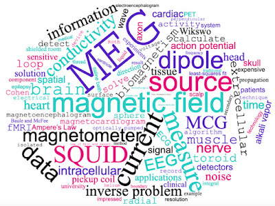
A word cloud derived from "Biomagnetism: The First Sixty Years."


Friday, April 21, 2023

The Magnetic Field Associated with a Plane Wave Front Propagating Through Cardiac Tissue

When I was on the faculty at Vanderbilt University, my student Marcella Woods and I examined the magnetic field produced by electrical activity in a sheet of cardiac muscle. I really like this analysis, because it provides a different view of the mechanism producing the magnetic field compared to that used by other researchers studying the magnetocardiogram. In another publication, here is how I describe our research. I hope you find it useful.
Roth and Marcella Woods examined an action potential propagating through a two-dimensional sheet of cardiac muscle [58]. In Fig. 6, a wave front is propagating to the right, so the myocardium on the left is fully depolarized and on the right is at rest. Cardiac muscle is anisotropic, meaning it has a different electrical conductivity parallel to the myocardial fibers than perpendicular to them. In Fig. 6, the fibers are oriented at an angle to the direction of propagation. The intracellular voltage gradient is in the propagation direction (horizontal in Fig. 6), but the anisotropy rotates the intracellular current toward the fiber axis. The same thing happens to the extracellular current, except that in cardiac muscle the intracellular conductivity is more anisotropic than the extracellular conductivity, so the extracellular current is not rotated as far. Continuity requires that the components of the intra- and extracellular current densities in the propagation direction are equal and opposite. Their sum therefore points perpendicular to the direction of propagation, creating a magnetic field that comes out of the plane of the tissue on the left and into the plane on the right (Fig. 6) [58–60].
Figure 6. The current and magnetic field produced by a planar wave front propagating in a two-dimensional sheet of cardiac muscle. The muscle is anisotropic with a higher conductivity along the myocardial fibers.

This perspective of the current and magnetic field in cardiac muscle is unlike that ordinarily adopted when analyzing the magnetocardiogram, where the impressed current is typically taken as in the same direction as propagation. Nonetheless, experiments by Jenny Holzer in Wikswo’s lab confirmed the behavior shown in Fig. 6 [61].

The main references are:

58. Roth, B.J.; Woods, M.C. The magnetic field associated with a plane wave front propagating through cardiac tissue. IEEE Trans. Biomed. Eng. 1999, 46, 1288–1292.

61. Holzer, J.R.; Fong, L.E.; Sidorov, V.Y.; Wikswo, J.P.; Baudenbacher, F. High resolution magnetic images of planar wave fronts reveal bidomain properties of cardiac tissue. Biophys. J. 2004, 87, 4326–4332. 

You can learn more about how magnetic fields are generated by cardiac muscle by reading about what happens at the apex of the heart. Or, solve homework problem 19 in Chapter 8 of Intermediate Physics for Medicine and Biology.

Friday, March 24, 2023

Three New Reviews

Over the last couple years, I’ve been writing lots of review articles. In the last few weeks three have been published. All of them are open access, so you can read them without a subscription.

Can MRI be Used as a Sensor to Record Neural Activity?

Can MRI be Used as a Sensor
to Record Neural Activity?
This review asks the question “Can MRI be Used as a Sensor to Record Neural Activity?” The article is published in the journal Sensors (Volume 23, Article Number 1337). The abstract is reproduced below.
Magnetic resonance provides exquisite anatomical images and functional MRI monitors physiological activity by recording blood oxygenation. This review attempts to answer the following question: Can MRI be used as a sensor to directly record neural behavior? It considers MRI sensing of electrical activity in the heart and in peripheral nerves before turning to the central topic: recording of brain activity. The primary hypothesis is that bioelectric current produced by a nerve or muscle creates a magnetic field that influences the magnetic resonance signal, although other mechanisms for detection are also considered. Recent studies have provided evidence that using MRI to sense neural activity is possible under ideal conditions. Whether it can be used routinely to provide functional information about brain processes in people remains an open question. The review concludes with a survey of artificial intelligence techniques that have been applied to functional MRI and may be appropriate for MRI sensing of neural activity.

Parts of the review may be familiar to readers of this blog. For instance, in June of 2016 I wrote about Yoshio Okada’s experiment to measure neural activation in a brain cerebellum of a turtle, in August 2019 I described Allen Song’s use of spin-lock methods to record brain activity, and in April 2020 I discussed J. H. Nagel’s 1984 abstract that may have been the first to report using MRI to image action currents. All these topics are featured in my review article. In addition, I analyzed my calculation, performed with graduate student Dan Xu, of the magnetic field produced inside the heart, and I reviewed my work with friend and colleague Ranjith Wijesinghe, from Ball State University, on MRI detection of bioelectrical activity in the brain and peripheral nerves. At the end of the review, I examined the use of artificial intelligence to interpret this type of MRI data. I don’t really know much about artificial intelligence, but the journal wanted me to address this topic so I did. With AI making so much news these days (ChatGPT was recently on the cover of TIME magazine!), I’m glad I included it.

Readers of Intermediate Physics for Medicine and Biology will find this review to be a useful extension of Section 18.12 (“Functional MRI”), especially the last paragraph of that section beginning with “Much recent research has focused on using MRI to image neural activity directly, rather than through changes in blood flow...”

Magneto-Acoustic Imaging in Biology

Magneto-Acoustic Imaging in Biology
Next is “Magneto-Acoustic Imaging in Biology,” published in the journal Applied Sciences (Volume 13, Article Number 3877). The abstract states

This review examines the use of magneto-acoustic methods to measure electrical conductivity. It focuses on two techniques developed in the last two decades: Magneto-Acoustic Tomography with Magnetic Induction (MAT-MI) and Magneto-Acousto-Electrical Tomography (MAET). These developments have the potential to change the way medical doctors image biological tissue.
The only place in IPMB where Russ Hobbie and I talked about these topics is in Homework Problem 31 in Chapter 8, which analyzes a simple example of MAT-MI.

A Mathematical Model of Mechanotransduction

A Mathematical Model of Mechanotransduction
Finally comes “A Mathematical Model of Mechanotransduction” in the new journal Academia Biology (Volume 1; I can’t figure out what the article number is?!).

This article reviews the mechanical bidomain model, a mathematical description of how the extracellular matrix and intracellular cytoskeleton of cardiac tissue are coupled by integrin membrane proteins. The fundamental hypothesis is that the difference between the intracellular and extracellular displacements drives mechanotransduction. A one-dimensional example illustrates the model, which is then extended to two or three dimensions. In a few cases, the bidomain equations can be solved analytically, demonstrating how tissue motion can be divided into two parts: monodomain displacements that are the same in both spaces and therefore do not contribute to mechanotransduction, and bidomain displacements that cause mechanotransduction. The model contains a length constant that depends on the intracellular and extracellular shear moduli and the integrin spring constant. Bidomain effects often occur within a few length constants of the tissue edge. Unequal anisotropy ratios in the intra- and extracellular spaces can modulate mechanotransduction. Insight into model predictions is supplied by simple analytical examples, such as the shearing of a slab of cardiac tissue or the contraction of a tissue sheet. Computational methods for solving the model equations are described, and precursors to the model are reviewed. Potential applications are discussed, such as predicting growth and remodeling in the diseased heart, analyzing stretch-induced arrhythmias, modeling shear forces in a vessel caused by blood flow, examining the role of mechanical forces in engineered sheets of tissue, studying differentiation in colonies of stem cells, and characterizing the response to localized forces applied to nanoparticles.

This review is similar to my article that I discussed in a blog post about a year ago, but better. I originally published it as a manuscript on the bioRxiv, the preprint server for biology, but it received little attention. I hope this version does better. If you want to read this article, download the pdf instead of reading it online. The equations are all messed up on the journal website, but they look fine in the file.

If you put these three reviews together with my previous ones about magnetic stimulation and the bidomain model of cardiac electrophysiology, you have a pretty good summary of the topics I’ve worked on throughout my career. Are there more reviews coming? I’m working feverishly to finish one more. For now, I’ll let you guess the topic. I hope it’ll come out later this year.

Friday, December 30, 2022

The Development of Transcranial Magnetic Stimulation

When I worked at the National Institutes of Health, I studied transcranial magnetic stimulation. In Chapter 8 of Intermediate Physics for Medicine and Biology, Russ Hobbie and I describe this technique to activate neurons in the brain.
Since a changing magnetic field generates an induced electric field, it is possible to stimulate nerve or muscle cells without using electrodes. The advantage is that for a given induced current deep within the brain, the currents in the scalp that are induced by the magnetic field are far less than the currents that would be required for electrical stimulation. Therefore transcranial magnetic stimulation (TMS) is relatively painless.

The method was invented in 1985 and when I arrived at NIH in 1988 the field was new and ripe for analysis. I spent the next seven years calculating electric fields in the brain and determining how the electric field couples to a nerve.

Roth, B. J. (2022) The Development of
Transcranial Magnetic Stimulation
,
BOHR International Journal of
Neurology and Neuroscience
,
Volume 1, Pages 8–20.
Recently, I wrote a review article telling the story of how transcranial magnetic stimulation began. You can get a copy at https://journals.bohrpub.com/index.php/bijnn/article/view/28; it is an open access article so everyone is free to download it. The abstract states
This review describes the development of transcranial magnetic stimulation in 1985 and the research related to this technique over the following 10 years. It not only focuses on work done at the National Institutes of Health but provides a survey of other related research as well. Key topics are the calculation of the electric field produced during magnetic stimulation, the interaction of this electric field with a long nerve axon, coil design, the time course of the magnetic stimulation pulse, and the safety of magnetic stimulation.

Readers of this blog will recognize some of the topics from earlier posts, such as the calculation of the induced electric field, determining the site of stimulation along a peripheral nerve, Paul Maccabee’s wonderful article, the four-leaf coil, the heating of metal electrodes, implantable microcoils, and Tony Barker's online interview. You could almost say I pre-wrote much of the review using this blog as my test bed. 

I like magnetic stimulation because it's a classic example of how a fundamental concept from physics can have a major impact in biology and medicine. If you combine this review of transcranial magnetic stimulation together with my earlier review of the bidomain model of cardiac tissue, you get a pretty good summary of my most important research.

Enjoy!

Friday, May 20, 2022

Using the Mechanical Bidomain Model to Analyze the Biomechanical Behavior of Cardiomyocytes

During the decade of 2010–2020, my research shifted from bioelectricity and biomagnetism to biomechanics and mechanotransduction. I took the bidomain model of cardiac electrophysiology—described in Chapter 7 of Intermediate Physics for Medicine and Biology— and adapted it to describe growth and remodeling in response to mechanical forces. In other words, I traded resistors for springs. This effort was not entirely successful, but I think it provided some useful insights.

In 2015 I described the mechanical bidomain model in a chapter of Cardiomyocytes: Methods and Protocols. This book was part of the series Methods in Molecular Biology, and each chapter had a unusual format. The research was outlined, with the details relegated to an extensive collection of endnotes. A second edition of the book was proposed, and I dutifully submitted an updated chapter. However, the new edition never come to pass. Rather than see my chapter go to waste, I offer it to you, dear reader. You can download a draft of my chapter for the second edition here. For those of you who have time only for a summary, below is the abstract.

The mechanical bidomain model provides a macroscopic description of cardiac tissue biomechanics, and also predicts the microscopic coupling between the extracellular matrix and the intracellular cytoskeleton of cardiomyocytes. The goal of this chapter is to introduce the mechanical bidomain model, to describe the mathematical methods required for solving the model equations, to predict where the membrane forces acting on integrin proteins coupling the intracellular and extracellular spaces are large, and to suggest experiments to test the model predictions.

The main difference between the chapter in the first edition and the one submitted for the second was a new section called “Experiments to Test the Mechanical Bidomain Model.” There I describe how the model can reproduce data obtained when studying colonies of embryonic stem cells, sheets of engineered heart tissue, and border zones between normal and ischemic regions in the heart. The chapter ends with this observation:

The most important contribution of mathematical modeling in biology is to make predictions that can be tested experimentally. The mechanical bidomain model makes many predictions, in diverse areas such as development, tissue engineering, and hypertrophy.
I particularly like a new figure in the second edition. It’s a revision of a figure created by Xavier Trepat and Jeffrey Fredberg that compares mechanobiology to a game of tug-of-war. I added the elastic properties of the extracellular space (the green arrows), saying “It is as if the game of tug-of-war is played on a flexible surface, such as a flat elastic sheet.” In other words, tug-of-war on a trampoline

Enjoy!

The “tug-of-war” model of tissue biomechanics, adapted from an illustration by Trepat and Fredberg.
The “tug-of-war” model of tissue biomechanics, adapted from an illustrationby Trepat and Fredberg. Top: the intracellular (yellow), extracellular (green) and integrin (blue) forces acting on a monolayer of cells. Middle: The analogous forces among the players of a game of tug-of-war. This figure is extended beyond that of Trepat and Fredberg by allowing both the intracellular and extracellular spaces to move. Bottom: Representation of the mechanical bidomain model by a ladder of springs.

Friday, November 12, 2021

Bidomain Modeling of Electrical and Mechanical Properties of Cardiac Tissue

This week Biophysics Reviews published my article “Bidomain Modeling of Electrical and Mechanical Properties of Cardiac Tissue” (Volume 2, Article Number 041301, 2021). The introduction states
This review discusses the bidomain model, a mathematical description of cardiac tissue. Most of the review covers the electrical bidomain model, used to study pacing and defibrillation of the heart. For a book-length analysis of this topic, consult the recently published second edition of Cardiac Bioelectric Therapy. In particular, one chapter in that book complements this review: it contains a table listing many bidomain predictions and their experimental confirmation, includes many original figures from earlier publications, and cites additional references. Near the end, the review covers the mechanical bidomain model, which describes mechanotransduction and the resulting growth and remodeling of cardiac tissue.

The review has several aims: to (1) introduce the bidomain model to younger investigators who are bringing new technologies from outside biophysics into cardiac physiology; (2) examine the interaction of theory and experiment in biological physics; (3) emphasize intuitive understanding by focusing on simple models and qualitative explanations of mechanisms; and (4) highlight unresolved controversies and open questions. The overall goal is to enable technologists entering the field to more effectively contribute to some of the pressing scientific questions facing physiologists.

My manuscript traveled a long and winding road. The initial version was a personal account of my career as I worked on the bidomain model (Russ Hobbie and I discuss the bidomain concept in Chapter 7 of Intermediate Physics for Medicine and Biology), and was organized around ten papers I published between 1986 and 2010, with an emphasis on the 1990s. My first draft (and all subsequent ones) benefited from thoughtful comments by my former graduate student, Dilmini Wijesinghe. After I fixed all the problems Dilmini found, I sent the initial version to the editor. He responded that the journal board wanted a more traditional, authoritative review article. That was fine, so I transformed the paper from a memoir into a review, and submitted it officially to the journal. Then the reviewers had a couple rounds of helpful comments, leading to more revisions. Next, there were changes in the page proofs to fulfill all the journal editorial rules. At last, it was published.

The final version is unlike the initial one. I changed the perspective from first person to third; added figures; increased the number of references by almost 50%; and deleted all the reminiscences, colorful anecdotes, and old war stories. 

I hope you enjoy the peer-reviewed, published article. If you want to read the original version (the one with the war stories), you can find it here.  

I made a word cloud based on the article. The giant “Roth” is embarrassing, but otherwise it provides a nice summary of what the paper is about.

Word Cloud of "Bidomain Modeling of Electrical and Mechanical Properties of Cardiac Tissue."

Biophysics Reviews is a new journal, edited by my old friend Kit Parker. Long-time readers of this blog may remember Parker as the guy who said “our job is to find stupid and get rid of it.” Listen to him describe his goals as Editor-in-Chief.

Kit Parker, Editor-in-Chief of Biophysics Reviews, introduces the journal.

https://www.youtube.com/watch?v=2V1fpskjJtM