Showing posts sorted by date for query 150. Sort by relevance Show all posts

Friday, March 25, 2011

Maxwell Equation Sesquicentennial

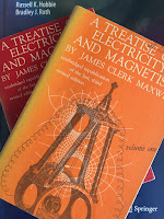
A Treatise on Electricity and Magnetism, by James Clerk Maxwell, superimposed on Intermeidate Physics for Medicine and Biology.
A Treatise on
Electricity and Magnetism,
by James Clerk Maxwell.
I am a big James Clerk Maxwell fan. In fact, I have made my living applying Maxwell’s equations to biology and medicine. Yes, I own one of those tee shirts with Maxwell’s equations written on it. I keep a copy of Maxwell’s A Treatise on Electricity and Magnetism in my office (although I have never read it in its entirety…Oh how I wish Maxwell had access to modern vector notation!). I have read The Maxwellians (outstanding) and The Man Who Changed Everything: The Life of James Clerk Maxwell (good). So, this month I am celebrating with gusto the sesquicentennial of the publication of Maxwell’s famous equations. The March 17 issue of the journal Nature has a special section containing four articles about Maxwell’s equation. In an editorial titled “A Bold Unifying Leap” (Volume 471, Page 265) the editor writes
In this issue we celebrate the first expression of those equations by Scottish physicist Maxwell in the Philosophical Magazine 150 years ago. There he drew together several strands of understanding about the behaviour of electricity, of magnetism, of light, and of the ways in which these fundamental aspects of nature behave in matter. As Albert Einstein remarked, “so bold was the leap” of this work that it took decades for physicists to grasp its full significance. And although it was a wonderful expression of science at its purest, it was forged in the thoroughly practical culture of intellects at that time.
Russ Hobbie and I mention Maxwell’s equations in the 4th edition of Intermediate Physics for Medicine and Biology. We added a new homework problem to the 4th edition in Chapter 8 (Biomagnetism).
Problem 22 Write down in differential form (a) the Faraday induction law, (b) Ampere’s law including the displacement current term, (c) Gauss’s law, and (d) Eq. 8.7. … These four equations together constitute “Maxwell’s equations.” Together with the Lorentz force law (Eq. 8.2), Maxwell’s equations summarize all of electricity and magnetism.
All four of Maxwell’s equations are discussed in our book. Section 6.3 is dedicated to Gauss’s law, governing the electric field produced by a collection of charges, and we analyze the usual suspects: a line of charge and a charged sheet. Ampere’s law appears in Section 8.2 (The Magnetic Field of a Moving Charge or Current), and—in one of my favorite homework problems—we show in Problem 13 of Chapter 8 how “one can obtain a very different physical picture of the source of a magnetic field using the Biot Savart law than one gets using Ampere’s law, even though the field is the same.” Faraday’s law is presented in Section 8.6 on Electromagnetic Induction, followed by a discussion of magnetic stimulation of the brain. Even Gauss’s law for a magnetic field (Eq. 8.7, stating that the magnetic field has no divergence) is introduced. Maxwell’s great insight was to add the displacement current term to Ampere’s law. We show how the charging of a capacitor implies the existence of this additional term on page 207, and explore its role in biomagnetism (slight).

The Feynman Lectures on Physics, by Richard Feynman, superimposed on Intermediate Physics for Medicine and Biology.
The Feynman Lectures on Physics,
by Richard Feynman.
Russ and I never analyze what may be the greatest prediction of Maxwell’s equations: the wave nature of light. We state in Section 14.1 that “the velocity of light traveling in a vacuum is given by electromagnetic theory as c = 1/√(ε0 μ0)”, but we never derive this result from Maxwell’s equations. Many of the applications of electromagnetic waves—such as wave guides, antennas, diffraction, radiation, and all of optics—are barely mentioned, if mentioned at all, in our text. For those who want to learn these topics (and all students of physics should want to learn these topics), I suggest Griffith’s Introduction to Electrodynamics (undergraduate) or Jackson’s Classical Electrodynamics (graduate). Richard Feynman introduces Maxwell’s equations in his celebrated book The Feynman Lectures on Physics. In Chapter 18 of Volume 2, he writes
It was not customary in Maxwell’s time to think in terms of abstract fields. Maxwell discussed his ideas in terms of a model in which the vacuum was like an elastic solid. He also tried to explain the meaning of his new equation in terms of the mechanical model. There was much reluctance to accept his theory, first because of the model, and second because there was at first no experimental justification. Today, we understand better that what counts are the equations themselves and not the model used to get them. We may only question whether the equations are true or false. This is answered by doing experiments, and untold numbers of experiments have confirmed Maxwell’s equations. If we take away the scaffolding he used to build it, we find that Maxwell’s beautiful edifice stands on its own. He brought together all of the laws of electricity and magnetism and made one complete and beautiful theory.
Anyone with a historical bent may want to read Maxwell’s original papers and accompanying commentary in Maxwell on the Electromagnetic Field: a Guided Study, by Thomas Simpson. The book contains a detailed analysis of Maxwell’s papers, including “On the Physical Lines of Force,” which is the publication we celebrate this month. Simpson’s book is the best place I know of to learn about the “scaffolding” Maxwell used to build his theory.

I will close with one of my favorite quotes, again from The Feynman Lectures. At the end of his first chapter introducing electromagnetism, Feynman writes
From a long view of the history of mankind—seen from, say, ten thousand years from now—there can be little doubt that the most significant event of the 19th century will be judged as Maxwell’s discovery of the laws of electrodynamics. The American Civil War will pale into provincial insignificance in comparison with this important scientific event of the same decade.

Friday, March 11, 2011

Retinal Injuries from a Handheld Laser Pointer

Are laser pointers safe? Apparently, it depends on the laser pointer. A recent article by Christine Negroni in the New York Times (Feb. 28, 2011) states that
Eye doctors around the world are warning that recent cases of teenagers who suffered eye damage while playing with high-power green laser pointers are likely to be just the first of many.
Negroni cites a letter that appeared last September in the New England Journal of Medicine (Wyrsch, Baenninger, and Schmid, “Retinal Injuries from a Handheld Laser Pointer,” N. Engl. J. Med., Volume 363, Pages 1089–1091, 2010), which says
In the past, laser pointers sold to the public had a maximal output of 5 mW, which is regarded as harmless because the human eye protects itself with blink reflexes. The measured output of the laser in [the case of a person who was injured] was 150 mW. The use of lasers that are threatening to the eye is normally restricted to occupational and military environments; laser accidents outside these fields are very rare. However, powerful laser devices, with a power of up to 700 mW, are now easily obtainable through the Internet, despite government restrictions. These high-power lasers are advertised as “laser pointers” and look identical to low-power pointers. The much higher power of such devices may produce immediate, severe retinal injury. Despite their potential to cause blinding, such lasers are advertised as fun toys and seem to be popular with teenagers. In addition, Web sites now offer laser swords and other gadgets that use high-power lasers.
I attended a talk just last week where the speaker waved his green laser pointer around like a light saber. I don’t know the power of his pointer, but I wonder if I was in danger.

One concern arises from the bozos who point lasers at airplanes. The U.S. Congress plans to toughen the laws on this sort of horseplay, making shining a laser at a plane a federal crime with up to five years imprisonment. I’m all for high school students learning science by hands-on activities, but do it right. Buy a 5 mW red helium-neon laser pointer and use it safely to do some optics experiments (I suggest observing Young’s double slit interference pattern). Don’t buy a 700 mW green laser pointer and start shining it up into the sky! Do you think I’m being a schoolmarm out to ruin your fun? Consider this: the website laserpointersafety.com reports that
A $5000 reward is being offered for information leading to the arrest of the person(s) who aimed a laser into the cockpit of a Southwest Airlines flight approaching Baltimore-Washington International Airport. The flight, which originated in Milwaukee, was 2000 feet over the town of Millersville, near Old Mill Road and Kenora Drive, when it was illuminated around 6:45 pm on Sunday, Feb. 20, 2011. Millersville is about 8 miles from BWI Airport.
You better be careful; someone may be watching.

How do you tell the difference between a safe, educational experience and a potentially disastrous prank? You begin by learning about light and its biological impact. Russ Hobbie and I discuss light in Chapter 14 of the 4th edition of Intermediate Physics for Medicine and Biology. We address topics related to light and safety, although we don’t analyze the particular concern of laser damage to the eye. For instance, we discuss how ultraviolet light damages the eye (Section 14.9.6) and how light can be used to heat tissue (Section 14.10), as well as a detailed discussion of radiometry (the measurement of radiant energy, Section 14.11) and the anatomy and optics of the eye (Section 14.12).

In another New York Times article, Negroni relates how high powered laser pointers can pose a risk to pilots. And on her blog, she explains why helicopters may be at a greater risk than airplanes.
A helicopter cockpit has glass extending below the level of the pilots' eyes toward the ground exactly where the lasers are. Rotor craft fly at low altitudes over residential areas and busy highways. They are not flying autopilot and they may be piloted by a single person. They hover and may make inviting targets. That was the case on Tuesday when a Los Angeles television station sent its chopper to follow and report on the police activity and it was hit by a laser.
The interaction of laser light and vision is one more example of why a firm understanding of physics applied to medicine and biology is so important.

Friday, February 4, 2011

Britton Chance (1913-2010)

Britton Chance died late last year. The website www.brittonchance.org states that
Britton Chance, M.D., Ph.D., D.Sc., for more than 50 years one of the giants of biochemistry and biophysics and a world leader in transforming theoretical science into useful biomedical and clinical applications, died on November 16, 2010, at age 97 in Philadelphia, PA. Dr. Chance had the rare distinction of being the recipient of a National Medal of Science (1974), a Gold Medal in the Olympics (1952, Sailing, Men’s 5.5 Meter Class), and a Certificate of Merit for his sensitive work during World War II.
His obituary in the New York Times describes his early work.
Over a lifetime of research, Dr. Chance focused on the observation and measurement of chemical reactions within cells, tissue and the body. But unlike most researchers, he also had expertise in mechanics, electronics and optics, and a great facility in instrument-building. His innovations helped transform theoretical science into biochemical and biophysical principles, the stuff of textbooks, and useful biomedical and clinical applications.

Early in his career he invented a tool, known as a stopped-flow apparatus, for measuring chemical reactions involving enzymes; it led to the establishment of a fundamental principle of enzyme kinetics, known as the enzyme-substrate complex.
Another obituary, in the December 17 issue of Science magazine, observed that
In his mid-70s, Chance (then emeritus) launched a new field of optical diagnostics that rests on the physics of light diffusion through scattering materials such as living tissue. He showed that scattered near-infrared light pulses could not only measure the dynamics of oxy- and deoxyhemoglobin levels in performing muscles, but also reveal and locate tumors and cancerous tissue in muscles and breast as well as injury in the brain. Because changing patterns of oxy- and deoxyhemoglobin in the brain reflect cognitive activity, the applications of this diagnostic approach widened to include assessing neuronal connectivity in premature babies.
Chance appears in the 4th edition of Intermediate Physics for Medicine and Biology because of his research on light diffusion. In Section 14.4 (Scattering and Absorption of Radiation), Russ Hobbie and I analyze the absorption and scattering coefficients of infrared light, and then give typical values that “are eyeballed from data from various tissues reported in the article by Yodh and Chance (1995),” with the reference being to Yodh, A. and B. Chance (1995) “Spectroscopy and Imaging with Diffusing Light,” Physics Today, March, Pages 34–40.

Then in Sec. 14.5 (The Diffusion Approximation to Photon Transport), we analyze pulsed measurements of infrared light.
A technique made possible by ultrashort light pulses from a laser is time-dependent diffusion. It allows determination of both [the scattering coefficient] and [the absorption coefficient]. A very short (150-ps) pulse of light strikes a small region on the surface of the tissue. A detector placed on the surface about 4 cm away records the multiply-scattered photons. A typical plot of the detected photon fluence rate is shown in Fig. 14.13.
Figure 14.13 is a figure from Patterson, M. S., B. Chance, and B. C. Wilson (1989) “Time Resolved Reflectance and Transmittance for the Noninvasive Measurement of Tissue Optical Properties,” Applied Optics, Volume 28, Pages 2331–2336, which has been cited over 1000 times in the scientific literature.

Finally, in Sec. 14.6 (Biological Applications of Infrared Scattering), we reproduce a figure from the Physics Today article by Yodh and Chance, which shows the absorption coefficient for water, oxyhemoglobin and deoxyhemoglobin.
The greater absorption of blue light in oxygenated hemoglobin makes oxygenated blood red…The wavelength 800 nm at which both forms of hemoglobin have the same absorption is called the isosbestic point. Measurements of oxygenation are made by comparing the absorption at two wavelengths on either side of this point.
This property of infrared absorption of light is the basis for pulse oximeters that measure oxygenation. Not all measurements of blood oxygen use pulsed light. Russ and I cite one of Chance’s papers that uses a continuous source: Liu, H., D. A. Boas, Y. Zhang, A. G. Yodh, and B. Chance (1995) “Determination of Optical Properties and Blood Oxygenation in Tissue Using Continuous NIR Light,” Physics in Medicine and Biology, Volume 40, Pages 1983–1993. A fourth of Chance’s paper that we include in our references is Sevick, E. M., B. Chance, J. Leigh, S. Nioka, and M. Maris (1991) “Quantitation of Time- and Frequency-Resolved Optical Spectra for the Determination of Tissue Oxygenation,” Analytical Biochemistry, Volume 195, Pages 330–351.

In 1987, Chance won the Biological Physics Prize (now known as the Max Delbruck Prize in Biological Physics) from the American Physical Society
for pioneering application of physical tools to the understanding of Biological phenomena. The early applications ranged from novel spectrometry that elucidated electron transfer processes in living systems to analog computation of nonlinear processes. Later contributions have been equally at the forefront.

Friday, May 28, 2010

Happy Birthday Laser!

Quantum Physics of Atoms, Molecules, Solids, Nuclei, and Particles, by Eisberg and Resnick, superimposed on Intermediate Physics for Medicine and Biology.
Quantum Physics of Atoms,
Molecules, Solids, Nuclei, and Particles,
by Eisberg and Resnick.
This month marks the 50th anniversary of the invention of the laser. In May 1960, Theodore Maiman built the first device to produce coherent light by the mechanism of “Light Amplification by Stimulated Emission of Radiation” at Hughes Research Laboratories in Malibu, making the laser just slightly older than I am. A special website, called laserfest, is commemorating this landmark event. Eisberg and Resnick discuss lasers in Section 11.7 of their textbook Quantum Physics of Atoms, Molecules, Solids, Nuclei, and Particles (quoted from the first edition, 1974).
In the solid state laser that operates with a ruby crystal, some Al atoms in the Al2O3 molecules are replaced by Cr atoms. These “impurity” chromium atoms account for the laser action… The level of energy E1 is the ground state and the level of energy E3 is the unstable upper state with a short lifetime (≈10−8 sec), the energy difference E3-E1 corresponding to a wavelength of about 5500 Å. Level E2 is an intermediate excited state which is metastable, its lifetime against spontaneous decay being about 3 x 10⁻³ sec. If the chromium atoms are in thermal equilibrium, the population number of the states are such that [n3 is less than n2 is less than n1]. By pumping in radiation of wavelength 5500 Å, however, we stimulate absorption of incoming photons by Cr atoms in the ground state, thereby raising the population number of energy state E3 and depleting energy state E1 of occupants. Spontaneous emission, bringing atoms from state 3 to state 2, then enhances the occupancy of state 2, which is relatively long-lived. The result of this optical pumping is to decrease n1 and increase n2, such that n2 is greater than n1 and population inversion exists. Now, when an atom does make a transition from state 2 to state 1, the emitted photon of wavelength 6943 Å will stimulate further transitions. Stimulated emission will dominate stimulated absorption (because n2 is greater than n1) and the output of photons of wavelength 6943 Å is much enhanced. We obtain an intensified coherent monochromatic beam.
Lasers are an important tool in biology and medicine. Russ Hobbie and I discuss their applications in Chapter 14 (Atoms and Light) the 4th edition of Intermediate Physics for Medicine and Biology. In Section 14.5 (The Diffusion Approximation to Photon Transport) we write
A technique made possible by ultrashort light pulses from a laser is time-dependent diffusion. It allows determination of both μs and μa [the scattering and absorption attenuation coefficients]. A very short (150-ps) pulse of light strikes a small region on the surface of the tissue. A detector placed on the surface about 4 cm away records the multiply-scattered photons… A related technique is to apply a continuous laser beam, the amplitude for which is modulated at various frequencies between 50 and 800 MHz. The Fourier transform of Eq. 14.29 gives the change in amplitude and phase of the detected signal. Their variation with frequency can also be used to determine μa and μs.
We also mention lasers in Section 14.10 (Heating Tissue with Light).
Sometimes tissue is irradiated in order to heat it; in other cases tissue heating is an undesired side effect of irradiation. In either case, we need to understand how the temperature changes result from the irradiation. Examples of intentional heating are hyperthermia (heating of tissue as a part of cancer therapy) or laser surgery (tissue ablation). Tissue is ablated when sufficient energy is deposited to vaporize the tissue.
Russ and I give many references about lasers in medicine in our Resource Letter (“Resource Letter MP-2: Medical Physics,” American Journal of Physics, Volume 77, Pages 967–978, 2009):
F. Lasers and optics

Lasers have introduced many medical applications of light, from infrared to the visible spectrum to ultraviolet.

150. Lasers in Medicine, edited by R. W. Waynant (CRC, Boca Raton, 2002). (I)

151. Laser-Tissue Interactions: Fundamentals and Applications, M. H. Niemz (Springer, Berlin, 2007). (I)

152. “Lasers in medicine,” Q. Peng, A. Juzeniene, J. Chen, L. O. Svaasand, T. Warloe, K.-E. Giercksky, and J. Moan, Rep. Prog. Phys. 71, Article 056701, 28 pages
(2008). (A)

A fascinating and fast-growing new technique to image biological tissue is optical coherence tomography “OCT.” It uses reflections like ultrasound but detects the reflected rays using interferometry.

153. Optical Coherence Tomography, M. E. Brezinski (Elsevier, Amsterdam, 2006). Overview of the physics of OCT and applications to cardiovascular medicine, musculoskeletal disease, and oncology. (I)

154. “Optical coherence tomography: Principles and applications,” A. F. Fercher, W. Drexler, C. K. Hitzenberger, and T. Lasser, Rep. Prog. Phys. 66, 239–303 (2003). (I)

With infrared light, scattering dominates over absorption. In this case, light diffuses through the tissue. Optical imaging in turbid media is difficult but not impossible.

155. “Recent advances in diffuse optical imaging,” A. P. Gibson, J. C. Hebden, and S. R. Arridge, Phys. Med. Biol. 50, R1–R43 (2005). (I)

156. “Pulse oximetry,” R. C. N. McMorrow and M. G. Mythen, Current Opinion in Critical Care 12, 269–271 (2006). The pulse oximeter measures the oxygenation of blood and is based on the diffusion of infrared light. (I)

One impetus for medical applications of light has been the development of new light sources, such as free-electron lasers and synchrotrons. In both cases, the light frequency is tunable over a wide range.

157. “Free-electron-laser-based biophysical and biomedical instrumentation,” G. S. Edwards, R. H. Austin, F. E. Carroll, M. L. Copeland, M. E. Couprie, W. E. Gabella, R. F. Haglund, B. A. Hooper, M. S. Hutson, E. D. Jansen, K. M. Joos, D. P. Kiehart, I. Lindau, J. Miao, H. S. Pratisto, J. H. Shen, Y. Tokutake, A. F. G. van der Meer, and A. Xie, Rev. Sci. Instrum. 74, 3207–3245 (2003). (I)

158. “Medical applications of synchrotron radiation,” P. Suortti and W. Thomlinson, Phys. Med. Biol. 48, R1– R35 (2003). (I)

Finally, photodynamic therapy uses light-activated drugs to treat diseases.

159. “The physics, biophysics and technology of photodynamic therapy,” B. C. Wilson and M. S. Patterson, Phys. Med. Biol. 53, R61–R109 (2008). (A)
Happy birthday, laser!

Friday, April 24, 2009

Proton Therapy

Section 16.11.3 in the 4th edition of Intermediate Physics for Medicine and Biology discusses proton therapy.
Protons are also used to treat tumors. Their advantage is the increase of stopping power at low energies. It is possible to make them come to rest in the tissue to be destroyed, with an enhanced dose relative to intervening tissue and almost no dose distally (“downstream”) as shown by the Bragg peak.
Proton therapy has become popular recently: see articles in US News and World Report and on MSNBC. There even exists a National Association for Proton Therapy. Their website explains the main advantage of protons over X-rays.
Both standard x-ray therapy and proton beams work on the principle of selective cell destruction. The major advantage of proton treatment over conventional radiation, however, is that the energy distribution of protons can be directed and deposited in tissue volumes designated by the physicians in a three-dimensional pattern from each beam used. This capability provides greater control and precision and, therefore, superior management of treatment. Radiation therapy requires that conventional x-rays be delivered into the body in total doses sufficient to assure that enough ionization events occur to damage all the cancer cells. The conventional x-rays lack of charge and mass, however, results in most of their energy from a single conventional x-ray beam being deposited in normal tissues near the body’s surface, as well as undesirable energy deposition beyond the cancer site. This undesirable pattern of energy placement can result in unnecessary damage to healthy tissues, often preventing physicians from using sufficient radiation to control the cancer.

Protons, on the other hand, are energized to specific velocities. These energies determine how deeply in the body protons will deposit their maximum energy. As the protons move through the body, they slow down, causing increased interaction with orbiting electrons.
Figure 16.51 of the 4th edition of Intermediate Physics for Medicine and Biology shows the dose versus depth from a 150 MeV proton beam, including the all-important Bragg peak located many centimeters below the tissue surface. If you want to understand better why proton energy is deposited in the Bragg peak rather than being spread throughout the tissue, solve Problem 31 in Chapter 16.

To learn more about the pros and cons of proton therapy, I suggest several
point/counterpoint articles from the journal Medical Physics: Within the Next Decade Conventional Cyclotrons for Proton Radiotherapy will Become Obsolete and Replaced by Far Less Expensive Machines using Compact Laser Systems for the Acceleration of the Protons, Chang-Ming Ma and Richard Maughan (Medical Physics, Volume 33, Pages 571–573, 2006), Proton Therapy is the Best Radiation Treatment Modality for Prostate Cancer, Michael Moyers and Jean Pouliot (Medical Physics, Volume 34, Pages 375378, 2007), and Proton Therapy is Too Expensive for the Minimal Potential Improvements in Outcome Claimed, Robert Schulz and Alfred Smith (Medical Physics, Volume 34, Pages 1135–1138, 2007).