Friday, December 30, 2016

The Story of the World in 100 Species

The Story of the World  in 100 Species,  by Christopher Lloyd, superimposed on Intermediate Physics for Medicine and BIology.
The Story of the World
in 100 Species,
by Christopher Lloyd.
I recently finished reading The Story of the World in 100 Species. The author Christopher Lloyd writes in the introduction
This book is a jargon-free attempt to explain the phenomenon we call life on Earth. It traces the history of life from the dawn of evolution to the present day through the lens of one hundred living things that have changed the world. Unlike Charles Darwin’s theory published more than 150 years ago, it is not chiefly concerned with the “origin of species,” but with the influence and impacts that living things have had on the path of evolution, on each other and on our mutual environment, planet Earth.
Of course, I began to wonder how many of the top hundred species Russ Hobbie and I mention in Intermediate Physics for Medicine and Biology. Lloyd lists the species in order of impact. The number 1 species is the earthworm. As Darwin understood, you would have little agriculture without worms churning the soil. The highest ranking species that was mentioned in IPMB is number 2, algae, which produces much of the oxygen in our atmosphere. According to Lloyd, algae might provide the food (ick!) and fuel we need in the future.

Number 6 is ourselves: humans. Although the species name Homo sapiens never appears in IPMB, several chapters—those dealing with medicine—discuss us. Number 8 yeast (specifically, S. cerevisiae) is not in IPMB, although it is mentioned previously in this blog. Number 15 is the fruit fly Drosophila melanogaster, which made the list primarily because it is an important model species for research. IPMB mentions D. melanogaster when discussing ion channels.

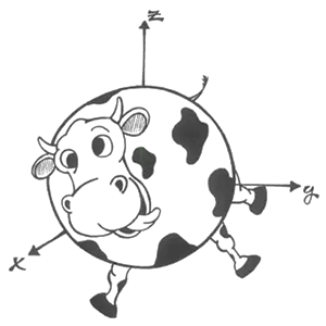
Cows are number 17; a homework problem in IPMB contains the phrase “consider a spherical cow.” The flea is number 18, and is influential primarily for spreading diseases such as the Black Death. In IPMB, we analyze how fleas survive high accelerations. Wheat reaches number 19 and is one of several grains on the list. In Chapter 11, Russ and I write: “Examples of pairs of variables that may be correlated are wheat price and rainfall, ….” I guess that wheat is in IPMB, although the appearance is fairly trivial. Like yeast, number 20 C. elegans, a type of roundworm, is never mentioned in IPMB but does appear previously in this blog because it is such a useful model. I am not sure if number 21, the oak tree, is in IPMB. My electronic pdf of the book has my email address, roth@oakland.edu, as a watermark at the bottom of every page. Oak is not in the appendix, and I am pretty sure Russ and I never mention it, but I haven’t the stamina to search the entire pdf, clicking on each page. I will assume oak does not appear.

Number 24, grass, gets a passing mention: in a homework problem about predator-prey models, we write that “rabbits eat grass…foxes eat only rabbits.” When I searched the book for number 25 ant, I found constant, quantum, implant, elephant, radiant, etc. I gave up after examining just a few pages. Let’s say no for ant. Number 28 rabbit is in that predator-prey problem. Number 32 rat is in my favorite J. B. S. Haldane quote “You can drop a mouse down a thousand-yard mine shaft; and arriving at the bottom, it gets a slight shock and walks away. A rat is killed, and man is broken, a horse splashes.” Number 33 bee is in the sentence “Bees, pigeons, and fish contain magnetic particles,” and number 38 shark is in the sentence “It is possible that the Lorentz force law allows marine sharks, skates, and rays to orient in a magnetic field.” My favorite species, number 42 dog, appears many times. I found number 44 elephant when searching for ant. I am not sure about number 46 cat (complicated, scattering, indicate, cathode, … you search the dadgum pdf!). It doesn’t matter; I am a dog person and don’t care for cats.

Number 53 apple; IPMB suggests watching Russ Hobbie in a video about the program MacDose at the website https://itunes.apple.com/us/itunes-u/photon-interactions-simulation/id448438300?mt=10. No way am I counting that; you gotta draw the line somewhere. Number 58 horse; “…horse splashes…”. Number 59 sperm whale; we mention whales several times, but don’t specify the species—I’m counting it. Number 61 chicken appears in one of my favorite homework problems: “compare the mass and metabolic requirements…of 180 people…with 12,600 chickens…” Number 65 red fox; see predator-prey problem. Number 67 tobacco; IPMB mentions it several times. Number 71 tea; I doubt it but am not sure (instead, steady, steam, ….). Number 77 HIV; see Fig. 1.2. Number 85 coffee; see footnote 7, page 258.

Altogether, IPMB includes twenty of the hundred species (algea, human, fruit fly, cow, flea, wheat, grass, rabbit, rat, bee, shark, dog, elephant, horse, whale, chicken, fox, tobacco, HIV, coffee), which is not as many as I expected. We will have to put more into the 6th edition (top candidates: number 9 influenza, number 10 penicillium, number 14 mosquito, number 26 sheep, number 35 maize aka corn).

Were any important species missing from Lloyd’s list? He includes some well-known model organisms (S. cerevisiae, D. melanogaster, C. elegans) but inexplicably leaves out the bacterium E. coli (Fig. 1.1 in IPMB). Also, I am a bioelectricity guy, so I would include Hodgkin and Huxley’s squid with its giant axon. Otherwise, I think Lloyd’s list is pretty complete.

If you want to get a unique perspective on human history, learn some biology, and better appreciate evolution, I recommend The Story of the World in 100 Species.

Friday, December 23, 2016

Implantable microcoils for intracortical magnetic stimulation

When I worked at the National Institutes of Health in the 1990s, I studied transcranial magnetic stimulation. Russ Hobbie and I discuss magnetic stimulation in Chapter 8 of Intermediate Physics for Medicine and Biology. Pass a pulse of current through a coil held near the head; the changing magnetic field of the coil induces an electric field in the brain that stimulates neurons. Typical magnetic stimulation coils are constructed from several turns of wire, each turn carrying kiloamps of current in pulses that last for a tenth of a millisecond. Most are a few centimeters in size. Researchers have tried to make smaller coils, but these typically require even larger currents, resulting in magnetic forces that tear the coil apart as well as prohibitive Joule heating.

Imagine my surprise when Russ told me about a recently published paper describing magnetic stimulation using microcoils, written by Seung Woo Lee and his colleagues (“Implantable microcoils for intracortical magnetic stimulation,” Science Advances, 2:e1600889, 2016). Frankly, I am not sure what to make of this paper. On the one hand, the authors describe a careful study in which they perform all the control experiments I would have insisted on had I reviewed the paper for the journal (I did not). On the other hand, it just doesn’t make sense. 

Lee et al. built a coil by bending a 50 micron insulated copper wire into a single tight turn having a diameter of about 100 microns (see figure). Their current pulse lasted a few tenths of a millisecond, and had a peak current of….drum roll, please….about fifty milliamps. Yes, that would be nearly a million times smaller than the kiloamp currents used in traditional transcranial magnetic stimulation. Can this be? If true, it is a breakthrough, opening up the use of magnetic stimulation with implanted coils at the single neuron level.

Figure from Lee et al. Science Advances, 2:e1600889, 2016.
 Figure from Lee et al. Science Advances, 2:e1600889, 2016.

Why am I skeptical? You can calculate the induced electric field E from the product of μo/4π times the rate of change of the current times an integral over the coil,
An equation to calculate the electric field during transcranial magnetic stimulation.
where R is the distance from a point on the coil to the point where you calculate E. The constant μo/4π is 10-7 V s/A m. The rate of change of the current is about 0.05 A/0.0001 s = 500 A/s. The product of these two factors is roughly 5 × 10⁻⁵ V/m. The difficult part of the calculation is the integral. However, it is dimensionless and if the coil size and distance to the field point are similar it should be on the order of unity. Maybe a strange geometry could provide a factor of two, or π, or even ten, but you don’t expect a dimensionless integral like this one to be orders of magnitude larger than one (Lee et al. derived an expression for this integral containing a logarithm, and we all know how slowly that function changes). So, the electric field induced by such a microcoil should on the order of 10-4 V/m. Hause has estimated an electric field threshold for a neuron of about 10 V/m. How do you account for the missing factor of 100,000?

Lee et al. focus on the gradient of the electric field, rather than on the electric field itself. The gradient of the electric field plays an important role when performing traditional magnetic stimulation of a long straight axon, as you might find in the median nerve of the arm. However, when the spatial extent over which the electric field varies is smaller than the length constant, the relationship between the transmembrane potential and the electric field gradient becomes complicated. Also, in the brain neurons bend, branch, and bulge, so that the electric field may be the more appropriate quantity to use when estimating threshold. Yet, the electric field induced by a microcoil is really small.

So what is going on? I don’t know. As I said, the authors do several control experiments, and their data is convincing. My hunch is that they stimulated by capacitive coupling, but they examined that possibility and claim it is not the mechanism. I don’t have an answer, but their results are too strange to believe and too important to ignore. One thing I know for sure: the experiments need to be consistent with the fundamental physical laws outlined in Intermediate Physics for Medicine and Biology.

Friday, December 16, 2016

Optical Magnetic Detection of Single-Neuron Action Potentials using Quantum Defects in Diamond

A figure from Barry et al., Optical Magnetic Detection of Single-Neuron Action Potentials Using Quantum Defects in Diamond. PNAS, 113:14133–14138, 2016.
A figure from Barry et al., PNAS, 113:14133-14138, 2016.
Last week in this blog, I discussed using a wire-wound toroid to measure the magnetic field of a nerve axon. In the comments to my November 25 post, my friend Raghu Parthasarathy (of the Eighteenth Elephant) pointed me to a recent article by Barry et al.: “Optical magnetic detection of single-neuron action potentials using quantum defects in diamond” (Proceedings of the National Academy of Sciences, 113:14133–14138, 2016). I liked the article, and not just because it cited five of my papers. It presents a new method for measuring biomagnetic fields that does not require toroids or SQUID magnetometers, the only two methods discussed in Chapter 8 of Intermediate Physics for Medicine and Biology.  You can read it online; it’s open access.

Barry et al.’s abstract states:
Magnetic fields from neuronal action potentials (APs) pass largely unperturbed through biological tissue, allowing magnetic measurements of AP dynamics to be performed extracellularly or even outside intact organisms. To date, however, magnetic techniques for sensing neuronal activity have either operated at the macroscale with coarse spatial and/or temporal resolution—e.g., magnetic resonance imaging methods and magnetoencephalography—or been restricted to biophysics studies of excised neurons probed with cryogenic or bulky detectors that do not provide single-neuron spatial resolution and are not scalable to functional networks or intact organisms. Here, we show that AP magnetic sensing can be realized with both single-neuron sensitivity and intact organism applicability using optically probed nitrogen-vacancy (NV) quantum defects in diamond, operated under ambient conditions and with the NV diamond sensor in close proximity (∼10 μm) to the biological sample. We demonstrate this method for excised single neurons from marine worm and squid, and then exterior to intact, optically opaque marine worms for extended periods and with no observed adverse effect on the animal. NV diamond magnetometry is noninvasive and label-free and does not cause photodamage. The method provides precise measurement of AP waveforms from individual neurons, as well as magnetic field correlates of the AP conduction velocity, and directly determines the AP propagation direction through the inherent sensitivity of NVs to the associated AP magnetic field vector.
Here is my poor attempt to explain how their technique works; I must confess I don’t understand it completely. Nitrogen-vacancy defects in a diamond create a spin system that you can use for optically detected electron spin resonance. You shine light onto the system and detect the fluorescence with a photodiode. The shift in the magnetic resonance spectrum contained in the fluoresced light provides a measurement of the magnetic field. For our purposes we can think of the system as a black box that measures the magnetic field; new type of magnetometer.

Barry et al.’s paper started me thinking about the relative advantages and disadvantages of toroids versus optical methods for measuring magnetic fields of nerves.
  1. A disadvantage of toroids is that you have to thread the nerve through the toroid center, which usually requires cutting the nerve. My PhD advisor John Wikswo created a “clip-on” toroid that avoids any cutting, but that technique never really caught on. In the optical method, you just drape the nerve over the detector like you would lie down on a bed. Winner: optical method.
  2. The toroid appears to provide a better signal-to-noise ratio than the optical method. Figure 3a in Roth and Wikswo (1985) shows that a signal of about 300 pT peak-to-peak can be detected with a signal-to-noise ratio of roughly 20 with no averaging (I don’t need to look at our 1985 paper to know this, I have a framed copy of this data on display in my office). Figure 2c of Barry et al. (2016) shows a 4000 pT peak-to-peak signal measured optically with a signal-to-noise ratio of about 10 after 600 averages. Perhaps this comparison is unfair because in 1985 we had spent several years optimizing our toroids, whereas this is a first measurement using the optical technique. Nevertheless, I think the toroids have a better signal-to-noise ratio. Winner: toroids.
  3. The optical technique appears to have better spatial resolution, although not dramatically so. Our toroids were typically one or two millimeters in size. In the optical method, the sensing layer is only 13 microns thick, but the length over which the detection occurs is two millimeters, so their method corresponds to a wide but small-diameter toroid. The spatial resolution of both methods could probably be improved, but once the size of the recording device is less than the length over which the action potential upstroke extends (about a millimeter) there is little to be gained by making the detector smaller. Both methods integrate the magnetic signal over the area of the device. Interestingly, the solution to last weeks new homework problem--integrating the biomagnetic field over the cross section of the toroid--is derived in the page 8 of the Barry et al.'s supplementary information. Winner: optical method.
  4. The optical method has better temporal resolution. Both, however, have  temporal resolution adequate to record action potentials with an upstroke of a tenth of a millisecond or longer. The toroid does not record the time course of the magnetic field directly, but rather a mixture of the magnetic field and its derivative, and the technique requires a calibration pulse to get the “frequency compensation” correct. As best I can tell, the optical method measures the magnetic field directly. However, I believe any errors arising from frequency compensation of the toroids are small, and that the temporal resolution of both methods is fine. Winner: optical method.
  5. The toroids are encapsulated in epoxy, so they are biocompatible. I don’t know how biocompatible the optical method is, but I suspect it could be made biocompatible if a thin insulating layer covered the detecting layer, with a very small and probably negligible reduction in spatial resolution. Winner: tie.
  6. The question of convenience is tricky. I am accustomed to using the toroids, so that to me they are not difficult to use, whereas I have never tried the optical method. Nevertheless, I think toroids are more convenient. The optical method requires a static bias magnetic field, and toroids do not. Toroid measurements are insensitive to the exact position of the nerve within the toroid, whereas the optical method appears to be sensitive to the exact placement of the nerve on or near the detector. The optical method requires a spectroscopic analysis of the light signal; the toroid only needs an amplifier to record the current induced in the toroid winding. Finally, the optical method is based on magnetic splitting of spin states and magnetic resonance, whereas toroids rely on good old Faraday induction—my kind of physics. Although I consider toroids more convenient, I would not want to defend that opinion against a skeptical scientist holding a different view, because it is more a matter of taste than science. Winner: toroids.
The result is 3 for the optical method, 2 for toroids, and 1 tie. However, the optical method victories for spatial and temporal resolution were close calls, whereas the toroid victory for signal-to-noise ratio was a landslide. This is not the electoral college: I declare toroids to be the overall winner. (It’s my blog!)

Perhaps a more interesting comparison is for situations where you want to make two-dimensional measurements of current distributions. Barry et al. discuss how the optical method might be extended to do 2D imaging. The traditional method that would be the main competition would be Wikswo’s microSQUID or nanoSQUID: an array of small superconducting coils placed over a sample. But in that case you need cryogenics to keep the coils cold, and I could easily see how a room-temperature array of quantum defect detectors in diamond, recorded optically with a camera, might be simpler.

Both the optical method and toroids require placing a detector near the nerve, so both are invasive. However, if (IF!) the biomagnetic field can be used as the gradient field for magnetic resonance imaging (see my June 3 post) then that technique becomes totally noninvasive. Winner: MRI.

Friday, December 9, 2016

Capabilities of a Toroid-Amplifier System for Magnetic Measurement of Current in Biological Tissue

In Section 8.9 of Intermediate Physics for Medicine and Biology, Russ Hobbie and I discuss the detection of weak magnetic fields.
If the [magnetic] signal is strong enough, it can be detected with conventional coils and signal-averaging techniques that are described in Chap. 11. Barach et al. (1985) used a small detector through which a single axon was threaded. The detector consisted of a toroidal magnetic core wound with many turns of fine wire... Current passing through the hole in the toroid generated a magnetic field that was concentrated in the ferromagnetic material of the toroid. When the field changed, a measurable voltage was induced in the surrounding coil. This neuromagnetic current probe has been used to study many nerve and muscle fibers (Wijesinghe 2010).
I have discussed the neuromagnetic current probe before in this blog. One of the best places to learn more about it is a paper by Frans Gielen, John Wikswo, and me in the IEEE Transactions on Biomedical Engineering (Volume 33, Pages 910–921, 1986). The paper begins
In one-dimensional tissue preparations, bioelectric action currents can be measured by threading the tissue through a wire-wound, ferrite-core toroid that detects the associated biomagnetic field. This technique has several experimental advantages over standard methods used to measure bioelectric potentials. The magnetic measurement does not damage the cell membrane, unlike microelectrode recordings of the internal action potential. Recordings can be made without any electrical contact with the tissue, which eliminates problems associated with the electrochemistry at the electrode-tissue interface. While measurements of the external electric potential depend strongly on the distance between the tissue and the electrode, measurements of the action current are quite insensitive to the position of the tissue in the toroid. Measurements of the action current are also less sensitive to the electrical conductivity of the tissue around the current source than are recordings of the external potential.
Figure 1 of this paper shows the toroid geometry
A illustration of a toroidal coil from Capabilities of a Toroid-Amplifier System for Magnetic Measurement of Current in Biological Tissue, by Gielen et al. (IEEE Trans Biomed Eng, 33:910-921, 1986)
When I was measuring biomagnetic fields back in graduate school, I wanted to relate the magnetic field in the toroid to the current passing through it. For simplicity, assume the current is in a wire passing through the toroid center. The magnetic field B a distance r from a wire carrying current i is (Eq. 8.7 in IPMB)
An equation giving the magnetic field produced by a current-carrying wire.
where μ is the magnetic permeability. The question is, what value should I use for r? Should I use the inner radius, the outer radius, the width, or some combination of these? The answer can be found by solving this new homework problem.
Section 8.2
Problem 11 1/2. Suppose a toroid having inner radius c, outer radius d, and width e is used to detect current i in a wire threading the toroids center. The voltage induced in the toroid is proportional to the magnetic flux through its cross section.
(a) Integrate the magnetic field produced by the current in the wire across the cross section of the ferrite core to obtain the magnetic flux.
(b) Calculate the average magnetic field in the toroid, which is equal to the flux divided by the toroid cross-sectional area.
(c) Define the “effective radius” of the toroid, reff, as the radius needed in Eq. 8.7 to relate the current in the wire to the average magnetic field. Derive an expression for reff in terms of the parameters of the toroid.
(d) If c = 1 mm, d = 2 mm, e = 1 mm, and μ=104μo, calculate reff.
The solution to this homework problem, the effective radius, appears on page 915 of our paper.

Finally, and just for fun, below I reproduce the short bios published with the paper, which appeared 30 years ago.

A brief bio of Frans Gielen, published in IEEE Trans Biomed Eng.

A brief bio of Brad Roth, published in IEEE Trans Biomed Eng.

A brief bio of John Wikswo, published in IEEE Trans Biomed Eng.

Friday, December 2, 2016

The Millikan Oil Drop Experiment

Selected Papers of Great American Physicists, superimposed on Intermediate Physics for Medicine and Biology.
Selected Papers of
Great American Physicists.
When I was in college, I was given a book published by the American Institute of Physics titled Selected Papers of Great American Physicists. Of the seven articles reprinted in this book, my favorite was “On the Elementary Electrical Charge and the Avogadro Constant” by Robert Millikan. Maybe I enjoyed it so much because I had performed the Millikan oil drop experiment as an undergraduate physics major at the University of Kansas. (I have discussed Millikan and his experiment previously in this blog.)

The charge of an electron is encountered often in Intermediate Physics for Medicine and Biology. Its one of those constants thats so fundamental to both physics and biology that its worth knowing how it was first measured. Below is a new homework problem requiring the student to analyze data like that obtained by Millikan. I have designated it for Chapter 6, right after the analysis of the force on a charge in an electric field and the relationship between the electric field and the voltage. I like this problem because it reinforces several concepts already discussed in IPMB (Stoke's law, density, viscosity, electrostatics), it forces the student to analyze data like that obtained experimentally, and it provides a mini history lesson.
Section 6.4

Problem 11 ½. Assume you can measure the time required for a small, charged oil drop to move through air (perhaps by watching it through a microscope with a stop watch in your hand). First, record the time for the drop to fall under the force of gravity. Then record the time for the drop to rise in an electric field. The drop will occasionally gain or lose a few electrons. Assume the drop’s charge is constant over a few minutes, but varies over the hour or two needed to perform the entire experiment, which consists of turning the electric field on and off so one drop goes up and down.

(a) When the drop falls with a constant velocity v1 it is acted on by two forces: gravity and friction given by Stokes’ law. When the drop rises at a constant velocity v2 it is acted on by three forces: gravity, friction, and an electrical force. Derive an expression for the charge q on your drop in terms of v1 and v2. Assume you know the density of the oil ρ, the viscosity of air η, the acceleration of gravity g, and the voltage V you apply across two plates separated by distance L to produce the electric field. These drops, however, are so tiny that you cannot measure their radius a. Therefore, your expression for q should depend on v1, v2, ρ, η, g, V, and L, but not a.
(b) You perform this experiment, and find that whenever the voltage is off the time required for the drop to fall 10 mm is always 12.32 s. Each time you turn the voltage on the drop rises, but the time to rise 10 mm varies because the number of electrons on the drop changes. Successive experiments give rise times of 162.07, 42.31, 83.33, 33.95, 18.96, and 24.33 s. Calculate the charge on the drop in each case. Assume η = 0.000018 Pa s, ρ = 920 kg m-3, V = 5000 V, L = 15 mm, and g = 9.8 m s-2.

(c) Analyze your data to find the greatest common divisor for the charge on the drop, which you can take as the charge of a single electron. Hint: it may be easiest to look at changes in the drops charge over time.
What impressed me most about Millikan’s paper was his careful analysis of sources of systematic error. He went to great pains to determine accurately the viscosity of air, and he accounted for small effects like the mean free path of the air molecules and the drop's buoyancy (effects you can neglect in the problem above). He worried about tiny sources of error such as distortions of the shape of the drop caused by the electric field. When I was a young graduate student, Millikans article provided my model for how you should conduct an experiment.