Tuesday, March 31, 2020

Art Winfree and Defibrillation

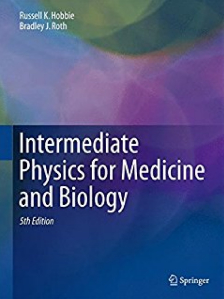
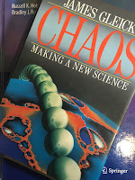
Chaos: Making a New Science, by James Gleick, superimposed on Intermediate Physics for Medicine and Biology.
Chaos: Making a New Science,
by James Gleick.
Yesterday I posted an excerpt from Chaos: Making a New Science. Here’s a dirty little secret: I haven’t read this book yet. I own a copy, and hope to read it soon (if we’re still locked down by the coronavirus once the semester is over, I’ll have plenty of time to read!). I did thumb through it, and found another interesting story. Regular readers of this blog know that the mathematical biologist Art Winfree played a key role in my development as a scientist. In Chaos, James Gleick tells how Winfree teamed up with Duke cardiologist Ray Ideker to study defibrillation.
Winfree told the story of an early researcher, George Mines, who in 1914 was twenty-eight years old. In his laboratory at McGill University in Montreal, Mines made a small device capable of delivering small, precisely regulated electrical impulses to the heart.

“When Mines decided it was time to begin work with human beings, he chose the most readily available experimental subject: himself,” Winfree wrote. “At about six o’clock that evening, a janitor, thinking it was unusually quiet in the laboratory, entered the room. Mines was lying under the laboratory bench surrounded by twisted electrical equipment. A broken mechanism was attached to his chest over the heart and a piece of apparatus nearby was still recording the faltering heartbeat. He died without recovering consciousness.”

One might guess that a small but precisely timed shock can throw the heart into fibrillation, and indeed even Mines had guessed it, shortly before his death. Other shocks can advance or retard the next beat, just as in circadian rhythms. But one difference between hearts and biological clocks, a difference that cannot be set aside even in a simplified model, is that a heart has a shape in space. You can hold it in your hand. You can track an electrical wave through three dimensions.

To do so, however, requires ingenuity. Raymond E. Ideker of Duke University Medical Center read an article by Winfree in Scientific American in 1983 and noted four specific predictions about inducing and halting fibrillation based on nonlinear dynamics and topology. Ideker didn’t really believe them. They seemed too speculative and, from a cardiologist’s point of view, so abstract. Within three years, all four had been tested and confirmed, and Ideker was conducting an advanced program to gather the richer data necessary to develop the dynamical approach to the heart. It was, as Winfree said, “the cardiac equivalent of a cyclotron.”

The traditional electrocardiogram offers only a gross one-dimensional record. During heart surgery a doctor can take an electrode and move it from place to place on the heart, sampling as many as fifty or sixty sites over a ten-minute period and thus producing a sort of composite picture. During fibrillation this technique is useless. The heart changes and quivers far too rapidly. Ideker’s technique, depending heavily on real-time computer processing, was to embed 128 electrodes in a web that he would place over a heart like a sock on a foot. The electrodes recorded the voltage field as each wave traveled through the muscle, and the computer produced a cardiac map.

Ideker’s immediate intention, beyond testing Winfree’s theoretical ideas, was to improve the electrical devices used to halt fibrillation. Emergency medical teams carry standard versions of defibrillators, ready to deliver a strong DC shock across the thorax of a stricken patient. Experimentally, cardiologists have developed a small implantable device to be sewn inside the chest cavity of patients thought to be especially at risk, although identifying such patients remains a challenge. An implantable defibrillator, somewhat bigger than a pacemaker, sits and waits, listening to the steady heartbeat, until it becomes necessary to release a burst of electricity. Ideker began to assemble the physical understanding necessary to make the design of defibrillators less a high-priced guessing game, more a science.
You can learn more about Winfree, nonlinear dynamics, the heart, and defibrillation in Intermediate Physics for Medicine and Biology.

James Gleick on Chaos: Making a New Science.
https://www.youtube.com/watch?v=3orIIcKD8p4

Ray Ideker discusses Mechanisms of
Ventricular Fibrillation and Defibrillation.
https://www.youtube.com/watch?v=94eU2ztM_uU

Monday, March 30, 2020

Chaos: Making a New Science


In Chapter 10 of Intermediate Physics for Medicine and Biology, Russ Hobbie and I include a homework problem about the Lorenz model.
Problem 36. Edward Lorenz (1963) published a simple, three-variable (x, y, z) model of Rayleigh–Bénard convection:

dx/dt = σ (yx)

dy/dt = x (ρz) − y

dz/dt = x yβ z

where σ = 10, ρ = 28, and β = 8/3.

(a) Which terms are nonlinear?

(b) Find the three equilibrium points for this system of equations.

(c) Write a simple program to solve these equations on the computer (see Sect. 6.14 for some guidance on how to solve differential equations numerically). Calculate and plot x(t) as a function of t for different initial conditions. Consider two initial conditions that are very similar, and compute how the solutions diverge as time goes by.

(d) Plot z(t) vs. x(t), with t acting as a parameter of the curve.
This model played a critical role in the history of nonlinear dynamics; it resulted in Lorenz discovering chaos. More specifically, his calculation revealed one of the hallmarks of chaotic behavior: sensitivity to initial conditions, also known as the butterfly effect.

Chaos: Making a New Science, by James Gleick, superimposed on Intermediate Physics for Medicine and Biology.
Chaos: Making a New Science,
by James Gleick.
I will let James Gleick, author of Chaos: Making a New Science, tell the story. Lorenz’s equations are a model for the weather, which he solved using one of the first computers.
One day in the winter of 1961, wanting to examine one sequence at greater length, Lorenz took a shortcut. Instead of starting the whole run over, he started midway through. To give the machine its initial conditions, he typed the numbers straight from the earlier printout. Then he walked down the hall to get away from the noise and drink a cup of coffee. When he returned an hour later, he saw something unexpected, something that planted the seed for a new science.

This new run should have exactly duplicated the old. Lorenz had copied the numbers into the machine himself. The program had not changed. Yet as he stared at the new printout, Lorenz saw his weather diverging so rapidly from the pattern of the last run that, within just a few months, all resemblance had disappeared. He looked at one set of numbers, then back at the other. He might as well have chosen two random weathers out of a hat. His first thought was that another vacuum tube had gone bad.

Suddenly he realized the truth. There had been no malfunction. The problem lay in the numbers he had typed. In the computer’s memory, six decimal places were stored: .506127. On the printout, to save space, just three appeared: .506. Lorenz had entered the shorter, rounded-off numbers, assuming that the difference—one part in a thousand—was inconsequential…

He decided to look more closely at the way two nearly identical runs of weather flowed apart. He copied one of the wavy lines of output onto a transparency and laid it over the other, to inspect the way it diverged. First, two humps matched detail for detail. Then one line began to lag a hairsbreadth behind. By the time the two runs reached the next hump, they were distinctly out of phase. By the third or fourth hump, all similarity had vanished.

It was only a wobble from a clumsy computer. Lorenz could have assumed something was wrong with his particular machine or his particular model—probably should have assumed. It was not as though he had mixed sodium and chlorine and got gold. But for reasons of mathematical intuition that his colleagues would begin to understand only later, Lorenz felt a jolt: something was philosophically out of joint. The practical import could be staggering. Although his equations were gross parodies of the earth’s weather, he had a faith that they captured the essence of the real atmosphere. That first day, he decided that long-range weather forecasting must be doomed.

Friday, March 27, 2020

Carl Woese, Biological Physicist

The Tangled Tree: A Radical New History of Life, by David Quammen, superimposed on Intermediate Physics for Medicine and Biology.
The Tangled Tree,
by David Quammen.
Recently I listened to the audiobook The Tangled Tree: A Radical History of Life, by David Quammen. The book is a wide-ranging history of molecular phylogenetics and its central character is Carl Woese. His landmark discovery was the place of the archaea in the history of life.
The discovery and identification of the archaea, which had long been mistaken for subgroups of bacteria, revealed the present-day life at the microbial scale is very different from what science had previously depicted, and that the early history of life was very different too.
Quammen writes
Carl Woese was a complicated man—fiercely dedicated and very private—who seized upon deep questions, cobbled together ingenious techniques to pursue those questions, flouted some of the rules of scientific decorum, make enemies, ignored niceties, said what he thought, focused obsessively on his own research program to the exclusion of most other concerns, and turned up at least one or two discoveries that shook the pillars of biological thought.
How does Woese’s career intersect with Intermediate Physics for Medicine and Biology? As an undergraduate at Amherst College, Woese was a physics major. Therefore, he represents yet another example of a scientist who made the switch from physics to biology. Quammen doesn’t explore this aspect of Woese’s career much, so I searched for what motivated his change, what challenges he faced, and what advantages his physics background provided. An article in the Amherst Magazine provided some insight. While at Amherst, Woese
fell in love with physics while studying under William M. Fairbank, who would go on from Amherst to become “one of the great low-temperature physicists in the world.” Fairbank inspired Woese to go on for his physics Ph.D. at Yale, and it was there that Woese became fascinated with biophysics: the study of biological processes at the molecular level. After earning his doctorate in 1953, Woese took a brief fling at medical school (“I couldn’t bear to treat sick children, so I quit”), then studied at the famed Louis Pasteur Institute in Paris and worked for a while in an experimental biophysics lab operated by General Electric. By 1964 he had signed on at the University of Illinois where, ever since, he has taught microbiology and studied the molecular processes that go on inside single-celled creatures.
Woese’s education parallels my own: a physics major in college, followed a physics PhD but an increasing emphasis on applying physics to biology, then post doctoral study at a leading research center: the Pasteur Institute for Woese and the National Institutes of Health for me. William Fairbank plays a role in both of our careers, as undergraduate mentor to Woese and as academic grandfather to me; my PhD advisor, John Wikswo, had Fairbank as his PhD advisor. You could say that Woese was my academic uncle.

The article continues
Soon after arriving in Urbana-Champaign, Woese dared to tackle a fundamental problem in microbiology—a key problem that had stumped both Stanford's C.B. van Niel and Roger Stanier of Cal-Berkeley, the leading microbiologists of the generation preceding Woese’s. The problem, in a phrase: How could you classify—or “phylogenetically order”—the vast ranks of bacterial and other one-celled organisms, given the fact that their small size and vast complexity made it extremely difficult to study and identify their anatomical and physiological features?

Years later, after gaining a worldwide reputation for solving the problem by making the key identifications at the molecular level by sequencing genetic macromolecules and then comparing one organism’s genetic inheritance to another’s, Woese realized that his training in physics at Amherst had played a major role in his discoveries. As he later told reporters: “I hadn’t been trained as a microbiologist, so I didn’t have their bias [against classifying micro-organic species]. And my physics background had taught me the vital importance of using ‘Occam’s Razor’ whenever I could, because it had taught me that most questions—no matter how seemingly complex—usually turn out to have rather simple, straightforward answers.”
Woese returned to his physics roots later in his career. In The Tangled Tree, Quammen writes
One day in September 2002 [Woese] reached out by email to a theoretical physicist in another corner of the University of Illinois campus. Nigel Goldenfeld was an Englishman, almost thirty years younger, who had arrived in Urbana as an assistant professor, risen to full professor, and spent his middle career studying the dynamics of complex interactive systems. That included topics such as crystal growth, the turbulent flow of fluids, structural transitions in materials, and how snowflakes take shape. The common element was patterns evolving over time. Goldenfeld had never met Woese but knew him by reputation. Later, he called that first ping “the most important email of my life”… In the email, Woese now explained that he wanted to discuss—with someone—the subject of complex dynamic systems. He felt that molecular biology had exhausted its vision, he wrote, and that it needed refocusing around drastic new insights…Woese wanted a partner who understood complex interactive systems and could quantify their dynamics with brilliant math. Whether that partner knew a bacterium from an archaeon, or Darwin from Dawkins, mattered less to him.
Goldenfeld and Woese wrote several papers together, including “Life is Physics: Evolution as a Collective Phenomenon Far From Equilibrium” (Annual Review of Condensed Matter Physics, Volume 2, Pages 375-399, 2011), in which they
discuss how condensed matter physics concepts might provide a useful perspective in evolutionary biology, the conceptual failings of the modern evolutionary synthesis, the open-ended growth of complexity, and the quintessentially self-referential nature of evolutionary dynamics.
In another paper about “Biology’s Next Revolution” (Nature, Volume 445, Page 369, 2007)  they begin
One of the most fundamental patterns of scientific discovery is the revolution in thought that accompanies the acquisition of an entirely new body of data. The new window on the Universe opened up by satellite-based astronomy has in the last decade overthrown our most cherished notions of Cosmology, especially related to the size, dynamics and composition of the Universe. Similarly, the convergence of new theoretical ideas in evolution together with the coming avalanche of environmental genomic data, especially from marine microbes and viruses, will fundamentally alter our understanding of the global biosphere, and is likely to cause a revision of such basic and widely-held notions as species, organism and evolution. Here’s why we foresee such a dramatic transformation on the horizon, and how biologists will need to join forces with quantitative scientists, such as physicists, to create a biology that embraces collective phenomena and supersedes the molecular reductionism of the twentieth century.
Woese and Goldenfeld are IPMB type of people.

Quammen concludes
In later years, as he grew more widely acclaimed, receiving honors of all kinds short of the Nobel Prize, Woese seems also to have grown bitter. He considered himself an outsider. He was elected to the National Academy of Sciences, an august body, but tardily, at age sixty, and the delay annoyed him…He was a brilliant crank, and his work triggered a drastic revision of one of the most basic concepts in biology: the idea of the tree of life, the great arboreal image of relatedness and diversification.
The Place of Carl Woese in Evolutionary Biology

 Listen to David Quammen discuss The Tangled Tree.

Thursday, March 26, 2020

The Goldman-Hodgkin-Katz Equation Including Calcium

In Section 9.6 of Intermediate Physics for Medicine and Biology, Russ Hobbie and I derive the Goldman-Hodgkin-Katz equation. It accounts for both diffusion and electrical forces acting on ions in the membrane (presumably passing through ion channels spanning the lipid bilayer). If only one ion were present, its concentration on each side of the membrane would determine the equilibrium, or reversal, potential. For instance more potassium is inside a cell than outside, so diffusion pushes the positively charged potassium ions out. As the outside becomes positive, the resulting electric field in the membrane pushes potassium back in. The reversal potential, vrev, is the potential across the membrane when diffusion and electrical forces balance.

Mathematically, we can derive the reversal potential for any ion C by starting with an expression for its current density, JC


where z is the valence, e is the elementary charge, v is the potential, ωC is the permeability, NA is Avogadro's number, kB is the Boltzmann constant, T is the absolute temperature, and [C1] and [C2] are the concentrations outside and inside the membrane. (See IPMB for a derivation of this complicated equation.) To find the reversal potential, we set JC to zero and solve for v.

When more than one ion can cross the membrane, the situation is more complicated. Russ and I examined a membrane that can pass three ions: sodium, potassium, and chloride. The resulting equation for the reversal potential—also known as the Goldman-Hodgkin-Katz equation—is

We then write
When ions have different valences, the GHK equation becomes more complicated. Lewis (1979) has derived an analogous equation for transport of sodium, potassium, and calcium.
The citation is to
Lewis CA (1979) “Ion-concentration dependence of the reversal potential and the single channel conductance of ion channels at the frog neuromuscular junction.” Journal of Physiology, Volume 286, Pages 417–445.
Below is a new homework problem, based on Appendix A of Lewis’s paper, analyzing a more complicated GHK equation that includes calcium along with sodium and potassium.
Section 9.6

Problem 20 ½. Derive an expression for the Goldman-Hodgkin-Katz equation when you have three ions that can pass through the membrane: sodium, potassium, and calcium.

(a) Write down an expression like Eq. 9.53 for the current density for each ion: JNa, JK, and JCa. Hint: be careful to include the valence z properly.

(b) Assume the amount of charge in the cell does not change with time, so JNa + JK + JCa = 0. Try to solve the resulting equation for the reversal potential, vrev. You should find it difficult, because the expression for JCa has a different denominator than do JNa and JK.

(c) Define a new permeability for calcium,
Now derive an expression for vrev. Your result should look similar to Eq. 9.55, except for some factors of four, and in the numerator the new calcium permeability will be multiplied by a voltage-dependent factor.
What’s the lesson to be learned from this homework problem? First, the GHK expression including calcium has the potential on the left side of the equation, but also on the right side, inside a logarithm. No simple way exists to calculate vrev. My first thought is to use an iterative method, but I haven’t looked into this in detail. Second, notice how a small modification to the problem—changing chloride to calcium—made a major change in how difficult the problem is to solve. Adding the negative chloride ion to positive sodium and potassium resulted in a trivial change to the GHK equation (the inside chloride concentration appears in the numerator rather than the outside concentration). However, adding the divalent cation calcium totally messes up the equation, making it difficult to solve except with numerical methods.

I advocate for simple models. They provide tremendous insight. However, the moral of this story is if you push a toy model too hard, it can become complicated; it’s no longer a toy.

Wednesday, March 25, 2020

A Severe Shortage of Blood!

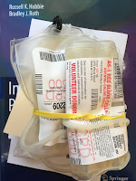
Blood donation bags sitting on top of Intermediate Physics for Medicine and BIology.
Blood donation bags
at a blood drive in
Rochester Michigan
on March 24, 2020.
Yesterday Michigan’s governor, Gretchen Whitmer, announced a shelter-in-place order. Now I need a legitimate reason to leave home. I have one: To donate blood! The Red Cross has a severe blood shortage. I encourage all readers of Intermediate Physics for Medicine and Biology to make an appointment.

What’s the purpose of blood, anyway? To carry oxygen. Let’s estimate the concentration of oxygen in blood (Fermi problem time). As a first step, Homework Problem 1 in Chapter 1 of IPMB asks the reader to estimate how much hemoglobin is in a red blood cell.
Problem 1. Estimate the number of hemoglobin molecules in a red blood cell. Red blood cells are little more than bags of hemoglobin, so it is reasonable to assume that the hemoglobin takes up all the volume of the cell.
My name tag at the blood drive.
Russ Hobbie and I don’t send IPMB’s solution manual to anyone except instructors, but because you all are going to make blood-donation appointments as soon as you’re done reading this post, I’ll share the solution to this problem.
1.1 An important skill for students to learn is order-of-magnitude estimation. The first four problems in this chapter require the students to estimate some quantity of biological interest.
Approximate the dimensions of a red blood cell as 8 μm × 8 μm × 2 μm. Approximate the dimensions of a hemoglobin molecule as 6 nm × 6 nm × 6 nm. The number N of hemoglobin molecules is equal to the volume of a red blood cell divided by the volume of a hemoglobin molecule: 
We do not expect a “back-of-the-envelope” estimate such as this one to be accurate to, say, a factor of 2 or π. But it should give a quick order of magnitude approximation.
A selfie of me giving blood, with Intermedaite Physics for Medicine and Biology balanced on my chest.
I had a difficult time taking
this selfie: one hand holding my
phone, the book balanced on my
chest, and a needle in the other arm.
Each hemoglobin molecule can bind with four oxygen molecules, so a red blood cell can contain 2400 million oxygen molecules. I’ll assume the hemoglobin isn’t packed too tightly, so let’s round that down to 2000. The volume of a red blood cell is 128 cubic microns. Inside a red blood cell the oxygen concentration is therefore 2000 million molecules per 128 cubic microns, or about 16,000,000/μm3. A typical hematocrit (fraction of blood volume occupied by red blood cells) is 40%. Therefore blood has an oxygen concentration of around 6 million per cubic micron.

I admit, those are strange units. A cubic micron is 10-15 liters, and 6 million molecules is 10-17 moles. So, the concentration of oxygen in blood is about 0.01 molar, or 10 mM.

You can estimate the concentration of oxygen in air using the ideal gas law, pV = nRT. Air is about 20% oxygen, so using p = 0.2 atm, T = 310 K, and R = 0.082 liter atm/(mole K), you get n/V = 0.008, or 8 mM. Within the uncertainty of our rough estimate, this result implies that the concentration of oxygen in blood is nearly the same as the concentration of oxygen in air. As it should be! The whole point of blood is to get oxygen from the air into the tissues.

The best part of blood donation.
Thanks to all the phlebotomists and volunteers for collecting blood, despite the risk; they’re heroes. I won’t be able to give blood again for another eight weeks. By that time I hope the @#$%&! coronavirus is gone and life has returned back to normal.

After giving blood. My daughter Stephanie,
who also donated, took the photo.

Tuesday, March 24, 2020

Bob Park’s What’s New has been Restored!

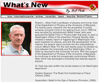
Screenshot of the What's New website, whatsnewbobpark.com.
Screenshot of the What's New website,
whatsnewbobpark.com.
From 1983–2006, physicist Bob Park was the director of public information in the Washington D. C. office of the American Physical Society. During this time, he wrote the delightful weekly column What’s New. When I was in graduate school, every Friday I’d look forward to a new post.

For years What’s New disappeared from the internet, but recently it’s been restored (at least partially) from internet archives. You can find it at http://whatsnewbobpark.com. [Note added October 9, 2020: the link no longer works properly. However, here is one that does: https://web.archive.org/web/20140124195058/http://bobpark.physics.umd.edu/archives.html] If you click on “About Bob” you’ll see:
Robert L. (Bob) Park is professor of physics and former chair of the Department of Physics at the University of Maryland. For twenty years, research into the properties of crystal surfaces occupied most of his waking hours, but in 1983 he was recruited by astrophysicist Willie Fowler (who was awarded the Nobel Prize in Physics later that year) to open a Washington Office of the American Physical Society. Bob initiated a weekly report of happenings in Washington that were important to science, and with the development of the internet, the weekly report evolved into the news/editorial column What’s New. For the next twenty years he divided his time between the University and the Washington Office. In 2003 he returned to the University full time. With the support of the Department of Physics of the University of Maryland, he continues to write the occasionally controversial What’s New, which has developed a following that extends beyond physics.

Dr. Park has also written two books based on his Washington experience:

Voodoo Science: The Road from Foolishness to Fraud (Oxford, 2000)

Superstition: Belief in the Age of Science (Princeton, 2008)
In What’s New, Park would return to certain topics again and again; for example, cell phones and cancer, creationism, climate change, and cold fusion. Often he would debunk alternative medicine, such as homeopathy, biomagnetic therapy, and therapeutic touch. Readers of Intermediate Physics for Medicine and Biology will enjoy how he applied physics reasoning to medicine. Below are excerpts from 2011, so you can sample Park’s writing style. 

Friday, June 10, 2011

1. ET TU TARA? “PIERCING THE FOG AROUND CELLPHONES AND CANCER.” The WELL blog by Tara Parker-Pope was the top story in Tuesdays NYT Health Section. Her story is not wrong, but its told in the wrong context. Science is a search for cause and effect, not an epidemiologic majority. To settle the question, WHO invited 31 experts to spend a week in Lyon, the culinary capital of France, strategically located between the two best wine regions. Meanwhile, much had been made of a study showing that the brain is “activated” by microwave radiation. Of course, it is. The effect of microwaves on the human brain, as on cold pizza, is to cause chemical bonds to vibrate, which we sense as heat. Unlike cold pizza, however, the human brain resists being heated. Deep within the brain, the hypothalamus, the thing below the thalamus, senses any increase in blood temperature. It calls on blood vessels in the heated area to expand, and increases the heart rate. The fresh blood is a coolant, but incidentally, also increases the rate of metabolism. “Microwaves have activated the brain,” the human observers shouted. The shout was heard in Lyon. Amidst the clinking of glasses, the vote of the expert panel tipped from “no effect” to “possibly carcinogenic to humans.” What could it matter? No one is going to stop using cell phones anyway. Does anyone care? One enormously powerful group cares, the tort industry.

Friday, August 19, 2011

1. HOMEOPATHY: THE DILUTION LIMIT AND THE CULTURE OF CREDULITY. Based in France, Boiron, a huge multinational maker of homeopathic-remedies, is suing an Italian blogger, Samuele Riva, for saying oscillococcinum, the companys featured flu medication, has no active ingredient. Congratulations Sam, I gave up trying to get Boiron to sue me, years ago but the Center for Inquiry, of which I’m a member, is pleading with Boiron to sue us. “Anas barbariae hepatis et cordis extractum,” is listed as the active ingredient by the company. Its prepared at a concentration of 200CK HPUS from the liver of the Barbary duck. The 200CK means the solution has been diluted 1 part in 100, shaken, and repeated sequentially 200 times. HPUS means the medication is listed in the Homeopathic Pharmacopeia of the United States, and prepared according to 1938 federal guidelines. Its a national disgrace that the antiquated law sanctioning homeopathy, introduced by Sen. Royal Copeland, himself a homeopathist, is still be on the books. The dilution claim is totally meaningless. Somewhere around the 30th of the 200 sequential dilutions, the dilution limit of Earth would be reached, with the entire Earth becoming the solute. That is, the possibility of even one molecule of the duck-liver extract remaining in the solution beyond that point would be negligible. Long before the 200th dilution, the dilution limit of the entire visible universe would have been reached. This is all quite meaningless. Astronomers put the number of atoms in the visible universe at about 10 to the 80th power. It would take many universes to get to a dilution of 200 C.

Friday, September 16, 2011

1. WI-FI REFUGE: UNITED STATES NATIONAL RADIO QUIET ZONE. A 34,000 km2 rectangle of land straddling the border of Virginia and West Virginia surrounds The Robert C. Byrd Green Bank Telescope, the world’s largest fully steerable radio telescope. The site was chosen partly because the Allegheny Mountains block the horizontal propagation of radio signals, but mostly because Robert C Byrd (D-WV) was one powerful US Senator. Radio transmission in the zone is either limited or banned outright. In addition to radio astronomers, the quiet zone has also attracted a colony of people who say they suffer from Electromagnetic Hypersensitivity (EHS). They certainly suffer from something, but EHS is not medically recognized in the US. In a BBC News interview last week I suggested that the appropriate treatment for a non-ailment such as EHS would be homeopathic medicine.

Sunday, October 23, 2011

2. CELL PHONEYS: BRAIN CANCER LINK IS REJECTED AGAIN. Ten years ago, a brilliant Danish epidemiological study found no link between mobile phone use and brain cancer (JNCI 2001, 93: 203-7). A decadal reexamination by Denmarks Institute of Cancer Epidemiology, released last week, again found no link. The object of the new study was to look for any evidence of latent cancer that had not yet shown up in 2001; none was found. In a 2001 JNCI editorial I pointed out that none would be expected, since microwave radiation is non-ionizing, Park, Robert L, JNCI 2001, 93: 166-167. Can we now put the damned cell-phone/cancer scare behind us?

Wednesday, November 16, 2011

2. CANCER AND CAUSALITY: EINSTEIN DIDNT HAVE A CELL-PHONE. Of the worlds 7 billion people, an incredible 5 billion have cell phones (mobiles in most countries). The safe use of mobiles is therefore a global health concern. The response of the World Health Organization was to conduct a huge epidemiologic study aimed at demonstrating a link between cell-phone radiation and brain cancer. The effort was seriously misguided no such link exists. The study served only to raise widespread public alarm over a nonexistent hazard. Epidemiology, which is the study of health patterns in populations; is important, but its not a substitute for science. Science is the organization of knowledge into testable laws and theories. It has been known for more than 100 years that electromagnetic radiation at frequencies below the ultraviolet is non-ionizing, and thus cannot create the mutant strands of DNA that constitute incipient cancers. In 1905, Einsteins miracle year, he theorized that electromagnetic radiation consists of discrete units of energy, now called photons, which are equal in energy to the frequency multiplied by Planck’s constant. It marked the origin of wave-particle duality and earned Einstein his 1921 Physics Nobel Prize. His theory is verified every time a cell phone works.
I miss Bob Park. We still need him. His mantle has been taken up by people like the Skep Doc Harriet Hall. We must expose quackery and embrace evidence-based science and medicine.

What’s New was hosted by a University of Maryland website. At the bottom of the page was the disclaimer:
Opinions are the author’s and are not necessarily shared by the University, but they should be.

Bob Park is featured in the video 
“You Don't Have to Be a Scientist to Spot 
the Fraudulent Science that Swirls Around Us (2000)” 

 Part 1 of Superstition: Belief in the Age of Science
featuring Bob Park (you can find the other six parts on YouTube).

Monday, March 23, 2020

Practice Problems in Bioelectricity and Biomagnetism

It’s funny how your memory can deceive you. I thought my interest in writing homework problems began with my work on Intermediate Physics for Medicine and Biology. Recently, however, I was rummaging through some old papers and discovered that I’ve been writing homework problems for a lot longer. This habit traces back to my graduate school days at Vanderbilt University, when I worked for John Wikswo. Among my old documents, I found a brittle yellowed copy of “The Magnetic Field of a Single Axon: Practice Problems.” It begins
These problems are presented to help someone to become familiar with the analytic volume conduction models of electric potentials and magnetic fields produced by nerve axons or bundles of nerve or muscle fibers developed between 1982 and 1988 in the Living State Physics Group. The problems vary in difficulty, with the very difficult ones marked by a *.
The problems are drawn from eight publications I helped write back in the day. If you need copies of these articles so you can solve the problems, just email me: roth@oakland.edu. (Technically the journal owns the copyright, so I won’t link to the pdfs in this blog. 😞)
J. K. Woosley, B. J. Roth, and J. P. Wikswo, Jr. (1985) “The Magnetic Field of a Single Axon: A Volume Conductor Model.” Mathematical Biosciences, Volume 75, Pages 1-36.

B. J. Roth and J. P. Wikswo, Jr. (1985)  “The Magnetic Field of a Single Axon: A Comparison of Theory and Experiment.” Biophysical Journal, Volume 48, Pages 93-109.

B. J. Roth and J. P. Wikswo, Jr. (1985) “The Electrical Potential and Magnetic Field of an Axon in a Nerve Bundle.” Mathematical Biosciences, Volume 76, Pages 37-57.

B. J. Roth and J. P. Wikswo, Jr. (1986) “A Bidomain Model for the Extracellular Potential and Magnetic Field of Cardiac Tissue.” IEEE Transactions on Biomedical Engineering, Volume 33, Pages 467-469.

B. J. Roth and J. P. Wikswo, Jr. (1986) “Electrically Silent Magnetic Fields.” Biophysical Journal, Volume 50, Pages 739-745.

B. J. Roth and F. L. H. Gielen (1987) “A Comparison of Two Models for Calculating the Electrical Potential in Skeletal Muscle.” Annals of Biomedical Engineering, Volume 15, Pages 591-602.

J. P. Wikswo, Jr. and B. J. Roth (1988) “Magnetic Determination of the Spatial Extent of a Single Cortical Current Source: A Theoretical Analysis.” Electroencephalography and Clinical Neurophysiology, Volume 69, Pages 266-276.

B. J. Roth (1987) “Longitudinal Resistance in Strands of Cardiac Muscle.” Ph.D. Thesis, Vanderbilt University, Nashville, Tennessee.
Many of these problems require analyzing Bessel functions and Fourier transforms; I was enamored by those mathematical methods in the 80’s. You can get a hint of what these old homework problems are like by looking at Problem 16 in Chapter 8 of IPMB, where the reader must use these techniques to calculate the magnetic field of a nerve axon.

Let me give you another example. Problem 10 in this ancient collection is based on the  paper by Woosley et al.:
Prove that Eq. (36) and (45) are equal.
Equation (36) is the magnetic field of a nerve axon derived using the law of Biot and Savart, and Equation (45) is the magnetic field derived using Ampere’s law. I’ve discussed before in this blog how I could not prove these two equations were equivalent until I found a Wronskian relating Bessel functions.

I’ve also analyzed the IEEE TBME paper in a blog post from 2018.

These practice problems are not for the faint of heart. Nevertheless, most of what you need to solve them is in IPMB. If you want to learn some advanced methods in theoretical bioelectricity and biomagnetism, download the practice problems and give them a try. If nothing else, they will provide insight into what I used to work on as a graduate student. Besides, with the coronavirus pandemic holding you in quarantine, what else do you have to do?

Friday, March 20, 2020

Traveling Waves and Standing Waves

Section 13.2 of Intermediate Physics for Medicine and Biology discusses waves. Russ Hobbie and I note that two solutions to the wave equation exist: traveling waves and standing waves.

Traveling Waves

We write that the pressure distribution p(x,t) = f(xct), where f is any function,
obeys the wave equation... It is called a traveling wave. A point on f(xct), for instance its maximum value, corresponds to a particular value of the argument xct. To travel with the maximum value of f(xct), as t increases, x must also increase in such a way as to keep xct constant. This means that the pressure distribution propagates to the right with speed c... Solutions p(x,t) = g(x + ct), where g is any function, also are solutions to the wave equation, corresponding to a wave propagating to the left.

Standing Waves

We then discuss standing waves.
Standing waves such as p(x,t) = p cos(ωt) sin(kx) are also solutions to the wave equation… [This] standing wave… has nodes fixed in space where sin(kx) is zero… A standing wave can also be written as the sum of two sinusoidal traveling waves, one to the left and one to the right. Conversely, two standing waves can be combined to give a traveling wave.

Converting Traveling Waves to Standing Waves

IPMB includes a homework problem asking the reader to show analytically that two traveling waves combine to make a standing wave, and vice versa.
Problem 8. Use the trigonometric identity sin(a ± b) = sin a cos b ± cos a sin b to show that a traveling wave can be written as the sum of two out-of phase standing waves, and that a standing wave can be written as the sum of two oppositely-propagating traveling waves.

Visualizations

Russ and I also include figures illustrating the difference between a traveling wave (our Fig. 13.4) and a standing wave (Fig. 13.5). To gain insight, however, nothing can replace a dynamic visualization. Fortunately, the internet is full of such visualizations. One appears in the Wikipedia article about standing waves. The Physics Hypertextbook also has traveling and standing wave animations.

This Youtube video shows trigonometry in action: the sum of two oppositely going traveling waves (blue wave propagating right, and green left) add to form a single standing wave (red).

Two traveling waves adding to form a standing wave.

I like the next video because it shows a traveling wave turning into a standing wave when it reflects off a boundary.

A traveling wave turning into a standing wave when it reflects off a boundary.

Here’s a nice video showing how standing waves can be created experimentally.


Standing waves created experimentally on a string fastened at both ends.

Finally, here’s a Flipping Physics video comparing standing and traveling waves. It’s a little corny, but I like it that way.

A lecture about waves from Flipping Physics.

Enjoy!

Thursday, March 19, 2020

Physics Girl

Because of the coronavirus, I had to transform my introductory physics course from in-person to online (in two days!). I thought: If I’m going to teach remotely, I might as well use some of the excellent resources that are available on the internet. This led me to Physics Girl.

Dianna Cowern produces funny and informative videos about physics. Some even deal with medical and biological physics. Below I have embedded a few about biomechanics, sound perception, sun screen, color vision, magnetic resonant imaging, and bioelectricity.

If you’re studying from Intermediate Physics for Medicine and Biology, consider these videos as supplementary material. If you like them, plenty more are at the Physics Girl YouTube channel.

Happy Physicsing!

Testing what exercise actually does to your butt.

What stretching actually does to your body.

Can you guess this note? Perfect pitch and physics.

Sunscreen in the UV.

Does this look like white to you?

The projector illusion.

Wednesday, March 18, 2020

Videos for PHY 3250, Biological Physics

Last fall, I recorded my lectures for my PHY 3250 (Biological Physics) class, and posted them on YouTube. The videos are not great; they are nowhere near professional quality, and often the chalkboard is difficult to read. I originally recorded them as a backup for my students, in case they missed a class or wanted to review something they heard me say in a lecture. Nevertheless, I think that students and instructors may find these videos useful.

My Biological Physics class covers the first ten chapters in Intermediate Physics for Medicine and Biology. Topics include biomechanics, fluid dynamics, the exponential function, biothermodynamics, diffusion, osmotic pressure, bioelectricity, biomagnetism, and feedback.

Some videos are missing: Monday, September 30 was Exam 1; Wednesday, October 30 was Exam 2; Wednesday, November 27 the class played Trivial Pursuit IPMB; and Friday, November 29 was the day after Thanksgiving.

If you are sitting at home self-quarantining with nothing to do, feel free to binge.

Enjoy!

Wednesday, September 4, 2019. Introduction.

Friday, September 6, 2019. Biomechanics.
Monday, September 9, 2019. Hydrostatics.

Wednesday, September 11, 2019. Fluid Dynamics.

Friday, September 13, 2019.  The exponential function.

Monday, September 16, 2019. Scaling.

Wednesday, September 18, 2019. Boltzmann factor.

Friday, September 20, 2019. Heat capacity.

Monday, September 23, 2019. Heat transfer.

Wednesday, September 25, 2019. Review for Exam 1.
Friday, September 27, 2019. Review for Exam 1 (cont.).

Wednesday, October 2, 2019. Heat conduction.

Friday, October 4, 2019. Diffusion.

Monday, October 7, 2019. Diffusion and convection.

Wednesday, October 9, 2019. Osmotic pressure.

Friday, October 11, 2019. Countercurrent exchange.

Monday, October 14, 2019. Bioelectricity.

Wednesday, October 16, 2019. Hodgkin & Huxley model.

Friday, October 18, 2019. Hodgkin & Huxley model (cont.).

Monday, October 21, 2019. The cable equation.

Wednesday, October 23, 2019. Action potential propagation.

Friday, October 25, 2019. Review for Exam 2.

Monday, October 28, 2019. Review for Exam 2 (cont.).

Friday, November 1, 2019. Extracellular stimulation of nerves.

Monday, November 4, 2019. Extracellular potentials and the dipole.

Wednesday, November 6, 2019. The heart.

Friday, November 8, 2019. The electrocardiogram.

Monday, November 11, 2019. Pacemakers and defibrillators.

Wednesday, November 13, 2019. The electroencephalogram.

Friday, November 15, 2019. Biomagnetism.

Monday, November 18, 2019. Transcranial magnetic stimulation.

Wednesday, November 20, 2019. Cardiac restitution.

Friday, November 22, 2019. Cellular automata.

Monday, November 25, 2019. Feedback.


Monday, December 2, 2019. Feedback (cont.).
Monday, December 4, 2019. Review for Exam 3.

 Wednesday, December 6, 2019. Review for Exam 3 (cont.).

Tuesday, March 17, 2020

The Ophthalmoscope

The First Steps in Seeing, by Robert Rodieck, superimposed on Intermediate Physics for Medicine and Biology.
The First Steps in Seeing,
by Robert Rodieck.
In The First Steps in Seeing, Robert Rodieck describes the ophthalmoscope.
Light passes into the eye through the pupil, and continues through its mainly transparent interior to reach the retina. The portion of the light that is not caught by the photoreceptors is either absorbed or scattered in all directions by the underlying tissues. Some of the scattered light passes back through the pupil and out of the eye. But when we look into another person’s pupil, the back of the eye, or fundus, appears black. This is because the optical pathway of the light that enters the eye and falls on a given region of the fundus is the same as that of the light scattered from that region, which leaves the eye through the pupil. In effect, in order to see the interior of the eye under ordinary conditions, one has to place one’s head into this common pathway of the light.

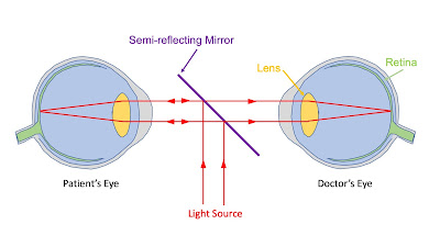
A brilliant young clinician, Hermann von Helmholtz (1821-1894), grasped this issue, and realized that all he needed to do to see the interior of another person’s eye was to devise an optical device by which he could get both his head and the light into the pathway. He did so by placing a piece of glass between his eye and the patient’s and angling the glass so that it partially reflected the light from a lamp into the patient’s eye… The piece of glass and the lamp formed a device termed an ophthalmoscope (Greek opthalmos = eye + skopion, from skopein = to see). Modern ophthalmoscopes have a built-in light source, colored filters to emphasize some aspect of the view, and lenses to correct for any error in the optics of the clinician or patient (i.e., lenses of the same power that they might use in spectacles.)
The picture below shows a simple ophthalmoscope, which consists of just a light source, a semi-reflecting mirror, and two eyes.
An ophthalmoscope.
An ophthalmoscope.
An image of the retina, as might be seen using an ophthalmoscope, is shown below. The dark patch in the center is the fovea, where the cone density is greatest. The light patch to its right is the optic disc where the optic nerve enters the blood vessels converge.

An image of the retina.
From Häggström, Mikael (2014). “Medical Gallery of Mikael Häggström 2014.”
WikiJournal of Medicine 1 (2). DOI:10.15347/wjm/2014.008
Learn more about the ophthalmoscope and its history from a website maintained by the College of Optometrists. Learn more about Helmholtz in one of my previous posts. Learn more about the physics of the eye in Chapter 14 of Intermediate Physics for Medicine and Biology.

The ophthalmoscope is yet one more example of how physics contributes of medicine and biology.