Friday, April 8, 2022

Intergovernmental Panel on Climate Change Sixth Assessment Report

IPCC Sixth Assessment Report
This week, the Intergovernmental Panel on Climate Change (IPCC) released the full version of its Sixth Assessment Report. A news article posted by the Union of Concerned Scientists begins with the headline
New IPCC Report Finds Sharp Cuts in Fossil Fuels and Emissions Urgently Needed, Policymakers’ Failures Putting Climate Goals at Risk
What, you may ask, does the IPCC report have to do with physics applied to medicine and biology? Everything. The medical and biological consequences of ignoring the physics of climate change will be catastrophic. The advances in medical physics and biological physics that Russ Hobbie and I outline in Intermediate Physics for Medicine and Biology are insignificant compared to the disastrous health risks that could result from unchecked global warming.

Fighting the climate crisis is not new. Below are excerpts from a 1999 position statement published in the American Journal of Physics (my favorite journal).
Climate Change and Greenhouse Gases

The following position statement was released on 28 January 1999 by the American Geophysical Union. The Executive Board of the American Association of Physics Teachers endorsed this statement at its meeting on 20 March 1999. 
Atmospheric concentrations of carbon dioxide and other greenhouse gases have substantially increased as a consequence of fossil fuel combustion and other human activities. These elevated concentrations of greenhouse gases are predicted to persist in the atmosphere for times ranging to thousands of years. Increasing concentrations of carbon dioxide and other greenhouse gases affect the Earth-atmosphere energy balance, enhancing the natural greenhouse effect and thereby exerting a warming influence at the Earth’s surface…

The world may already be committed to some degree of human-caused climate change, and further buildup of greenhouse gas concentrations may be expected to cause further change. Some of these changes may be beneficial and others damaging for different parts of the world. However, the rapidity and uneven geographic distribution of these changes could be very disruptive. AGU recommends the development and evaluation of strategies such as emissions reduction, carbon sequestration, and adaptation to the impacts of climate change. AGU believes that the present level of scientific uncertainty does not justify inaction in the mitigation of human-induced climate change and/or the adaptation to it.

Alas, this statement was followed by two decades of inaction. The IPCC Sixth Assessment Report describes the danger we now face. We know the science; now we must act. I urge readers of IPMB to study the IPCC Report and to consider the issue of climate change when deciding who to vote for in future elections. 

Act Now on Climate Change

Friday, April 1, 2022

Diffusion with a Buffer

The Mathematics of Diffusion, by John Crank, superimposed on Intermediate Physics for Medicine and Biology.
The Mathematics of Diffusion,
by John Crank.
Homework Problem 27 in Chapter 4 of Intermediate Physics for Medicine and Biology examines diffusion in the presence of a buffer. The problem shows that the buffer slows diffusion and introduces the idea of an effective diffusion constant. I like this problem but I admit it’s rather long-winded. Recently, when thumbing through John Crank’s book The Mathematics of Diffusion (doesn’t everyone thumb through The Mathematics of Diffusion on occasion?), I found an easier way to present the same basic idea. Below is a simplified version of Problem 27.

Section 4.8

Problem 27½. Calcium ions with concentration C diffuse inside cells. Assume that this free calcium is in instantaneous local equilibrium with calcium of concentration S that is bound to an immobile buffer, such that

S = RC ,          (1)

where R is a dimensionless constant. Calcium released from the buffer acts as a source term in the diffusion equation

   C/∂t = D2C/∂x2 − ∂S/∂t .        (2)

(a) Explain in words why ∂S/∂t is the correct source term. Be sure to address why there is a minus sign.

(b) Substitute Eq. (1) into Eq. (2), derive an equation of the form

C/∂t = Deff2C/∂x2 ,         (3)

and obtain an expression for the effective diffusion constant Deff.

(c) If R is much greater than one, describe the physical affect the buffer has on diffusion.

(d) Show that this problem corresponds to the case of Problem 27 when [B] is much greater than [CaB]. Explain physically what this means.

To do part (d), you will need to look at the problem in IPMB

The bottom line: an immobile buffer hinders diffusion. The stronger the buffer (the larger the value of R), the slower the calcium diffuses. The beauty of the homework problem is that it illustrates this property with only a little mathematics.

Enjoy!

Friday, March 25, 2022

Physics of Life

Decadal Survey of Biological Physics/
Physics of Living Systems
.
A report by the National Academies.
On Wednesday I attended a National Academies webinar about the release of its Decadal Survey of Biological Physics/Physics of Living Systems. You can download a free pdf copy of this report from the website nap.edu/physicsoflife. I urge all readers of Intermediate Physics for Medicine and Biology—and, indeed, anyone interested in the interface between physics and biology—to read this report. It’s surprisingly well written for a product of a committee. In this post I’ll outline its contents and explore how IPMB fits with its conclusions and recommendations.

If you absolutely don’t have time to read the entire 315-page report, at least look at the 2-page Executive Summary. It begins with this delightful sentence:
“Biological physics, or the physics of living systems, brings the physicist’s style of inquiry to bear on the beautiful phenomena of life.”
Russ Hobbie and I try to capture the “physicist’s style” in IPMB, using toy models, quantitative analysis, and connections to simple physics principles. When I taught biology students in my Biological Physics and Medicine Physics classes at Oakland University, one of the greatest challenges was in getting these students comfortable with the physics style.

The Introduction and Overview of the report emphasizes how biological physicists “turn qualitative impressions into quantitative measurements, taming the complexity and organizing the diversity of life.” I think that “taming the complexity” lies at the heart of what physicists offer to biology. I know some biologists think we go to far in our quest to simplify, but I view this effort as essential to understanding the unity of life.

Part I explores four “big questions” facing biological physics
1. What physics problems do organisms need to solve? 
2. How do living systems represent and process information? 
3. How do macroscopic functions of life emerge from interactions among many microscopic constituents? 
4. How do living systems navigate parameter space?
The view that organisms need to solve physics problems is extraordinarily useful. Perhaps we don’t focus enough on information, but often consideration of information takes you into discussions of the genome, a topic that neither Russ nor I had any expertise in (although we do cover bioelectricity in detail, which is how organisms transmit information through the nervous system). Emergent behavior is another area IPMB doesn’t explore in great detail, but we do show how a simple cellular automaton model provides insight into complex heart arrhythmias. At first I wasn’t sure what “navigating parameter space” meant, but then I thought of the mathematical models describing cardiac membrane behavior, with their dozens of parameters, and I realized how crucial it is to characterize such a complex system. I was particularly struck by how the report related “navigating parameter space” to evolution. One can think of evolution as a way to optimize the values of these many parameters in order to produce the desired emergent behaviors.

Part II explores connections between the physics of living systems and other fields of physics; biology and chemistry; and health, medicine, and technology. Russ and I focus on the connection to health and medicine, which means IPMB bridges the boundary between biological physics and medical physics. I believe one of the strengths of our book, and one of the strengths of biological physics as a discipline, is the integrated view it provides of many diverse scientific fields. I was particularly fascinated by the report’s discussion of how “results and methods from the biological physics community have been central in the world’s response to the COVID-19 pandemic.” The report concludes that “biological physics now has emerged fully as a field of physics, alongside more traditional fields of astrophysics and cosmology, atomic, molecular and optical physics, condensed matter physics, nuclear physics, particle physics, and plasma physics.”

Part III addresses challenges that the physics of living systems faces in the future. The one most relevant to IPMB is in educating the next generation of biological physicists. The report states
Although teaching physics of course involves teaching particular things, there is a unique physics culture at the core of our teaching. This culture emphasizes general principles, and the use of these principles to predict the behavior of specific systems; the importance of numerical facts about the world, and how these facts are related to one another through the general principles; the value of idealization and simplification, sometimes even to the point of over-simplification; and the deep connections between distant subfields of physics. It is vital that this unifying culture is transmitted to students in biological physics.

That paragraph summarizes what Russ and I have tried to do in our book: develop a culture that emphasizes general principles. I don’t know how well we have succeeded, but it’s a crucial goal of Intermediate Physics for Medicine and Biology.

I can only begin to scratch the surface of what is in this Decadal Survey. Congratulations to the committee, and in particular the chair William Bialek, for this effort. I’ll end with the lovely last sentence of the executive summary:

Ultimately, a mature physics of life will change our view of ourselves as humans.

Friday, March 18, 2022

Otto Schmitt and the Bidomain Model

In Chapter 7 of Intermediate Physics for Medicine and Biology, Russ Hobbie and I discuss the bidomain model.
Myocardial cells are typically about 10 μm in diameter and 100 μm long. They have the added complication that they are connected to one another by gap junctions… This allows currents to flow directly from one cell to another without flowing in the extracellular medium. The bidomain (two-domain) model is often used to model this situation… It considers a region, small compared to the size of the heart, that contains many cells and their surrounding extracellular fluid. It simplifies the problem by assuming that each small volume element contains two domains, intracellular and extracellular.

The bidomain model has become the state-of-the-art representation of the electrical properties of cardiac tissue, and much of my research was focused on it. Les Tung’s 1979 PhD dissertation was one of the first publications to use the model (“A Bi-Domain Model for Describing Ischemic Myocardial DC Potentials,” Massachusetts Institute of Technology). I read his dissertation in graduate school and it had a huge impact on my research. Tung writes

The bidomain structure developed here is a detailed, quantitative realization of the concept of interpenetrating domains, described qualitatively by Schmitt (1969).
At about the same time, David Geselowitz and his student Tom Miller developed a similar model. In a 1983 paper (“A Bidomain Model for Anisotropic Cardiac Tissue,” Annals of Biomedical Engineering, Volume 11, Pages 191–206), they also cite the same source.
Schmitt (20) introduced the concept of “interpenetrating domains” based on a consideration of the electrical properties of a region containing many cells. He proposed that each point in the muscle be represented by an intracellular resistivity “representing cytoplasmic impedance of a neighborhood of like cells on a volume normalized basis,” and by a similar extracellular resistivity. The two would be connected at each point by a distributed nonlinear admittivity simulating active cell membrane.

In 1984, Robert Plonsey and Roger Barr published an early paper about the bidomain model (“Current Flow Patterns in Two-Dimensional Anisotropic Bisyncytia with Normal and Extreme Conductivities,” Biophysical Journal, Volume 45, Pages 557–571). They wrote 

Because the viewpoint is global rather than cellular (discrete) it is convenient to consider both intracellular space and interstitial space to be continuous and described by the same coordinates (both spaces are necessarily congruent, or, as described by Schmitt (2), “interpenetrating domains”).
All three publications cite the same book chapter by Otto Schmitt titled “Biological Information Processing Using the Concept of Interpenetrating Domains” (in Information Processing in The Nervous System, Leibovic, K. N., editor, Springer, Berlin, Pages 325–331). I decided that if this chapter is the true source of the bidomain concept, then I should read it (or reread it, as I remember looking at it decades ago). The interesting feature about the chapter is not what’s in it, but what isn’t. Schmitt never uses these words: bidomain, cardiac, heart, myocardium, syncytium, gap junction, or cable. Instead, the chapter focuses entirely on the nervous system, and never even hints at cardiovascular applications. So, what did Schmitt write that was so influential?

Let us introduce the notion of a local regional electrical vector impedivity representing cytoplasmic impedance of a neighborhood of like cells on a volume normalized basis and similarly represent regional interstitial fluid as an external impedivity with similar normalization and vectorial properties. Connect these two at every point by a distributed, scalar, nonlinear admittivity, simulating typical active cell membrane.
Schmitt then presents a more visual description of his idea.
If there is difficulty in comprehending this triple interpenetration of two impedivity and one admittivity domains, think of the following homely illustration. Imagine a three dimensional cubic fly screen of resistance wire as the first impedivity. Notice that another identical screen of perhaps different conductivity could be fitted completely within the first fly screen without touching it. A moderately conductive fluid poured into the fly screen system would, for all practical purposes, connect the two screens everywhere but only in a very limited neighborhood around each paired mesh cell would this conductivity be important.

Schmitt called this the interpenetrating domain model, but nowadays we call it the bidomain model (or sometimes, the bisyncytial model). Ironically, this idea is rarely used to model the nervous system, which was what Schmitt had in mind. It’s most applicable to syncytial tissues: when the cytoplasm of an individual cell is coupled to neighboring cells through gap junctions. Without such intercellular channels, the intracellular domain is not coupled like a “fly screen” but rather consists of uncoupled individual cells. Cardiac muscle is the classic example of a tissue in which all the cells are coupled via gap junctions, so it acts like a syncytium. 

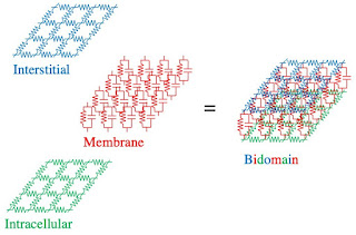
Interestingly, the fly screen analogy looks similar to this bidomain resistor illustration, versions of which I've used in many publications.

The Bidomain Model, represented as grids of resistors and capacitors.
The Bidomain Model (two interpenetrating domains).

I think it’s a stretch to say that Schmitt is the father of the bidomain model. Perhaps grandfather would better characterize his contribution. He didn’t derive a mathematical formulation of his idea. But he certainly conceived an intuitive picture of two coupled interpenetrating domains, and that influenced later work by Tung, Geselowitz, and Plonsey.

I’ll end with the abstract from a short biography of Otto Schmitt by Jon Harkness (Physics in Perspective, Volume 4, Pages 456-490, 2002), written four years after Schmitt’s death.

A Lifetime of Connections: Otto Herbert Schmitt, 1913–1998 
Jon M. Harkness

Otto H. Schmitt was born in St. Louis, Missouri, in 1913. As a youth, he displayed an affinity for electrical engineering but also pursued a wide range of other interests. He applied his multi-disciplinary talents as an undergraduate and graduate student at Washington University, where he worked in three departments: physics, zoology, and mathematics. For his doctoral research, Schmitt designed and built an electronic device to mimic the propagation of action potentials along nerve fibers. His most famous invention, now called the Schmitt trigger, arose from this early research. Schmitt spent most of his career at the University of Minnesota, where he did pioneering work in biophysics and bioengineering. He also worked at national and international levels to place biophysics and bioengineering on sound institutional footings. His years at Minnesota were interrupted by World War II. During that conflict—and the initial months of the Cold War to follow—Schmitt carried out defense-related research at the Airborne Instruments Laboratory in New York. Toward the end of his career at Minnesota, Schmitt coined the term biomimetics. He died in 1998.

Otto Schmitt discussing his work during World War II.

https://www.youtube.com/watch?v=lMCJG2C2_CY

Friday, March 11, 2022

Numerical Recipes is Online

Numerical Recipes, by Press, Teukolsky, Vetterling, and Flannery, superimposed on Intermediate Physics for Medicine and Biology.
Numerical Recipes,
by Press, Teukolsky, Vetterling, and Flannery.
In Intermediate Physics for Medicine and Biology, Russ Hobbie and I often refer to Numerical Recipes: The Art of Scientific Computing, by William Press, Saul Teukolsky, William Vetterling, and Brian Flannery. We usually cite the second edition of the book with programs written in C (1995), but the copy on my bookshelf is the second edition using Fortran 77 (1992). For those of you who don’t own a copy of this wonderful book, did you know you can read it online?

The address of the Numerical Recipes website is easy to remember: numerical.recipes. There you will find free copies of the second editions of Numerical Recipes for Fortran 77, Fortran 90, C, and C++ (2002). If you want easy, quick access to the third edition (2007), you will have to pay a fee. But if you are willing to put up with brief delays and annoying messages (which the authors call “nags”), you also can read the third edition for free.

The text below is from the Preface to the third edition.
“I was just going to say, when I was interrupted...” begins Oliver Wendell Holmes in the second series of his famous essays, The Autocrat of the Breakfast Table. The interruption referred to was a gap of 25 years. In our case, as the autocrats of Numerical Recipes, the gap between our second and third editions has been “only” 15 years. Scientific computing has changed enormously in that time.

The first edition of Numerical Recipes was roughly coincident with the first commercial success of the personal computer. The second edition came at about the time that the Internet, as we know it today, was created. Now, as we launch the third edition, the practice of science and engineering, and thus scientific computing, has been profoundly altered by the mature Internet and Web. It is no longer difficult to find somebody’s algorithm, and usually free code, for almost any conceivable scientific application. The critical questions have instead become, “How does it work?” and “Is it any good?” Correspondingly, the second edition of Numerical Recipes has come to be valued more and more for its text explanations, concise mathematical derivations, critical judgments, and advice, and less for its code implementations per se.

Recognizing the change, we have expanded and improved the text in many places in this edition and added many completely new sections. We seriously considered leaving the code out entirely, or making it available only on the Web. However, in the end, we decided that without code, it wouldn’t be Numerical Recipes. That is, without code you, the reader, could never know whether our advice was in fact honest, implementable, and practical. Many discussions of algorithms in the literature and on the Web omit crucial details that can only be uncovered by actually coding (our job) or reading compilable code (your job). Also, we needed actual code to teach and illustrate the large number of lessons about object-oriented programming that are implicit and explicit in this edition.
Russ and I cited Numerical Recipes in IPMB when we discussed integration, least squares fitting, random number generators, partial differential equations, the fast Fourier transform, aliasing, the correlation function, the power spectral density, and bilinear interpolation. Over the years, in my own research I have consulted the book about other topics, including solving systems of linear equations, evaluation of special functions, and computational analysis of eigensystems.

I highly recommend Numerical Recipes to anyone doing numerical computing. I found the book to be indispensable.

Friday, March 4, 2022

The Annotated Hodgkin & Huxley: A Readers Guide

The Annotated Hodgkin & Huxley, by Indira Raman and David Ferster, superimposed on Intermediate Physics for Medicine and Biology.
The Annotated Hodgkin & Huxley,
by Indira Raman and David Ferster.
I have always loved the classic set of five papers published by Hodgkin and Huxley in the Journal of Physiology. I often assigned the best of these, the fifth paper, when I taught my Biological Physics class. But reading the original papers can be a challenge. I was therefore delighted to discover The Annotated Hodgkin & Huxley: A Readers Guide, by Indira Raman and David Ferster. In their introduction, they write
After nearly seventy years, Alan Hodgkin and Andrew Huxley’s 1952 papers on the mechanisms underlying the action potential seem more and more like the Shakespeare plays of neurophysiology, works of astounding beauty that become less accessible to each successive generation of scientists. Everyone knows the basic plot (the squid dies at the beginning), but with their upside-down and backwards graphs and records, unfamiliar terminology and techniques, now arcane scientific asides, and complex mathematical underpinnings, the papers become a major effort to read closely without guidance. It is our goal to provide such guidance, by translating graphs and terminology into the modern idiom, explaining the methods and underlying theory, and providing historical perspective on the events that led up to the experiments described. By doing so, we hope to bring the pleasure of reading these extraordinary papers to any physiologist inclined to read them.
Raman and Ferster then give seven reasons to be so inclined.
  • “The sheer pleasure of an exciting scientific saga...” 
  • “Coming to know the electrical principles that govern the operation of neurons...” 
  • “To see firsthand what it is like to be at a scientific frontier...” 
  • “The series of papers provide an exemplary…illustration of the scientific method at its best...” 
  • “To understand the purpose and power of quantification and computation in science...” 
  • “The papers teach us that rigorous science does not require the elimination of error and artifact,”
  • “To develop a sense of one’s place in history.”

After a brief chapter about the historical background, the fun really begins. Each of the five papers is presented verbatim on even-numbered pages, with annotations (notes, redrawn figures, explanations, comments) on facing odd-numbered pages. 

One annoying problem with the original papers is that Hodgkin and Huxley defined the transmembrane potential differently than everyone else; they took resting potential to be zero and denote depolarization as negative. Raman and Ferster have redrawn all the figures using the modern definition; rest is −65 mV and depolarization is positive. With this change, the plots of the m, h, and n gates (which control the opening and closing of the sodium and potassium channels) versus transmembrane potential look the same as they do in Fig. 6.37 of Intermediate Physics for Medicine and Biology. I sometimes wish I could take a time machine, go back to 1952, and say “Hey Al! Hey Andy! Don’t use that silly convention for specifying the transmembrane potential. It will be a blemish your otherwise flawless series of papers.”

One of my favorite annotations is in response to Hodgkin and Huxley’s sentence “the equations derived in Part II of this paper [the fifth article in the series] predict with fair accuracy many of the electrical properties of the squid giant axon.” Raman and Ferster write “This may be the greatest understatement of the entire series of five papers.” I would say it’s one of the greatest understatements in all of science.

The final annotation follows Hodgkin and Huxley’s final sentence of their fifth paper: “it is concluded that the responses of an isolated giant axon of Loligo to electrical stimuli are due to reversible alterations in sodium and potassium permeability arising from changes in membrane potential.” Raman and Ferster add “H&H end their tour de force with a mild but precise statement that electrical excitability in the squid giant axon results from voltage-gated conductances. The discoveries that underlie this simple conclusion, however, completely transformed the understanding of cellular excitability in particular and bioelectricity in general. As John W. Moore—a postdoc with Kenneth Cole in 1952—once quipped, it took the rest of the field about a decade to catch up.”

The appendices at the end of The Annotated Hodgkin & Huxley are useful, particularly Appendix Five about numerical methods for solving the Hodgkin & Huxley equations. Huxley performed his calculations on a mechanical calculator. Raman and Ferster write

The mechanical calculator Huxley used was a pre-war era Brunsviga Model 20… which is an adding machine with a few features that streamline the calculations. To multiply 1234 by 5678, for example, one must follow these steps:

  • Enter 1234 on the sliding levers (on the machine’s curved face), one digit at a time. 
  • Position the carriage (at the front of the machine) in the ones position. 
  • Turn the crank eight (at the far right) times. 
  • Slide the carriage to the tens position 
  • Turn the crank seven times. 
  • Slide the carriage to the hundreds position. 
  • Turn the crank six times. 
  • Slide the carriage to the thousands position. 
  • Turn the crank five times.

Thus, 30 individual operations are required to multiply two four-digit numbers.

Andrew Huxley, you are my hero.

Happy 70th anniversary of the publication of these landmark papers. I’ll end with a quote about them from Raman and Ferster’s epilogue

The following decades saw tremendous advances in physiology that built directly on the discoveries of H&H. Ultimately, what began as a basic scientific inquiry in a fragile invertebrate with a fortuitously oversized axon would provide the basis for the development of a vast array of biomedical research fields. The studies that the H&H papers made possible would not only yield immeasurable insights into brain and muscle function but also identify, explain, and alleviate medical conditions as diverse as epilepsies, ataxias, myotonias, arrhythmias, and pain.

Friday, February 25, 2022

Teaching Dynamics to Biology Undergraduates: the UCLA Experience

The goal of Intermediate Physics for Medicine and Biology, and the goal of this blog, is to explore the interface between physics, medicine, and biology. But understanding physics, and in particular the physics used in IPMB, requires calculus. In fact, Russ Hobbie and I state in the preface of IPMB that “calculus is used without apology.” Unfortunately, many biology and premed students don’t know much calculus. In fact, their general math skills are often weak; even algebra can challenge them. How can students learn enough calculus to make sense of IPMB?

A team from UCLA has developed a new way to teach calculus to students of the life sciences. The group is led by Alan Garfinkel, who appears in IPMB when Russ and I discuss the response of cardiac tissue to repetitive electrical stimulation (see Chapter 10, Section 12). An article describing the new class they’ve developed was published recently in the Bulletin of Mathematical Biology (Volume 84, Article Number 43, 2022).
There is a growing realization that traditional “Calculus for Life Sciences” courses do not show their applicability to the Life Sciences and discourage student interest. There have been calls from the AAAS, the Howard Hughes Medical Institute, the NSF, and the American Association of Medical Colleges for a new kind of math course for biology students, that would focus on dynamics and modeling, to understand positive and negative feedback relations, in the context of important biological applications, not incidental “examples.” We designed a new course, LS 30, based on the idea of modeling biological relations as dynamical systems, and then visualizing the dynamical system as a vector field, assigning “change vectors” to every point in a state space. The resulting course, now being given to approximately 1400 students/year at UCLA, has greatly improved student perceptions toward math in biology, reduced minority performance gaps, and increased students’ subsequent grades in physics and chemistry courses. This new course can be customized easily for a broad range of institutions. All course materials, including lecture plans, labs, homeworks and exams, are available from the authors; supporting videos are posted online.
Sharks and tuna, the predator-prey problem,
from Garfinkel et al.,
Bulletin of Mathematical Biology
,
84:43, 2022.

This course approaches calculus from the point of view of modeling. Its first example develops a pair of coupled differential equations (only it doesn’t use such fancy words and concepts) to look at interacting populations of sharks and tuna; the classical predator-prey problem analyzed as a homework problem in Chapter 2 of IPMB. Instead of focusing on equations, this class makes liberal use of state space plots, vector field illustrations, and simple numerical analysis. The approach reminds me of that adopted by Abraham and Shaw in their delightful set of books Dynamics: The Geometry of Behavior, which I have discussed before in this blog. The UCLA course uses the textbook Modeling Life: The Mathematics of Biological Systems, which I haven’t read yet but is definitely on my list of books to read.

My favorite sentence from the article appears when it discusses how the derivative and integral are related through the fundamental theorem of calculus.
We are happy when our students can explain the relation between the COVID-19 “New Cases per day” graph and the “total cases” graph.
If you want to learn more, read the article. It’s published open access, so anyone can find it online. You can even steal its illustrations (like I did with its shark-tuna picture above).

I’ll end by quoting again from Garfinkel et al.’s article, when they discuss the difference between their course and a traditional calculus class. If you replace the words “calculus” and “math” by “physics” in this paragraph, you get a pretty good description of the approach Russ and I take in Intermediate Physics for Medicine and Biology.
The course that we developed has a number of key structural and pedagogical differences from the traditional “freshman calculus” or “calculus for life sciences” classes that have been offered at UCLA and at many other universities. For one, as described above, our class focuses heavily on biological themes that resonate deeply with life science students in the class. Topics like modeling ecological systems, the dynamics of pandemics like COVID-19, human physiology and cellular responses are of great interest to life science students. We should emphasize that these examples are not simply a form of window dressing meant to make a particular set of mathematical approaches palatable to students. Rather, the class is structured around the idea that, as biologists, we are naturally interested in understanding these kinds of systems. In order to do that, we need to develop a mathematical framework for making, simulating and analyzing dynamical models. Using these biological systems not purely as examples, but rather as the core motivation for studying mathematical concepts, provides an intellectual framework that deeply interests and engages life science students.


Introduction to state variables and state space. Video 1.1 featuring Alan Garfinkel.

https://www.youtube.com/watch?v=yZWG0ALL3mI


Defining vectors in higher dimensions. Video 1.2 featuring Alan Garfinkel.

https://www.youtube.com/watch?v=2Rjk0O3yWc8

Friday, February 18, 2022

The Emperor of All Maladies: A Biography of Cancer

One topic that appears over and over again throughout Intermediate Physics for Medicine and Biology is cancer. In Section 8.8, Russ Hobbie and I discuss using magnetic nanoparticles to heat a tumor. Section 9.10 describes the unproven hypothesis that nonionizing electromagnetic radiation can cause cancer. In Section 13.8, we analyze magnetic resonance guided high intensity focused ultrasound (MRgHIFUS), which has been proposed as a treatment for prostate cancer. Section 14.10 includes a discussion of how ultraviolet light can lead to skin cancer. One of the most common treatments for cancer, radiation therapy, is the subject of Section 16.10. Finally, in Section 17.10 we explain how positron emission tomography (PET) can assist in imaging metastatic cancer. Despite all this emphasis on cancer, Russ and I don’t really delve deeply into cancer biology. We should.

Last week, I attended a talk (remotely, via zoom) by my colleague and friend Steffan Puwal, who teaches physics at Oakland University. Steffan has a strong interest in cancer, and has compiled a reading list: https://sites.google.com/oakland.edu/cancer-reading. These books are generally not too technical but informative. I urge you to read some of them to fill that gap between physics and cancer biology.

The Emperor of All Maladies, by Siddhartha Muknerjee, superimposed on Intermediate Physics for Medicine and Biology.
The Emperor of All Maladies,
by Siddhartha Mukherjee.
Steffan says that the best of these books is The Emperor of All Maladies: A Biography of Cancer, by Siddhartha Mukherjee. Ken Burns has produced a television documentary based on this book. You can listen to the trailer at the bottom of this post. If you are looking for a more technical review paper, Steffan suggests “Hallmarks of Cancer: The Next Generation,” by Douglas Hanahan and Robert Weinberg (Cell, Volume 4, Pages 646–674, 2011). It’s open access, so you don’t need a subscription to read it. He also recommends the websites for the MD Anderson Cancer Center (https://www.mdanderson.org) and the Dana Farber Cancer Institute (https://www.dana-farber.org). 

Thanks, Steffan, for teaching me so much about cancer.

Cancer: The Emperor of All Maladies, Trailer with special introduction by Dr. Siddhartha Mukherjee.
 https://www.youtube.com/watch?v=L9lIsNkfQsM

 
Siddhartha Mukherjee, The Cancer Puzzle

Friday, February 11, 2022

The Rest of the Story 3

Harry was born and raised in England and attended the best schools. After excelling at Summer Fields School, he won a King’s Scholarship to Eton College—the famous boarding school that produced twenty British Prime Ministers—where he won prizes in chemistry and physics. In 1906 he entered Trinity College at the University of Oxford, the oldest university in the English-speaking world, and four years later he graduated with his bachelor’s degree.

Next Harry went to the University of Manchester, where he worked with the famous physicist Ernest Rutherford. In just a few short years his research flourished and he made amazing discoveries. Rutherford recommended Harry for a faculty position back at Oxford. He might have taken the job, but after Archduke Franz Ferdinand of Austria was assassinated in Sarajevo in June 1914, the world blundered into World War I.

Like many English boys of his generation, Harry volunteered for the army. He joined the Royal Engineers, where he could use his technical skills as a telecommunications officer to support the war effort. Millions of English soldiers were sent to fight in France, where the war soon bogged down into trench warfare.

Page 2

First Lord of the Admiralty Winston Churchill devised a plan to break the deadlock. England would attack the Gallipoli peninsula in Turkey. If the navy could fight their way through the Dardanelles, they could take Constantinople, reach the Black Sea, unite with their ally Russia, and attack the “soft underbelly” of Europe. Harry was assigned to the expeditionary force for the Gallipoli campaign.

The plan was sound, but the execution failed; the navy could not force the narrows. The army landed on the tip of the peninsula and immediately settled into trench warfare like in France. There in Gallipoli, on August 10, 1915, a Turkish sniper shot and killed 27-year-old Second Lieutenant Henry Moseley—known as Harry to his boyhood friends.

Isaac Asimov wrote that Moseley’s demise “might well have been the most costly single death of the War to mankind.” Moseley’s research using x-rays to identify and order the elements in the periodic table by atomic number was revolutionary. He almost certainly would have received a Nobel Prize if that honor were awarded posthumously.

And now you know the REST OF THE STORY. Good Day!


-----------------------------------------------------------------------------------------------------

This blog entry was written in the style of Paul Harvey’s radio show “The Rest of the Story.” My February 5, 2016 and March 12, 2021 entries were also in this style. Homework Problem 3 in Chapter 16 of Intermediate Physics for Medicine and Biology explores Moseley’s work. Learn more about Henry Moseley in my March 16, 2012 blog entry.

Friday, February 4, 2022

Does a Nerve Axon Have an Inductance?

When I was measuring the magnetic field of a nerve axon in graduate school, I wondered if I should worry about a nerve’s inductance. Put another way, I asked if the electric field induced by the axon’s changing magnetic field is large enough to affect the propagation of the action potential.

Here is a new homework problem that will take you through the analysis that John Wikswo and I published in our paper “The Magnetic Field of a Single Axon” (Biophysical Journal, Volume 48, Pages 93–109, 1985). Not only does it answer the question about induction, but also it provides practice in back-of-the-envelope estimation. To learn more about biomagnetism and magnetic induction, see Chapter 8 of Intermediate Physics for Medicine and Biology.
Section 8.6

Problem 29½. Consider an action potential propagating down a nerve axon. An electric field E, having a rise time T and extended over a length L, is associated with the upstroke of the action potential.

(a) Use Ohm’s law to relate E to the current density J and the electrical conductivity σ
(b) Use Ampere’s law (Eq. 8.24, but ignore the displacement current) to estimate the magnetic field B from J and the permeability of free space, μ0. To estimate the derivative, replace the curl operator with 1/L
(c) Use Faraday’s law (Eq. 8.22, ignoring the minus sign) to estimate the induced electric field E* from B. Replace the time derivative by 1/T
(d) Write your result as the dimensionless ratio E*/E
(e) Use σ = 0.1 S/m, μ0 = 4 π × 10-7 T m/A, L = 10 mm, and T = 1 ms, to calculate E*/E
(f) Check that the units in your calculation in part (e) are consistent with E*/E being dimensionless. 
(g) Draw a picture of the axon showing E, J, B, E*, and L
(h) What does your result in part (e) imply about the need to consider inductance when analyzing action potential propagation along a nerve axon.

For those of you who don’t have IPMB handy, Equation 8.24 (Ampere’s law, ignoring the displacement current) is

∇×B = μ0 J

and Eq. 8.22 (Faraday’s law) is

∇×E = −∂B/∂t .

I’ll leave it to you to solve this problem. However, I’ll show you my picture for part (g).

Also, for part (e) I get a small value, on the order of ten parts per billion (10-8). The induction of a nerve axon is negligible. We don't need an inductor when modeling a nerve axon.