Friday, February 11, 2022

The Rest of the Story 3

Harry was born and raised in England and attended the best schools. After excelling at Summer Fields School, he won a King’s Scholarship to Eton College—the famous boarding school that produced twenty British Prime Ministers—where he won prizes in chemistry and physics. In 1906 he entered Trinity College at the University of Oxford, the oldest university in the English-speaking world, and four years later he graduated with his bachelor’s degree.

Next Harry went to the University of Manchester, where he worked with the famous physicist Ernest Rutherford. In just a few short years his research flourished and he made amazing discoveries. Rutherford recommended Harry for a faculty position back at Oxford. He might have taken the job, but after Archduke Franz Ferdinand of Austria was assassinated in Sarajevo in June 1914, the world blundered into World War I.

Like many English boys of his generation, Harry volunteered for the army. He joined the Royal Engineers, where he could use his technical skills as a telecommunications officer to support the war effort. Millions of English soldiers were sent to fight in France, where the war soon bogged down into trench warfare.

Page 2

First Lord of the Admiralty Winston Churchill devised a plan to break the deadlock. England would attack the Gallipoli peninsula in Turkey. If the navy could fight their way through the Dardanelles, they could take Constantinople, reach the Black Sea, unite with their ally Russia, and attack the “soft underbelly” of Europe. Harry was assigned to the expeditionary force for the Gallipoli campaign.

The plan was sound, but the execution failed; the navy could not force the narrows. The army landed on the tip of the peninsula and immediately settled into trench warfare like in France. There in Gallipoli, on August 10, 1915, a Turkish sniper shot and killed 27-year-old Second Lieutenant Henry Moseley—known as Harry to his boyhood friends.

Isaac Asimov wrote that Moseley’s demise “might well have been the most costly single death of the War to mankind.” Moseley’s research using x-rays to identify and order the elements in the periodic table by atomic number was revolutionary. He almost certainly would have received a Nobel Prize if that honor were awarded posthumously.

And now you know the REST OF THE STORY. Good Day!


-----------------------------------------------------------------------------------------------------

This blog entry was written in the style of Paul Harvey’s radio show “The Rest of the Story.” My February 5, 2016 and March 12, 2021 entries were also in this style. Homework Problem 3 in Chapter 16 of Intermediate Physics for Medicine and Biology explores Moseley’s work. Learn more about Henry Moseley in my March 16, 2012 blog entry.

Friday, February 4, 2022

Does a Nerve Axon Have an Inductance?

When I was measuring the magnetic field of a nerve axon in graduate school, I wondered if I should worry about a nerve’s inductance. Put another way, I asked if the electric field induced by the axon’s changing magnetic field is large enough to affect the propagation of the action potential.

Here is a new homework problem that will take you through the analysis that John Wikswo and I published in our paper “The Magnetic Field of a Single Axon” (Biophysical Journal, Volume 48, Pages 93–109, 1985). Not only does it answer the question about induction, but also it provides practice in back-of-the-envelope estimation. To learn more about biomagnetism and magnetic induction, see Chapter 8 of Intermediate Physics for Medicine and Biology.
Section 8.6

Problem 29½. Consider an action potential propagating down a nerve axon. An electric field E, having a rise time T and extended over a length L, is associated with the upstroke of the action potential.

(a) Use Ohm’s law to relate E to the current density J and the electrical conductivity σ
(b) Use Ampere’s law (Eq. 8.24, but ignore the displacement current) to estimate the magnetic field B from J and the permeability of free space, μ0. To estimate the derivative, replace the curl operator with 1/L
(c) Use Faraday’s law (Eq. 8.22, ignoring the minus sign) to estimate the induced electric field E* from B. Replace the time derivative by 1/T
(d) Write your result as the dimensionless ratio E*/E
(e) Use σ = 0.1 S/m, μ0 = 4 π × 10-7 T m/A, L = 10 mm, and T = 1 ms, to calculate E*/E
(f) Check that the units in your calculation in part (e) are consistent with E*/E being dimensionless. 
(g) Draw a picture of the axon showing E, J, B, E*, and L
(h) What does your result in part (e) imply about the need to consider inductance when analyzing action potential propagation along a nerve axon.

For those of you who don’t have IPMB handy, Equation 8.24 (Ampere’s law, ignoring the displacement current) is

∇×B = μ0 J

and Eq. 8.22 (Faraday’s law) is

∇×E = −∂B/∂t .

I’ll leave it to you to solve this problem. However, I’ll show you my picture for part (g).

Also, for part (e) I get a small value, on the order of ten parts per billion (10-8). The induction of a nerve axon is negligible. We don't need an inductor when modeling a nerve axon.

Friday, January 28, 2022

How Far Can Bacteria Coast?

Random Walks in Biology, by Howard Berg, superimposed on Intermediate Physics for Medicine and Biology.
Random Walks in Biology,
by Howard Berg.
In last week’s blog post, I told you about the recent death of Howard Berg, author of Random Walks in Biology. This week, I present a new homework problem based on a topic from Berg’s book. When discussing the Reynolds number, a dimensionless number from fluid dynamics that is small when viscosity dominates inertia, Berg writes
The Reynolds number of the fish is very large, that of the bacterium is very small. The fish propels itself by accelerating water, the bacterium by using viscous shear. The fish knows a great deal about inertia, the bacterium knows nothing. In short, the two live in very different hydrodynamic worlds.

To make this point clear, it is instructive to compute the distance that the bacterium can coast when it stops swimming.
Here is the new homework problem, which asks the student to compute the distance the bacterium can coast.
Section 1.20

Problem 54. When a bacterium stops swimming, it will coast to a stop. Let us calculate how long this coasting takes, and how far it will go.

(a) Write a differential equation governing the speed, v, of the bacterium. Use Newton’s second law with the force given by Stokes law. Be careful about minus signs.

(b) Solve this differential equation to determine the speed as a function of time.

(c) Write the time constant, τ, governing the decay of the speed in terms of the bacterium’s mass, m, its radius, a, and the fluid viscosity, η.

(d) Calculate the mass of the bacterium assuming it has the density of water and it is a sphere with a radius of one micron.

(e) Calculate the time constant of the decay of the speed, for swimming in water having a viscosity of 0.001 Pa s.

(f) Integrate the speed over time to determine how far the bacterium will coast, assuming its initial speed is 20 microns per second.
I won’t solve all the intermediate steps for you; after all, it’s your homework problem. However, below is what Berg has to say about the final result.
A cell moving at an initial velocity of 2 × 10⁻³ cm/sec coasts 4 × 10⁻¹⁰ cm = 0.04 , a distance small compared with the diameter of a hydrogen atom! Note that the bacterium is still subject to Brownian movement, so it does not actually stop. The drift goes to zero, not the diffusion.

Berg didn’t calculate the deceleration of the bacterium. If the speed drops from 20 microns per second to zero in one time constant, I calculate the acceleration to be be about 91 m/s2, or nearly 10g. This is similar to the maximum allowed acceleration of a plane flying in the Red Bull Air Race. That poor bacterium.

Friday, January 21, 2022

Howard Berg (1934–2021)

Random Walks in Biology,
by Howard Berg.
Look up at the picture of books at the top of this blog, showing my ideal bookshelf. You see Intermediate Physics for Medicine and Biology towering in the center. The small volume two books to the right of IPMB is Random Walks in Biology, by Howard Berg.

Berg died on December 30, 2021. He was the Herchel Smith Professor of Physics in the Rowland Institute at Harvard University. He was known for his studies of flagellar motility and sensory transduction in bacteria, as described in his 2004 book E. coli in Motion.

Berg obtained his bachelor’s degree from Cal Tech, and a PhD from Harvard. He was on the faculty at the University of Colorado, then at Cal Tech, and finally at Harvard. He was a fellow of the American Physical Society and a member of the National Academy of Sciences. In 1984 he and Edward Purcell received the Max Delbrück Prize in Biological Physics from the American Physical Society “for the elucidation of complex biological phenomena, in particular chemotaxis and bacterial locomotion, through simple but penetrating physical theories and brilliant experiments.”

I had the pleasure of meeting Berg at a 2014 Gordon Research Conference about Physics Research and Education: The Complex Intersection of Biology and Physics, held at Mount Holyoke College in South Hadley, Massachusetts. He was a quiet, thoughtful, kind man. I wish I knew him better.

Purcell mentioned Berg in his influential article “Life at Low Reynolds Number” (American Journal of Physics, Volume 45, Pages 3–11, 1977).
I might say what got me into this. To introduce something that will come later, I’m going to talk partly about how microorganisms swim. That will not, however, turn out to be the only important question about them. I got into this through the work of a former colleague of mine at Harvard, Howard Berg. Berg got his Ph.D. with Norman Ramsey, working on a hydrogen maser, and then he went back into biology, which had been his early love, and into cellular physiology. He is now at the University of Colorado at Boulder, and has recently participated in what seems to me one of the most astonishing discoveries about the questions we're going to talk about. So it was partly Howard's work, tracking E. coli and finding out this strange thing about them, that got me thinking about this elementary physics stuff.
Section 4.10 of Intermediate Physics for Medicine and Biology analyzes chemotaxis, and cites Berg’s 1977 paper with Purcell “Physics of Chemoreception” (Biophysical Journal, Volume 20, Pages 119–136). Below is the abstract.
Statistical fluctuations limit the precision with which a microorganism can, in a given time T, determine the concentration of a chemoattractant in the surrounding medium. The best a cell can do is to monitor continually the state of occupation of receptors distributed over its surface. For nearly optimum performance only a small fraction of the surface need be specifically adsorbing. The probability that a molecule that has collided with the cell will find a receptor is Ns/(Ns + πa), if N receptors, each with a binding site of radius s, are evenly distributed over a cell of radius a. There is ample room for many independent systems of specific receptors. The adsorption rate for molecules of moderate size cannot be significantly enhanced by motion of the cell or by stirring of the medium by the cell. The least fractional error attainable in the determination of a concentration c is approximately (TcaD)−1/2, where D is the diffusion constant of the attractant. The number of specific receptors needed to attain such precision is about a/s. Data on bacteriophage adsorption, bacterial chemotaxis, and chemotaxis in a cellular slime mold are evaluated. The chemotactic sensitivity of Escherichia coli approaches that of the cell of optimum design.

To learn more about Berg's life, education, and career, read his interview with Current Biology.

I will end with Berg’s introduction to his masterpiece Random Walks in Biology. If you want to learn about diffusion, start with Berg’s book.
Biology is wet and dynamic. Molecules, subcellular organelles, and cells, immersed in an aqueous environment, are in continuous riotous motion. Alive or not, everything is subject to thermal fluctuations. What is this microscopic world like? How does one describe the motile behavior of such particles? How much do they move on the average? Questions of this kind can be answered only with an intuition about statistics that very few biologists have. This book is intended to sharpen that intuition. It is meant to illuminate both the dynamics of living systems and the methods used for their study. It is not a rigorous treatment intended for the expert but rather an introduction for students who have little experience with statistical concepts.

The emphasis is on physics, not mathematics, using the kinds of calculations that one can do on the back of an envelope. Whenever practical, results are derived from first principles. No reference is made to the equations of thermodynamics. The focus is on individual particles, not moles of particles. The units are centimeters (cm), grams (g), and seconds (sec).

Topics range from the one-dimensional random walk to the motile behavior of bacteria. There are discussions of Boltzmann’s law, the importance of kT, diffusion to multiple receptors, sedimentation, electrophoresis, and chromatography. One appendix provides an introduction to the theory of probability. Another is a primer on differential equations. A third lists some constants and formulas worth committing to memory. Appendix A should be consulted while reading Chapter 1 and Appendix B while reading Chapter 2. A detailed understanding of differential equations or the methods used for their solution is not required for an appreciation of the main theme of this book.


Howard Berg. Marvels of Bacterial Behavior. Part 1.

Howard Berg. Marvels of Bacterial Behavior. Part 2.

Friday, January 14, 2022

The Chain of Reason vs. the Chain of Thumbs

Bully for Brontosaurus, by Stephen Jay Gould, superimposed on Intermediate Physics for Medicine and Biology.
Bully for Brontosaurus,
by Stephen Jay Gould.
I have written previously in this blog about my admiration for evolutionary biologist Stephen Jay Gould and his essays published in his monthly column “This View of Life” in the magazine Natural History. Today, I focus on one of these essays, “The Chain of Reason vs. the Chain of Thumbs,” that is related to a topic in Intermediate Physics for Medicine and Biology. You can find this essay reprinted in Gould’s book Bully for Brontosaurus.

IPMB has a chapter on biomagnetism (the production of magnetic fields by the body) and a section on the possible effects if weak external electric and magnetic fields. Much nonsense has been written about using magnetic fields to treat diseases, including to relieve pain. When did all this silliness begin? Over two hundred years ago.

Gould’s essay describes the fascinating story of the Franz Mesmer, who operated a clinic in Paris in the seventeenth century to treat various illnesses using “animal magnetism.” Gould writes
Franz Anton Mesmer was a German physician who had acquired wealth through marriage to a well endowed widow; connections by assiduous cultivation;… and renown with a bizarre, if fascinating, theory of “animal magnetism” and its role in human health.

Mesmer, insofar as one can find coherence in his ideas at all, claimed that a single (and subtle) fluid pervaded the universe, uniting and connecting all bodies. We give different names to this fluid according to its various manifestations: gravity when we consider planets in their courses; electricity when we contemplate a thunderstorm; magnetism when we navigate by compass. The fluid also flows through organisms and may be called animal magnetism. Disease results from a blockage of this flow, and cure of disease requires a reestablishment of the flux and a restoration of equilibrium.
Cure of illness requires the aid of an “adept,” a person with unusually strong magnetism who can locate the “poles” of magnetic flow on the exterior of a human body and, by massaging these areas, break the blockage within and reestablish the normal flux…

Mesmer's treatments were quite dramatic.

Within a few minutes of mesmerizing, sensitive patients would fall into the characteristic “crisis” taken by Mesmer as proof of his method. Bodies would begin to shake, arms and legs move violently and involuntarily, teeth chatter loudly. Patients would grimace, groan, babble, scream, faint, and fall unconscious.

Gould then tells the story of a Royal Commission established in 1784 by French king Louis XVI to investigate Mesmer’s claims. The commission was headed by American Benjamin Franklin, and included chemist Antoine Lavoisier and medical doctor Joseph Guillotin.

In a clever series of experiments, designed mainly by Lavoisier and carried out at Franklin’s home in Passy, the commissioners made the necessary separations and achieved a result as clear as any in the history of debunking: crises are caused by suggestion; not a shred of evidence exists for any fluid, and animal magnetism, as a physical force, must be firmly rejected.
Gould was impressed by the quality of the commission’s work.
Never in history has such an extraordinary and luminous group been gathered together in the service of rational inquiry by the methods of experimental science. For this reason alone, the Rapport des commissaires chargés par le roi de l’examen du magnétisme animal (Report of the Commissioners Charged by the King to Examine Animal Magnetism) is a key document in the history of human reason. It should be rescued from its current obscurity, translated into all languages, and reprinted by organizations dedicated to the unmasking of quackery and the defense of rational thought.

Nowadays we see a lot of ridiculous claims that magnetic fields can alleviate pain and have other health effects. Julian Whitaker and Brenda Adderly, in their book The Pain Relief Breakthrough, assert that magnets can cure backaches, arthritis, menstrual cramps, carpal tunnel syndrome, sports injuries, and more. Mexican surgeon Isaac Goiz Duran says that his “biomagnetic therapy” can cure diabetes, AIDS, cancer, and Covid-19. All these therapies are based on static magnetic fields, a type of 21st century animal magnetism.

I highly recommend all of Gould’s essays, including this one. Remember the efforts of Franklin, Lavoisier, and Guillotin before you start believing that static magnetic fields can improve your health.

Friday, January 7, 2022

Cells, Gels and the Engines of Life

Cells, Gels and the Engines of Life, by Gerald Pollack, superimposed on Intermediate Physics for Medicine and Biology.
Cells, Gels and the Engines of Life,
by Gerald Pollack.
I recently read Gerald Pollack’s book Cells, Gels and the Engines of Life: A New, Unifying Approach to Cell Function. I’ve always taken a simple, physicist’s view of a cell: salt water inside, salt water outside, with a membrane between; the membrane being where all the action is. Pollack’s perspective is entirely different, and challenges the standard dogma in cell biology. He focuses on the how the inside of a cell resembles a gel.

The gel-like nature of the cytoplasm forms the foundation of this book. Biologists acknowledge the cytoplasm’s gel-like character, but textbooks nevertheless build on aqueous solution behavior. A gel is quite different from an aqueous solution—it is a matrix of polymers to which water and ions cling. That’s why gelatin desserts retain water, and why a cracked egg feels gooey.

The concept of a gel-like cytoplasm turns out to be replete with power. It accounts for the characteristic partitioning of ions between the inside and outside of the cell... It also explains the cell’s electrical potential: potentials of substantial magnitude can be measured in gels as well as in demembranated cells... Thus, the gel-like character of the cytoplasm accounts for the basic features of cell biophysics.

What do I think of Pollack’s radical attitude toward biology? I’m not sold on his ideas, but his book certainly made me rethink my fundamental assumptions about biology in general, and electrophysiology in particular.


Pumps and Channels

In Intermediate Physics for Medicine and Biology, Russ Hobbie and I describe the standard view of how ion gradients across the cell membrane are created by a pump (page 155).

To maintain the ion concentrations a membrane protein called the sodium-potassium pump uses metabolic energy to pump potassium into the cell and sodium out.
On page 193, we discuss ion channels.
Selective ion channels are responsible for the initiation and propagation of the action potentials.
Pollack doesn’t believe pumps and channels are important. How can this be? What about patch clamping? Section 9.7 of IPMB talks this revolutionary technique (page 251).
The next big advance was patch-clamp recording (Neher and Sakmann 1976). Micropipettes were sealed against a cell membrane that had been cleaned of connective tissue by treatment with enzymes. A very-high-resistance seal resulted [(2–3)×107 Ω] that allowed one to see the opening and closing of individual channels.
Pollack says
The existence of single ion channels appeared to be confirmed by ground-breaking experiments using the patch-clamp technique… This dazzling result has so revolutionized the field of membrane electrophysiology that the originators of this technique, Erwin Neher and Bert Sakmann, were awarded the Nobel Prize. The observation of discrete events would seem to confirm beyond doubt that the ions flow through discrete channels.

Results from the laboratory of Fred Sachs, on the other hand, make one wonder. Sachs found that when the patch of membrane was replaced by a patch of silicon rubber, the discrete currents did not disappear… they remained essentially indistinguishable from those measured when the membrane was present.
Yikes! Pollack’s arguments against pumps don’t terrify me quite as much.
Pumping faces obstacles of space and energy. The membrane’s size is fixed but the number of pumps will inevitably continue to grow. At some stage the demand for space could exceed the supply, and what then? Pumping also requires energy. The Na/K pump alone is estimated to consume an appreciable fraction of the cell’s energy supply, and that pump is one of very many, including those in internal membranes. How is the cell to cope with the associated energy requirement?


 Membranes

Pollack goes on to renounce the importance of the cell membrane.
Continuing to move boldly, we took it upon ourselves in this chapter to reconsider the notion of the continuous ion barrier [the membrane]. If the barrier were continuous, we reasoned, violating its continuity by tearing large holes should allow ions to surge across the cell boundary and solutes to leak out, dramatically altering the cell’s makeup, shutting down cell function, and eventually killing the cell.

But that did not happen. Whether created by shoving a micropipette into the cell, plucking a patch from the membrane, riddling the membrane with an electrical barrage [electroporation], or slicing the cell into two, the wounds seemed to matter little; the cell could often continue to function as though there had been no violation. It was as though function could be sustained by the cytoplasm alone.


The Resting Transmembrane Potential

Russ and I explain the resting potential of a cell in the conventional way: the membrane is selectively permeable to potassium, and the potassium ion concentration is higher inside the cell than outside. The concentrations will be in equilibrium when the resting potential is negative, with a magnitude given by the Nernst equation. Pollack’s explanation is completely different, and focuses on the structuring of water near the hydrophilic surface of proteins.
Cell water excludes ions because it is structured. Exclusion is more pronounced for sodium than for potassium because sodium’s hydration shell is larger and hence more difficult to accommodate in the structured water lattice. Thus, intracellular sodium concentration remains low, whereas potassium can partition more easily into the cytoplasm.
No pumps, no channels, no membranes, no Nernst equation, and no metabolic ATP. Just water.


 The Action Potential

Pollack’s story may sound plausible, if not convincing, so far, but what about the action potential? This is where gels come in the forefront of the story. According to Pollack, the cytoplasm is like a gel that can undergo a polymer-gel phase transition. Normally the cytoplasmic polymers are cross linked by calcium, allowing in little water. If the calcium is not there to do the cross-linking, water gets sucked in, loosening the structure.
A plausible way in which the action potential could be initiated, then, is by replacing calcium with a monovalent [singly charged ion such as sodium or potassium]. Classically, sodium is thought to enter the cytoplasm through a localized, receptor-mediated permeability increase. In the proposed [Pollack’s] model, sodium ions flow into the peripheral cytoskeleton and begin displacing calcium. Replacement loosens the network, enabling it to adsorb water and expand. As it expands, permeability is increased, allowing for more sodium entry, further Ca displacement, additional expansion, etc.—like ripping open a zipper.

It sure doesn’t sound much like Hodgkin and Huxley.


Internal Perfusion

For a moment, I thought I had a devastating counter-example that would demolish Pollack’s theory. As mentioned before in this blog, you can squeeze the cytoplasm out of a squid giant axon and replace it by salt water, and the axon still works. But Pollack must have seen me coming; he shot down my counter-example in advance.
Lying just inside the cell membrane is a dense polymer-gel matrix known as the peripheral cytoskeleton… The presence of such a matrix had been unknown during the Hodgkin-Huxley era when experimental axons were routinely “rolled” to extrude the cytoplasm and presumably leave only the membrane. What in fact remains is the combination of membrane plus contiguous cytoskeleton.


Conclusion

I have only begun to cover the ideas discussed in Cells, Gels and the Engines of Life. Pollack provides us with a wonderfully written, beautifully illustrated, carefully argued, and well cited alternative view of biology. It was a joy to read, but I remain skeptical. I can think of many arguments in support of the “standard model” of cell physiology that Pollack doesn’t address. My “salt water inside, salt water outside” assumption may be too simplistic, and Pollack’s book is useful for pointing out its many limitations, but Pollack’s ideas have limitations too. Cells, Gels and the Engines of Life is an interesting read, but think long and hard before you start believing it.

Friday, December 31, 2021

Adrianus Kalmijn (1933–2021)

Adrianus Kalmijn, a biophysicist known for his studies of electroreception in sharks, died December 7, 2021, at the age of 88.

In Chapter 9 of Intermediate Physics for Medicine and Biology, Russ Hobbie and I discuss Kalmijn’s work.

Kalmijn et al. discovered that the ocean flounder generates a current dipole of 3 × 10⁻⁷ A m. Sea water with resistivity of 0.23 Ω m gives an electric field of 2 × 10⁻⁵ V m−1 at a distance 10 cm in front of the flounder. They were able to show in a beautiful series of behavioral experiments that dogfish (a small shark) could detect the electric field 0.4 m from a current dipole of 4 × 10⁻⁷ A m, corresponding to an electric field of 5 × 10⁻⁷ V m−1. The fish would bite at the electrodes, ignoring a nearby odor source. A field of 10−4 V m−1 would elicit the startle response. A field ⅒ as large caused a physiologic response. 
The first page of Kalmijn, A. J. (1977) The electric and magnetic sense of sharks, skates, and rays. Oceanus 20:45–52, superimposed on Intermediate Physics for Medicine and Biology.
Kalmijn, A. J. (1977) The electric and
magnetic sense of sharks, skates,
and rays. Oceanus 20:45–52.

 My favorite paper by Kalmijn is

Kalmijn, A. J. (1977) The electric and magnetic sense of sharks, skates, and rays. Oceanus 20:45–52.
Below is an excerpt.
During the summer of 1976, we learned from longline fishing off Cape Cod that the smooth dogfish Mustelus regularly frequents the shallow, inshore waters of Vineyard Sound on its nightly feeding excursions. This predatory shark is a warm-season visitor, arriving at Woods Hole in May and leaving for the South again in late October or shortly thereafter. It is an active bottom hunter, preying on small fish as well as crustaceans and other invertebrate animals. The females reach an average length of 115 centimeters; the males are slightly smaller. The smooth dogfish is truly live-bearing; the new-born measure 29 to 37 centimeters.

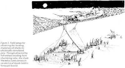
To observe the sharks’ feeding behavior, we worked from an inflatable rubber raft (Zodiac Mark II) free of any metal under the waterline. On station in 2.5 to 3.0-meter-deep water over a sand patch devoid of seaweed, we attracted the sharks by squeezing liquified herring through a long Tygon tube that ran from the raft to the bottom of the sea. The Tygon chumming tube was attached to a polypropylene line, suspended from a Styrofoam float and stretched over the ocean floor between two polyvinyl pipes anchored in low-profile cinder blocks (Figure 3). Starting after dark, we illuminated the area with a 100-watt, battery-operated underwater light. To break the water surface, we used a glass-bottom viewing box secured behind the stern of the raft. 
Figure 3 from Kalmijn, A. J. (1977) Oceanus 20:45–52.
With permission from the Woods Hole Oceanographic Institution.
Two pairs of agar-filled, salt-bridge electrodes were tied to the polypropylene line and positioned on the sand, one on either side of the odor source and 30 centimeters from it. Mekka underwater plugs with stainless steel pins and integral cables connected the thin, 30 to 90-centimeter-long Silastic salt-bridge tubes to the electrical equipment set up in the rubber raft. The use of a constant-current source virtually eliminated the adverse effects of polarization at the stainless steel/seawater interfaces. From the raft, we could conveniently vary the strength of the field and select the pair of electrodes to be energized, the other pair functioning as the control. The applied direct-current dipole moments ranged from 1 to 8 microamperes × 5 centimeters (dipole current × distance between electrodes), roughly corresponding to the bioelectric fields of small prey at a seawater resistivity of 20.0 to 20.5 ohm·centimeters and a temperature of 19 to 22 degrees Celsius.
After entering the area, the smooth dogfish began frantically searching over the sand, apparently trying to locate the odor source. Both young and mature sharks were observed, sometimes alone, sometimes in groups of two to five. Neither the raft nor the underwater light appeared to bother them. Most interestingly, when nearing the odor source, the animals did not bite at the opening of the chumming tube but from distances up to 25 centimeters turned sharply to the current-electrodes, viciously attacking the electrically simulated prey. After snapping the line with their teeth right at the position of the electrodes, the sharks usually attempted to rip them apart—and one night they succeeded. When the current was switched to the other pair of electrodes, the animals let go, circled around for awhile, and attacked again, but at the electrodes on the other side of the odor source. At the lower current levels, the sharks kept responding, though from increasingly shorter distances.

These observations convincingly demonstrate that odor-motivated sharks are capable of detecting and taking prey by the exclusive use of their electric sense, not only under well-controlled laboratory conditions, but also in their electrically more noisy, ocean habitat.
You can find an obituary of Kalmijn at the Scripps Institution of Oceanography website. It states
Family members remember Kalmijn as a renaissance man and a maverick. His work was his passion. He set a very high standard of integrity in his work and sought truth, accuracy, and scientific insight.

As a scientist, you can’t ask for more than that.

Friday, December 24, 2021

Michael Faraday and the Royal Institution Christmas Lectures

Tonight is Christmas Eve. 

At this time of the year, I think of the great English physicist Michael Faraday giving the Royal Institution Christmas Lectures. Below is a famous illustration of Faraday delivering one of his talks.

Professor Faraday lecturing at the Royal Institution, December 27, 1855, from a painting by Alexander Blaikley, commemorating the Attendance of HRH the Prince of Wales and HRH Prince Alfred, at the Juvenile Course of Lectures, 1855–1856.
https://commons.wikimedia.org/wiki/File:Faraday_Michael_Christmas_lecture.jpg

Here’s a photo of the statue of Michael Faraday at the Royal Institution in London.

Faraday was very closely associated with the Royal Institution. He was first appointed as laboratory assistant there in 1813, became director of the laboratory in 1825, and Fullerian Professor of Chemistry there from 1833 to 1867. It was there too that he conducted his electricity experiments; as superintendent of the institution, he also lived in a flat there with his wife Sarah, until the couple were given a house near Hampton Court in 1858. Photograph, caption, and commentary by Jacqueline Banerjee
https://victorianweb.org/sculpture/foley/4.html

I’m fond of Michael Faraday because he discovered electromagnetic induction. Induction is the process that underlies transcranial magnetic stimulation of the brain, a technique that I worked on in the 1990s at the National Institutes of Health. In Intermediate Physics for Medicine and Biology, Russ Hobbie and I write

In 1831 Michael Faraday discovered that a changing magnetic field causes an electric current to flow in a circuit. It does not matter whether the magnetic field is from a permanent magnet moving with respect to the circuit or from the changing current in another circuit. The results of many experiments can be summarized in the Faraday induction law: Eds = – d/dtBdS.
I usually associate the Royal Institution with Victorian scientists like Michael Faraday, John Tyndall, and James Dewar. But the Royal Institution—nowadays referred to as the Ri—is alive and well! I love its vision:

A world where everyone is inspired to think more deeply about science and its place in our lives.

The Ri continues to host the Christmas Lectures every year. This year, the lecture is—of course—about Covid-19. Its title is “Going Viral: How Covid Changed Science Forever.” England’s Deputy Chief Medical Officer, Jonathan Van-Tam, will be joined by top UK scientists to examine the science of viruses. Typically these lectures contain many demonstrations and are especially aimed at a young audience, but I like them too. Eventually, the Ri will post these lectures online for all to see. I can’t wait.

I learned on the Ri website that 2021 is the 200th anniversary of Faraday's invention of the electric motor.

The Royal Institution will begin a year-long series of activities from September, to mark the 200th anniversary of Michael Faraday’s development of the world’s first electric motor, the science charity announced today.

Activities will begin on Friday 3 September – 200 years to the day since Faraday’s world-shaping breakthrough – with an opportunity to name one of 200 seats in the very same theatre in which Faraday lectured on many occasions to an audience of Ri Members and the general public. The ‘200 seats for 200 years’ fundraising campaign is designed to help secure the future of the Ri, after it’s income was severely impacted by the Covid-19 pandemic.
The Royal Institution also has a lot of great videos. Here are some I enjoyed, which are related to Intermediate Physics for Medicine and Biology

Merry Christmas.

How do Medicine and Physics Overlap?

Celebrating Crystallography - An Animated Adventure

How Does Convection Work? - Christmas Lectures with George Porter

Michael Faraday's Electric Frogs

Rutherford, Radioactivity and the Future of Physics - with the Cosmic Shambles Network

Friday, December 17, 2021

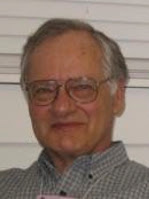
Russell Hobbie (1934–2021)

Russell Hobbie
Russell Hobbie
I am heartbroken to have to tell you that Russ Hobbie, my coauthor and friend, passed away this week, after a long battle with Parkinson’s disease. Russ was the sole author on the first three editions of Intermediate Physics for Medicine and Biology, and was the senior author with me on the fourth and fifth editions.

Below are excerpts from Russ’s preface to the first edition, describing how he came to write IPMB.
Between 1971 and 1973 I audited all of the courses medical students take in their first two years at the University of Minnesota. I was amazed at the amount of physics I found in those courses and how little of it is discussed in the general physics course.

I found a great discrepancy between the physics in some papers in the biological research literature and what I knew to be the level of understanding of most biology majors who had taken a year of physics. It was clear that an intermediate level physics course would help these students. It would provide the physics they needed and would relate it directly to the biological problems where it is useful. Making the connection with biology is something that we tend to leave for the student. When we do that, I think we overestimate the ability of most students to see the application and work out the details. Seeing few applications is also a powerful motivation for mastering difficult material.

This book is the result of my having taught such a course since 1973. It is intended to serve as a text for an intermediate course taught in a physics department and taken by a variety of majors… 

Here is a list from Google Scholar of some of his publications.

First ten listings in the Google Scholar entry for Russ Hobbie.

His most highly cited publication, by far, was IPMB (all editions are combined into one citation score). His most highly cited research article was a 2010 paper that he coauthored with his daughter Sarah. Some of the figures in that paper found their way, with slight modifications, into the 5th edition of IPMB: exponential decay assuming constant error bars (Fig. 2.6 in IPMB) or a constant percentage error bars (Fig. 2.7). He has articles going back to 1960, and the most recent edition of IPMB was in 2015, implying an amazing 55 year publication history. His earliest papers in nuclear physics were published during his time at the Harvard cyclotron laboratory

You can learn more about Russ by reading the transcript of his 1994 interview as part of an oral history project at the University of Minnesota. Some excerpts:

I grew up as a college brat. My parents both taught at Skidmore College in upstate New York. I was born in 1934. One of my earliest recollections is at age three or four falling in the college fishpond and being fished out by some of the Skidmore students. My father taught physics there...
[My high school had] a standard college preparatory course except that they had machine shop and mechanical drawing. I don’t know how I decided it, but I decided that I wanted to go to college at MIT [Massachusetts Institute of Technology], which I did and I thoroughly enjoyed that experience. I liked Cambridge so much that I wanted to stay in Cambridge. Everybody told me that I ought to change schools for graduate school; so, I went up the river to Harvard as a graduate student...
[At Harvard] my TA [teaching assistant] assignment was to work with Ed[ward Mills] Purcell, who is a Nobel Prize winning physicist, a very wonderful and humble person, redesigning the junior electricity and magnetism lab. That was a very great experience...

I became an RA [research assistant] at the Harvard cyclotron and drifted into doing my thesis in experimental nuclear physics on the cyclotron. I took my final Ph.D. oral exam on April Fool's Day in 1960, which means that I had done my Ph.D. in slightly under four years because I graduated from college in 1956...

I had met my wife [Cynthia], a public health nurse, in Boston. She had grown up in Iowa, had gone to the University of Iowa, and had then come out to work for the Visiting Nurses’ Association. She had some interest in moving closer to her home and I had seen this about Minnesota...
I got out here [an interview at the University of Minnesota] and discovered that I was actually being interviewed by Morris Blair to be a post-doc on the old Van de Graaff generator. I spent the day with him. About two o’clock that afternoon, he offered me a job and I accepted. All of this is a little bit different from the way things are done now...

I was attracted by the fact that it appeared to me at that time that at the University of Minnesota, research was important. It was a research university. Having grown up seeing a small college, I thought that that was a bit stifling and I didn’t want that. I thought that one could combine teaching and a research career here...
I also saw coming up the fact that nuclear physics was going to change and that the experiments were going to have to be done at national laboratories because individual universities could not afford to keep these things going; so, I made a conscious decision about that time—I was now an associate professor—that I wanted not to continue in nuclear physics. The department was fairly supportive of that and I became the director of undergraduate studies in physics for a few years...

I’d come out here [Minnesota] and happened, at some point along here, to be invited to the neighbors for a dinner party at which I met a pathologist named Richard Riess [spelled Reece], who was a pathologist at, then, St. Barnabas Hospital, a principal in Lufkin Medical Laboratories, and who was interested in using computers for interpreting lab test results...
So, Riess was making things that he called diagnotes, which were just lists of what could cause an elevated uric acid, or a low calcium, or a high calcium, and so on. At that dinner party, he started asking, "Was there any way that one could computerize this?" Having just put in this online computer at the Tandem Lab, I started using it to try to do some pattern matching. This, then, led to, for several years, my working with Riess as a collaborator and Lufkin Medical Labs having a research contract with the University of Minnesota that supported a couple of students over the years. We did a lot of work on developing automatic interpretation of the clinical laboratory results and published several papers in this area. I can remember still being the director of undergraduate studies in Physics and Mort[on] Hamennesh, who was the department head, coming in one day to tell me that they were promoting me to full professor based on the work that I had done at the Tandem in nuclear physics and the online computer...
[Reece] got me to thinking that it might be interesting to put some [medical] examples in the pre-med physics course; so, I wrote Al Sullivan who was the assistant dean of the Medical School asking if it was possible to snoop around over there. Al asked me to have lunch with him one day—it was in October—and said, "What you really ought to do is to attend Medical School." I said, "I can’t. I’m director of undergraduate studies in Physics. I’m teaching a full load, which is a course each quarter. There’s just no time to do that." He said, "You could just audit things and skip the labs." So, for two years, I did that....

Probably around 1972 or 1973, I started teaching that course, developing it as I went. That turned into a book [Intermediate Physics/or Medicine and Biology] that was published by Wiley in 1978 with a second edition about 1988. I’m trying, without much success, to do a third edition right now [Russ was ultimately successful]....

Included in the interview was a story about Russ’s daughter Ann.

It is now time to do the second edition of my book. I have the solutions manual and the new problems, I have already put into the computer. The new problems are there, but the old ones aren’t; so, Wiley agreed to hire my youngest daughter, Anne [Ann], to type all of the old solutions manual into the computer. She thought she had a summer job. She was done in two and half weeks.

If you still want to know more about Russ, check out the December 2006 issue of The Biological Physicist, the newsletter of the Division of Biological Physics in the American Physical Society. Russ and I (mostly Russ) answered questions about IPMB. Here is what I said about becoming Russ’s coauthor.

THE BIOLOGICAL PHYSICIST (to Brad Roth): Tell us a little about how you first became acquainted with Hobbie’s text, and how you see it has having influenced the field. 

Brad Roth: I used the first edition of Hobbie’s book for a class taught by John Wikswo when I was a graduate student at Vanderbilt University. This was a very crucial time in my education, when I was changing from a physics undergraduate student to a biological physics graduate student. The book had a huge impact on me and my career.

When visitors come by my office at OaklandUniversity, they sit politely and listen to me describe my research. Then I mention “Oh, by the way, I am also going to be second author on the 4th edition of Hobbie’s book Intermediate Physics in Medicine and Biology.” At this point, their eyes usually light up and they say, almost with disbelief, “Really? I know that book.”

At the end of the preface to the first edition of IPMB, Russ wrote

Every list of acknowledgments seems to close with thanks to a long-suffering family. I never knew what those words really meant, nor how deep the indebtedness, until I wrote this book.

 Below is the obituary prepared by his family.

Russell Klyver Hobbie, 87, died peacefully at home surrounded with love on December 16. Russ was born on November 3, 1934. His parents were Eulin Klyver and John Remington Hobbie. He grew up in Saratoga Springs, NY and Springfield, MA. He graduated from MIT with a BS in physics, and earned a PhD in physics from Harvard University. In 1960, he and his wife Cynthia moved with their young family to Minneapolis, where he joined the faculty at the University of Minnesota. Over his 38-year career, he was a wonderful professor and stalwart advocate for students. 
After auditing two years of medical school, Russ changed his specialty from nuclear physics to biophysics. He developed a new medical physics course which led to the first edition of his text book, Intermediate Physics for Medicine and Biology. For 12 years, Russ served as Associate Dean of Student Affairs in the Institute of Technology. After retirement, despite having been diagnosed with Parkinson’s disease, Russ completed the fourth and fifth edition of the book. 
Russell had many interests, among them canoeing and fishing in the BWCA, the Quetico and the Canadian Arctic. He enjoyed working on genealogy, and developed his skills as a woodworker, making beautiful furniture for his children and grandchildren. He and Cynthia took many trips in the US and abroad and loved spending time at the family cabin on Burntside Lake. They enjoyed theater, music, and visual arts in the Twin Cities. 
Russ is survived by his wife of 64 years, and his children, Lynn (Kevin Little), Erik (Pam Gahr), Sarah (Jacques Finlay) and Ann (Jeff Benjamin), and by his six grandchildren, Henry Benjamin, Grace Little, William Benjamin, Owen Finlay, Phoebe Finlay, and Rosie Hobbie. His sister Jane Bacon, two nieces and several grand nieces and nephews also survive him. 
Russ was a most wonderful and kind person and we will miss him very much. There will be a Memorial Service over Zoom from First Congregational Church in SE Minneapolis on Sunday January 2 at 2PM. We invite people to join us on-line. In lieu of flowers, Memorials may be directed to Save the Boundary Waters or Friends of the Boundary Waters.

Russ, I will miss you. Your work left a legacy that will influence how physics is taught to medical and biology students for years to come.

Russ Hobbie demonstratg the computer program MacDose. Part 1.
https://www.youtube.com/watch?v=eZGfHBYMuHg

Russ Hobbie demonstrating the computer program MacDose. Part 2.
https://www.youtube.com/watch?v=Rwp8eeKFycQ

Russ Hobbie demonstrating the computer program MacDose. Part 3.

Biology’s Built-In Faraday Cages

Klee (2014) "Biology's Built-In Faraday Cage," American Journal of Physics, 82:451–459, superimposed on the cover of Intermediate Physics for Medicine and Biology.
Klee, M. (2014)
Amer
. J. Phys. 82:451-459.
In Chapter 9 of Intermediate Physics for Medicine and Biology, Russ Hobbie and I discuss how well an external electric field penetrates the human body. We examine a sinusoidally varying field, but another problem is an field that turns on suddenly and then remains constant (a step function). This case is analyzed by Maurice Klee in his American Journal of Physics article “Biology’s Built-In Faraday Cages” (Volume 82, Pages 451-459, 2014). Klee obtained a PhD in Biomedical Engineering from Case Western Reserve University, under the direction of Robert Plonsey, and a law degree from George Washington University. His abstract states
Biological fluids are water-based, ionic conductors. As such, they have both high relative dielectric constants and substantial conductivities, meaning they are lossy dielectrics. These fluids contain charged molecules (free charges), whose movements play roles in essentially all cellular processes from metabolism to communication with other cells. Using the problem of a point source in air above a biological fluid of semi-infinite extent, the bound charges in the fluid are shown to perform the function of a fast-acting Faraday cage, which protects the interior of the fluid from external electric fields. Free charges replace bound charges in accordance with the fluid’s relaxation time, thereby providing a smooth transition between the initial protection provided by the bound charges and the steady state protection provided by the free charges. The electric fields within the biological fluid are thus small for all times just as they would be inside a classical Faraday cage.
The most interesting part of this article is the interplay between bound and free charges at the tissue surface. Klee assumes that bound charge, arising primarily from the rotation of the polarized water molecules, responds to the external electric field instantaneously, while free charge responds with a delay. The rapid bound charge shields the tissue immediately, but incompletely. Over time the free charge replaces the bound charge, eventually providing complete shielding of the tissue from the external electric field.

Klee addresses two questions: how completely does the bound charge shield the tissue, and how fast does the free charge replace the bound charge? (1) He shows that the initial bound charge is less than the final free charge by a factor of (κ – 1)/(κ + 1), where κ is the dielectric constant. Klee uses the value of 80 for κ, and finds that the bound charge provides 97.5% of the shielding that the free charge ultimately contributes. Russ and I point out that the dielectric constant of tissue can be much greater than water, and suggest a value of one million might be more appropriate. (2) The time constant for free charge to replace bound charge is κεo/σ, where εo is the permittivity of free space and σ is the conductivity. If you use σ = 0.5 S/m and κ = 80, the time constant is 1.4 nanoseconds. If you use κ = 1,000,000, the time required for complete shielding by the free charge is much longer (about 2 microseconds) but the initial shielding from the bound charge is 99.9998% complete.

Klee concludes that
Through a combination of bound and free charges, a biological fluid surrounded by a non-conductor of low relative dielectric constant does not develop large internal electric fields as a result of charges located outside the fluid. The fluid is thus its own robust Faraday cage, thereby ensuring that biological molecules within the fluid do not experience large electric fields due to outside sources.