Friday, December 17, 2010

Subtracting Large Numbers

One of the most notorious difficulties in numerical computations is the loss of precision when subtracting two similar, large numbers to obtain a smaller one. Russ Hobbie and I illustrate this hazard in Chapter 11 of the 4th edition of Intermediate Physics for Medicine and Biology. We begin this chapter with a discussion of the method of least squares, and we derive the formulas (Eqs. 11.5a and 11.5b) for fitting data to a straight line, y = ax + b. We then add “In doing computations where the range of data is small compared to the mean, better numerical accuracy can be obtained from…” and then present alternative formulas (Eqs. 11.5c, 11.5d, and 11.5e). Homework Problem 7 in Chapter 11 (one of the many new problems in the 4th edition) illustrates the advantage of the second set of equations.
Problem 7 Consider the data

   x       y
100   4004
101   4017
102   4039
103   4063

(a) Fit these data with a straight line y=ax+b using Eqs. 11.5a and 11.5b to find a.
(b) Use Eq. 11.5c to determine a. Your result should be the same as in part (a).
(c) Repeat parts (a) and (b) while rounding all the intermediate numbers to 4 significant figures. Do Eqs. 11.5a and 11.5b give the same result as Eq. 11.5c? If not, which is more accurate?
(Spoiler alert: Don’t continue reading if you want to solve the problem yourself first, as you should.) If you solve this problem, you will find that Eqs. 11.5a and 11.5b do not work very well at all for this problem. Their flaw is that they require you to subtract two really big numbers to get a much smaller one.

Numerical Methods That Work,  by Forman Acton, superimposed on Intermediate Physics for Medicine and Biology.
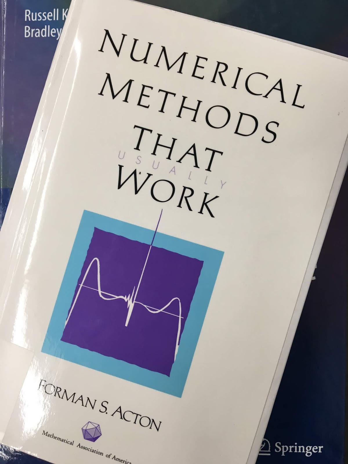
Numerical Methods That Work,
by Forman Acton.
A good discussion of this issue can be found in Forman Acton’s book Numerical Methods that Work.
The following problem often appears as a puzzle in Sunday Supplements. The difficulties are numerical rather than formulative and hence it is an especially appropriate challenge to the aspiring numerical analyst. We strongly urge that the reader solve it in his own way before turning to the “official” solution.

A railroad rail 1 mile long is firmly fixed at both ends. During the night some prankster cuts the rail and welds in an additional foot, causing the rail to bow up in the arc of a circle. The classical question concerns the maximum height this rail now achieves over its former position. To put it more precisely: We are faced…with the chord of a circle AB that is exactly 1 mile long and the corresponding arc AB that is 1 mile plus 1 foot and our question concerns the distance d between the chord and the arc at their midpoints. [See Acton’s book for the accompanying figure]

The relationships available are the simple ones from trigonometry involving the subtended half angle, θ, and the Pythagorean relationship. The student at this point should attempt to solve the problem before turning to the solution given in Chapter 2. He should attempt to find the distance d to an accuracy of three significant figures. In his effort he will probably be faced with subtracting two large and nearly equal numbers, which will cause a horrendous loss of significant figures. He can live with this process by shear brute force, but it will involve use eight-significant-figure trigonometric tables to preserve three figures in his answer. The point of the problem here is to find another method of calculating d, one that does not require such extreme measures. The three-figure answer can, indeed, be obtained rather easily using nothing more than pencil, paper, and a slide rule. The student should seek such a method.
If you find numerical methods interesting (as I do), you will love Acton’s delightfully written book. Originally published in 1970, it is all the more charming for its now-quaint references to slide rules and trigonometric tables. Yet, the concepts are not out-of-date. Even with powerful computers, errors can arise from subtracting nearly equal numbers. I’ve run into the issue myself when using the finite difference method and relaxation to solve Laplace’s equation with a fine grid and only single precision arithmetic.

Real Computing Made Real, by Forman Acton, superimposed on Intermediate Physics for Medicine and Biology.
Real Computing Made Real,
by Forman Acton.
Unfortunately, Acton’s book is not cited in the 4th edition of Intermediate Physics for Medicine and Biology (we’ll have to fix that in later editions), although I have mentioned it before in this blog. Acton is an emeritus professor in the Department of Computer Science at Princeton University (a department with an illustrious history). Also interesting is his more recent book Real Computing Made Real: Preventing Errors in Scientific and Engineering Calculations.

Friday, December 10, 2010

Robert Millikan

One fundamental constant that appears repeatedly in the 4th edition of Intermediate Physics for Medicine and Biology is the charge of the electron (the elementary charge, e), equal to 1.6 × 10⁻¹⁹ C. The first appearance of e that I can find is in Section 3.8 on the Nernst Equation. It appears in another context in Section 8.9, The Detection of Weak Magnetic Fields, when discussing Superconducting Quantum Interference Device (SQUID) magnetometers and the quantum of flux, equal to Planck’s constant divided by two times e. It shows up repeatedly in Chapter 9 on Electricity and Magnetism at the Cellular Level, and then again in Chapter 14 when discussing the energy levels of the hydrogen atom. It appears in Chapter 15 in the Klein-Nishina formula and in the expression for the classical radius of the electron.

Understanding Physics: The Electron, Proton, and Neutron, by Isaac Asimov, suuperimposed on Intermediate Physics for Medicine and Biology.
Understanding Physics:
The Electron, Proton, and Neutron,
by Isaac Asimov.
How was the charge of the electron first measured? Isaac Asimov tells the story in Understanding Physics: The Electron, Proton, and Neutron.
The experiments that determined the size of the electric charge on the electron were conducted by the American physicist Robert Andrews Millikan (1868–1953) in 1911.

Millikan made use of two horizontal plates, separated by about 1.6 centimeters, in a closed vessel containing air at low pressure, The upper plate had a number of fine holes in it and was connected to a battery that could place a positive charge upon it. Millikan sprayed fine drops of nonvolatile oil into the closed vessel above the plates. Occasionally, one droplet would pass through one of the holes in the upper plate and would appear in the space between the plates. There it could be viewed through a magnifying lens because it was made to gleam like a star through its reflection of a powerful beam of light entering from one side.

Left to itself, the droplet of oil would fall slowly, under the influence of gravity. The rate of this fall in response to gravity, against the resistance of air (which is considerable for so small and light an object as an oil droplet), depends on the mass of the droplet. Making use of an equation first developed by the British physicist George Gabriel Stokes (1819–1903), Millikan could determine the mass of the oil droplets.

Millikan then exposed the container to the action of X rays. This produced ions in the atmosphere within (see page 110). Occasionally, one of these ions attached itself to the droplet. If it were a positive ion, the droplet, with a positive charge suddenly added, would be repelled by the positively-charged plate above, and would rush downward at a rate greater than could be accounted for by the action of gravity alone. If the ion were negative, the droplet would be attracted to the positively-charged plate and might even begin to rise in defiance of gravity.

The change in velocity of the droplet would depend on the intensity of the electric field (which Millikan knew) and the charge on the droplet, which he could now calculate.

Millikan found that the charge on the droplet varied according to the nature of the ion that was adsorbed and on the number of ions that were adsorbed. All the charges were, however, multiples of some minimum unit, and this minimum unit could reasonably be taken as the smallest possible charge on an ion and therefore, equal to the charge on the electron. Millikan's final determination of this minimum charge was quite close to the value now accepted, which is 4.80298 × 10⁻¹⁰ electrostatic units (“esu”), or 0.000000000480298 esu.
We don’t use electrostatic units in Intermediate Physics for Medicine and Biology (although they appear briefly in homework problem 3 in Chapter 6), but this is equivalent to 1.6 × 10⁻¹⁹ Coulombs.

Selected Papers of Great American Physicists superimposed on Intermediate Physics for Medicine and Biology.
Selected Papers of
Great American Physicists.
I remember doing Millikan’s oil drop experiment as an undergraduate physics major at the University of Kansas. It required several hours in a dark room staring at small oil drops through a microscope. When in graduate school, I read one of Millikan’s papers in the book Selected Papers of Great American Physicists: The Bicentennial Commemorative Volume of The American Physical Society. I was particularly impressed by Millikan’s careful analysis of sources of systematic error in his experiment. In fact, I used that paper as a model for one of my few experimental papers: “The Magnetic Field of a Single Axon: A Comparison of Theory and Experiment” (Roth and Wikswo, Biophysical Journal, Volume 48, Pages 93–109, 1985). Some have claimed that Millikan committed scientific fraud by an improper selection of data to use in his analysis, but that claim has been debunked (see “Data Selection and Responsible Conduct: Was Millikan a Fraud?” by Richard Jennings, Science and Engineering Ethics, Volume 10, Pages 639–653, 2004).

I have a personal reason for being interested in the work of Robert Millikan. According to his Nobel Prize biography, he was born in Morrison Illinois, a small town 120 miles west of Chicago, about 15 miles from the Mississippi River. This is the town I grew up in, from an age of just a few months until I was 12 years old. At the time, I didn’t realize who Robert Millikan was, or that Morrison was the home to a Nobel Prize winning physicist. But over the years I have become a big fan of “Millikan from Morrison.” According to the Morrison chamber of commerce, there is now a downtown park named after Millikan. I must go visit.

Friday, December 3, 2010

Physical Biology of the Cell

Physical Biology of the Cell, by Phillips, Kondev, and Theriot, superimposed on Intermediate Physics for Medicine and Biology.
Physical Biology of the Cell,
by Phillips, Kondev, and Theriot.
I spent some time this week looking over the recently published textbook Physical Biology of the Cell, by Rob Phillips, Jane Kondev, and Julie Theriot. In some ways this book is a competitor of the 4th edition of Intermediate Physics for Medicine and Biology (it is always good to know your competition). Bernard Chasan reviewed Physical Biology of the Cell in the November 2010 issue of the American Journal of Physics.
The authors of this book are, in a very real sense, missionaries. They want to convince a wide audience to share their enthusiasm for and commitment to a more quantitative and scientifically rigorous approach to cell biology than is normally encountered in the teaching literature.

To achieve this goal, they set out a program of quantitative model building based on physical principles…. What the authors describe (awkwardly but evocatively) as the mathematizing of the semiqualitative models of cell biology (referred to as “cartoons” in some circles) has now become central to cell biology—as evidenced by a half a dozen recent texts and the relatively new and thriving discipline of systems biology. The work being reviewed is the latest and most comprehensive attempt to foster and advocate for this approach…

At the center of their approach is the art of model making—well presented with the aid of some excellent figures, which show the choices needed to model proteins, as one example. The main point is that modeling requires a simplifying choice, which emphasizes one view of the protein and essentially ignores others. If it suits your purposes to model the protein as a collection of hydrophobic and hydrophilic amino acid residues—a good model for protein folding—then you cannot at the same time consider the protein as a two state system.
After skimming through Physical Biology of the Cell (I wish I had time to read it thoroughly), I have several observations.
  1. The second half of Intermediate Physics for Medicine and Biology (IPMB) is about clinical medical physics: imaging and therapy. None of this appears in Physical Biology of the Cell (PBC). Also, in IPMB Russ Hobbie and I steer clear of molecular biology, saying in the preface that “molecular biophysics has been almost completely ignored: excellent texts already exist, and this is not our area of expertise.” PBC is all molecular and cellular. The main overlap between the two books is several chapters in PBC that cover similar topics as are in the first half of IPMB. So, I guess IPMB and PBC are not really in direct competition. However, if I was Phil Nelson, author of Biological Physics: Energy, Information, Life, I might be concerned about market share.
  2. PBC is illustrated by Nigel Orme. Let me be frank; Orme’s drawings are much better than what we have in IPMB. One thing I like about PBC is that you can skip the text altogether and just look at the pictures, and still learn the gist of the subject. Figure 1.4 showing the genetic code reminds me of the sort of graphics that Edward Tufte promotes in The Visual Display of Quantitative Information. The authors of PBC state in the acknowledgments “this book would never have achieved its present incarnation without the close and expert collaboration of our gifted illustrator, Nigel Orme, who is responsible for the clarity and visual appeal of the more than 550 figures found in these pages, as well as the overall design of the book.” As generous as this tribute is, it may be an understatement. Then, just when I thought the artwork couldn’t get any better, I found that PBC contains several beautiful figures contributed by David Goodsell, author of The Machinery of Life.
  3. In the 4th edition of IPMB, Russ and I added an initial section exploring the relative size of biological objects. In PBC, a similar discussion fills the entire Chapter 2. There is lots of numerical estimating in this chapter, reminding me of the Bionumbers website. Chapter 3 looks at different temporal scales, which is more difficult to show visually than spatial scales (Russ and I didn’t try), although Orme’s drawings do a pretty good job. Chapter 4 of PBC looks at the many model systems used in biology, with an eye toward history (Mendel’s pea plants, hemoglobin and the structure of a protein, the bacteriophage in genetics, etc.). Great reading.
  4. Some subjects—such as diffusion, fluid dynamics, thermodynamics, and bioelectricity—are covered in both PBC and IPMB. Which book explains these topics better? Obviously I am biased, but I suggest that Russ and I develop the physics in a more detailed and systematic way, starting from the fundamentals, whereas Phillips, Kondev and Theriot present the physics rather quickly, and then apply it to many interesting biological applications. I would say that PBC does for molecular and cellular biology what Air and Water by Mark Denny does for physiology: use physics and math to explain biological concepts quantitatively. Russ and I, on the other hand, teach physics using biological examples. The difference is more about approach, tone, and point-of-view than about substance. The reader can look at both books and draw their own conclusions.
  5. PBC has a few nice homework problems, but I prefer IPMB’s more extensive collection. The student learns more by doing than by reading.
  6. The final chapter in PBC, “Wither Physical Biology,” is an excellent summary of the “the role of quantitative analysis in the study of living matter.” Anyone working at the interface between physics and biology must read these ten pages.
Phillips, Kondev, and Theriot ought to have the last word, so I will finish this blog entry by quoting PBC’s eloquent closing paragraph.
The act of writing this book has convinced each of us that the study of living matter is one of the most exciting frontiers in human thought. Just as the makings of the large scale universe are being revealed by ever more impressive telescopes, living matter is now being viewed in ways that were once as unimaginable as was going to the Moon. Despite the muscle-enhancing weight of this book, we feel that we have only scratched the surface of the rich and varied applications of physical reasoning to biological problems. Our overall goal has been to communicate a style of thinking about problems where we have done our best to illustrate the power of the style using examples chosen from biological systems that are well defined and usually well studied from a biological perspective. As science moves forward into the twenty-first century, it is our greatest hope that synthetic approaches for understanding the natural world from biological, physical, chemical, and mathematical perspectives simultaneously will enrich all of these fields and illuminate the world around us. We can only hope the reader has at least a fraction of the pleasure in answering that charge as we have had in attempting to describe the physical biology of the cell.

Friday, November 26, 2010

Acetylcholine and Loewi’s Dream

In 1936, Otto Loewi was awarded the Nobel Prize in Physiology or Medicine for the discovery of the role of acetylcholine and other chemicals in nerve and muscle transmission. Russ Hobbie and I don’t mention Loewi in the 4th edition of Intermediate Physics for Medicine and Biology, but we do discuss acetylcholine. In Chapter 6, we write
At the end of a nerve cell the signal passes to another nerve cell or to a muscle cell across a synapse or junction. A few synapses in mammals are electrical; most are chemical…In electrical synapses, channels connect the interior of one cell with the next. In the chemical case a neurotransmitter chemical is secreted by the first cell. It crosses the synaptic cleft (about 50 nm) and enters the next cell.

At the neuromuscular junction the transmitter is acetylcholine (ACh). ACh increases the permeability of nearby muscle to sodium, which then enters and depolarizes the muscle membrane. The process is quantized. Packets of acetylcholine of definite size are liberated.
In Homework Problem 20 in Chapter 4, we ask the student to calculate the time required for acetylcholine to diffuse across the synaptic cleft. The release of acetylcholine at the nerve-muscle junction in discrete quanta provides a nice example of Poisson Statistics described in Appendix J. In Chapter 7, when discussing the heart, we mention how acetylcholine, released by parasympathetic nerves, decreases the heart rate.

The Left Hand of the Electron, by Isaac Asimov, superimposed on Intermediate Physics for Medicine and Biology.
The Left Hand of the Electron,
by Isaac Asimov.
I can’t tell you about Otto Loewi and acetylcholine without mentioning the fascinating tale of Loewi’s dream. Since Isaac Asimov is a much better storyteller than I am, I will simply quote from his essay “The Eureka Phenomenon” published in The Left Hand of the Electron.
The German physiologist Otto Loewi was working on the mechanism of nerve action, in particular, on the chemicals produced by nerve endings. He woke at 3 A.M. one night in 1921 with a perfectly clear notion of the type of experiment he would have to run to settle a key point that was puzzling him. He wrote it down and went back to sleep. When he woke in the morning, he found he couldn't remember what his inspiration had been. He remembered he had written it down, but he couldn't read his writing.

The next night, he woke again at 3 A.M. with the clear thought once more in mind. This time, he didn't fool around. He got up, dressed himself, went straight to the laboratory and began work. By 5 A.M. he had proved his point and the consequences of his findings became important enough in later years so that in 1936 he received a share in the Nobel prize in medicine and physiology.

Friday, November 19, 2010

Viral Outbreak: The Science of Emerging Disease

Textbooks such as the 4th edition of Intermediate Physics for Medicine and Biology are essential for studying and learning a new topic, but other ways of learning can be equally effective (or, sometimes, even better). Today I want to mention two examples.

Each December, the Howard Hughes Medical Institute presents its Holiday Lectures on Science. These excellent seminars will be webcast live on December 2 and 3, starting at 10 A.M. This year, the lectures are about “Viral Outbreak: The Science of Emerging Disease.” Joseph DeRisi (University of California, San Francisco) and Eva Harris (University of California, Berkeley) will explain how to detect and fight infectious agents. The lectures will answer questions such as “Why is dengue fever becoming a worldwide health threat,” “What other epidemics are on the horizon,” and “How can we detect and counter emerging infectious diseases?” If you miss the live webcast, you can download an on-demand webcast starting December 6. I have watched these holiday lectures in the past, and they are very good. They are aimed at a serious high school student, or an undergraduate science major. They are also great for a physicist looking for a general introduction to a biological or medical topic.

Of course, the best way to learn science is to do science. For undergraduates (the main readers of Intermediate Physics for Medicine and Biology), the first exposure to doing science may come during a summer research project. Now is the time to start looking for summer research opportunities. One that I recommend is the Summer Internship Program in Biomedical Science at the National Institutes of Health. I worked at NIH for seven years, and it is a wonderful place to do scientific research. My advice is to apply for this internship today. You won’t regret it.

Friday, November 12, 2010

Bionumbers

One feature of the 4th edition of Intermediate Physics for Medicine and Biology that distinguishes it from many other medical or biological textbooks is its focus on analyzing biomedical topics quantitatively. This point of view is also promoted at the BIONUMB3R5 (bionumbers) website, established by researchers in the systems biology department at Harvard. There is also a BIONUMB3R5 wiki where many researchers are coming together to provide new insights into key numbers in biology.

I particularly like the “Bionumber of the Month” feature. The March 2010 entry (“What are the Time Scales for Diffusion in Cells”) could easily be made into a homework problem for Chapter 4 of Intermediate Physics for Medicine and Biology. The January 2010 entry (“What is Faster, Transcription or Translation?”) is fascinating.
Transcription, the synthesis of mRNA from DNA, and translation, the synthesis of protein from mRNA, are the main pillars of the central dogma of molecular biology. How do the speeds of these two processes compare? …

Transcription of RNA by RNA polymerase in E. coli cells proceeds at a maximal speed of about 40–80 bp/sec… Translation by the ribosome in E. coli proceeds at a maximal speed of about 20 aa/sec… Interestingly, since every 3 base pairs code for one amino acid, the rates of the two processes are quite similar…
The “collection of fundamental numbers in molecular biology” found at the bionumbers website has the same tone as the first section of Chapter 1 in Intermediate Physics for Medicine and Biology, in which Russ Hobbie and I look at the relative size of biological objects. The collection contains this gem: “concentration of 1 nM in a cell the volume of E. coli is ~ 1 molecule/cell.”

The bionumbers website arose from an article by Rob Phillips and Ron Milo in the Proceedings of the National Academy of Sciences (Volume 106, pages 21465–21471, 2009), “A Feeling for the Numbers in Biology.” The abstract of their paper is given below:
Although the quantitative description of biological systems has been going on for centuries, recent advances in the measurement of phenomena ranging from metabolism to gene expression to signal transduction have resulted in a new emphasis on biological numeracy. This article describes the confluence of two different approaches to biological numbers. First, an impressive array of quantitative measurements make it possible to develop intuition about biological numbers ranging from how many gigatons of atmospheric carbon are fixed every year in the process of photosynthesis to the number of membrane transporters needed to provide sugars to rapidly dividing Escherichia coli cells. As a result of the vast array of such quantitative data, the BioNumbers web site has recently been developed as a repository for biology by the numbers. Second, a complementary and powerful tradition of numerical estimates familiar from the physical sciences and canonized in the so-called “Fermi problems” calls for efforts to estimate key biological quantities on the basis of a few foundational facts and simple ideas from physics and chemistry. In this article, we describe these two approaches and illustrate their synergism in several particularly appealing case studies. These case studies reveal the impact that an emphasis on numbers can have on important biological questions.
Russ and I introduce similar order-of-magnitude estimates (Fermi problems) in Chapter 1 of our book (for example, see homework problems 1–4, which are new in the 4th edition). One of my favorite Fermi problems, which I first encountered in the book Air and Water by Mark Denny, is to calculate the concentration of oxygen molecules in blood and in air, and compare them. Not too surprisingly, they are nearly the same (about 8 mM). I suspect the bionumbers folks would enjoy Air and Water. (I hope they would enjoy Intermediate Physics for Medicine and Biology, too.)

For those of you who find all of this interesting but prefer video over text, see the bionumbers video on YouTube.

Bionumbers: The data base of useful biological numbers. 

Friday, November 5, 2010

Seeing the Natural World with a Physicist’s Lens


One theme of this blog—and indeed, one theme of the 4th edition of Intermediate Physics for Medicine and Biology—is the role of physics in the biological sciences. So imagine my delight when Russ Hobbie sent me a similarly themed article from the November 1 issue of the New York Times (a publication that, alas, has more readers than does my blog). Natalie Angier, who studied for two years at that little college down the road in Ann Arbor, wrote an article titled Seeing the Natural World With a Physicist’s Lens. Its thesis is that many biological systems have evolved to perfection, in the sense that physical laws don’t let them get any better. Angier writes
Yet for all these apparent flaws, the basic building blocks of human eyesight turn out to be practically perfect. Scientists have learned that the fundamental units of vision, the photoreceptor cells that carpet the retinal tissue of the eye and respond to light, are not just good or great or phabulous at their job. They are not merely exceptionally impressive by the standards of biology, with whatever slop and wiggle room the animate category implies. Photoreceptors operate at the outermost boundary allowed by the laws of physics, which means they are as good as they can be, period. Each one is designed to detect and respond to single photons of light—the smallest possible packages in which light comes wrapped…

Photoreceptors exemplify the principle of optimization, an idea, gaining ever wider traction among researchers, that certain key features of the natural world have been honed by evolution to the highest possible peaks of performance, the legal limits of what Newton, Maxwell, Pauli, Planck et Albert will allow. Scientists have identified and mathematically anatomized an array of cases where optimization has left its fastidious mark… In each instance, biophysicists have calculated, the system couldn’t get faster, more sensitive or more efficient without first relocating to an alternate universe with alternate physical constants.
Angier has written a lot of articles for the NYT, and has published several books, that will be of interest to readers of Intermediate Physics for Medicine and Biology. Enjoy!

Friday, October 29, 2010

Iatrogenic Problems in End-Stage Renal Failure

In Section 5.7 of the 4th edition of Intermediate Physics for Medicine and Biology, where Russ Hobbie and I discuss the artificial kidney, we say
The artificial kidney provides an example of the use of the transport equations to solve an engineering problem… The reader should also be aware that this “high-technology” solution to the problem of chronic renal disease is not an entirely satisfactory one. It is expensive and uncomfortable and leads to degenerative changes in the skeleton and severe atherosclerosis

The alternative treatment, a transplant, has it own problems, related primarily to the immunosuppressive therapy. Anyone who is going to be involved in biomedical engineering or in the treatment of patients with chronic disease should read the account by Calland (1972), a physician with chronic renal failure who had both chronic dialysis and several transplants.
The paper by Chad Calland, in the New England Journal of Medicine (“Iatrogenic Problems in End-Stage Renal Failure,” Volume 287, Pages 334–336, 1972), was published on the same day that Calland took his own life. Wikipedia defines “iatrogenic” as “inadvertent adverse effects or complications caused by or resulting from medical treatment or advice.” It is a problem we must constantly be aware of as we seek to improve medical care through technology. Calland wrote
The physician is more often a voyeur than a partaker in human suffering. I am a physician who has undergone chronic renal failure, dialysis and multiple transplants. As a physician-partaker, I am distressed by the controversial dialogue that separates the nephrologist from the transplant surgeon, so that, in the end, it is the patient who is given short shrift. I have observed that both nephrologist and transplant surgeon work alone in their own separate fields, and that the patient becomes lost in a morass of professional role playing and physician self-justification. As legitimate as their altruistic but differing opinions may be, the nephrologist and the transplant surgeon must work together for the patient, so that therapy is tailored to suit the individual patient, his circumstances, his needs and the quality of his life.

Friday, October 22, 2010

Glimpses of Creatures in Their Physical Worlds

Glimpses of Creatures in their Physical Worlds, by Steven Vogel, superimposed on Intermediate Physics for Medicine and Biology.
Glimpses of Creatures
in their Physical Worlds,
by Steven Vogel.
I am a loyal member of Sigma Xi, the Scientific Research Society, and am a regular reader of its marvelous magazine American Scientist. One of the best parts of this bimonthly periodical is its book reviews. In the November-December 2010 issue of American Scientist, Mark Denny (author of Air and Water) reviews the new book by Steven Vogel: Glimpses of Creatures in Their Physical Worlds (Princeton University Press, 2009). Both Denny and Vogel appear in the 4th edition of Intermediate Physics for Medicine and Biology. Denny writes
Vogel’s contributions to biomechanics have had two admirable objectives. In Life in Moving Fluids (1981), Life’s Devices (1988), Vital Circuits (1992), Prime Mover (2001) and Comparative Biomechanics (2003), his goal is to explain the mechanics of biology to a general audience. If you want to know how fish swim, fleas jump and bats fly, or why hardening of your arteries is a bad thing, them dip into these sources; you will come away both informed and amused…

All too often, biologists observe only what they are prepared to see. Vogel’s second objective is therefore to expand their perspectives by conjuring up and carefully analyzing systems that might be… For example, dogs don’t sweat as humans do. Instead, they pant, evaporating water from their respirator tracts and expelling the resulting warm, moist air with each breath. But panting requires the repeated contraction of chest muscles, which adds to the heat the animal desires to loss. Could there be a better way?...

To find out, read Glimpses of Creatures in Their Physical Worlds. Here, as in Cats’ Paws and Catapults (1998), Vogel takes a decidedly nontraditional look at biology, unleashing his talent for unbridled speculation. The 12 chapters of Glimpses, which began as a series of essays in the Journal of Biosciences, have been revised and updated. They cover topics that range from the ballistics of seeds (plants use both catapults and cannons to launch their propagules) to the breathing apparatus of diving spiders (tiny hairs on the body take advantage of surface tension to maintain an airspace into which oxygen can flow), with stops along the way to explore the efficiency of man-made and natural pumps, the twist-to-bend ratios of daffodils in the breeze, and the physics of cow tipping…

If what you desire in a readable science book is food for thought, Glimpses of Creatures in Their Physical Worlds provides a feast. Biologists, engineers and physicists—indeed, anyone with curiosity about the natural world—will revel in this smorgasbord of biomechanical ideas.
I’ll put reading Glimpses on my to do list, maybe during the semester break.

If you get a copy of American Scientist so you can read Denny’s entire review, don’t miss another review in the same issue about a new edition (with notes and commentary) of the classic Flatland by Edwin Abbott. Flatland is a favorite of mine, and I agree with Colin Adams who says in his review: “In the pantheon of popular books about mathematics, one would be hard-pressed to name another that has lasted so long in popularity or had such a dramatic impact.”

Friday, October 15, 2010

Michael Faraday, Biological Physicist?

Last week in this blog I discussed the greatest physicist of all time, Isaac Newton. However, if we narrow consideration to only experimental physicists, I would argue that the greatest is Michael Faraday (with apologies to Ernest Rutherford, who is a close second). In Section 8.6 of the 4th edition of Intermediate Physics for Medicine and Biology, Russ Hobbie and I discuss Faraday’s greatest discovery: electromagnetic induction.
In 1831 Faraday discovered that a changing magnetic field causes an electric current to flow in a circuit. It does not matter whether the magnetic field is from a permanent magnet moving with respect to the circuit or from the changing current in another circuit. The results of many experiments can be summarized in the Faraday induction law.
I have always admired the 19th century Victorian physicists, such as Faraday, Maxwell and Kelvin. Michael Faraday, in particular, is a hero of mine (it is good to have heroes; they help you stay inspired when the mundane chores of life distract you). I had the pleasure of quoting from Faraday’s Experimental Researches in Electricity in an editorial I wrote in 2005 for the journal Heart Rhythm:  “Michael Faraday and Painless Defibrillation.” I tried to get a picture of Faraday included as part of the editorial, but alas the journal editor removed it. The article described a heart defibrillator having a design that included a type of Faraday cage.
Michael Faraday, arguably the greatest experimental physicist who ever lived, first demonstrated the shielding effect of a hollow conductor in 1836 by building a 12 ft × 12 ft × 12 ft cubic chamber out of metal. We would now call it a “Faraday cage.”

“I went into the cube and lived in it, and using lighted candles, electrometers, and all other tests of electrical states, I could not find the least influence upon them, or indication of anything particular given by them, though all the time the outside of the cube was powerfully charged, and large sparks and brushes were darting off from every part of its outer surface.” [Faraday M. Experimental Researches in Electricity. Paragraph 1174. Reprinted in: Hutchins RM, editor. Great Books of the Western World, Volume 45. Encyclopedia Britannica, Chicago, 1952.]

Faraday cages are used to shield sensitive electronic equipment. The metal skin of an airplane, acting as a Faraday cage, protects passengers from injury by lightning. Researchers perform electrophysiology experiments inside a Faraday cage to prevent external noise from contaminating the data. A rather spectacular example of shielding can be seen in the Boston Museum of Science, where a van de Graaff generator of over one million volts produces a dramatic display of lightning, while the operator stands nearby—safe inside a Faraday cage.
Why this little physics lesson? In this issue of Heart Rhythm, Jayam et al. [Jayam V, Zviman M, Jayanti V, Roguin A, Halperin H, Berger RD. “Internal Defibrillation with Minimal Skeletal Muscle Activation: A New Paradigm Toward Painless Defibrillation,” Heart Rhythm, Volume 2, Pages 1108–1113, 2005] describe a new electrode system for internal defibrillation that eliminates the skeletal muscle activation and pain associated with a shock. The central feature of their design is a Faraday cage: a conducting sock fitted over the epicardial surface of the heart…
In Section 8.7, Russ and I describe what may be the most important biomedical application of Faraday’s work: magnetic stimulation.
Since a changing magnetic field generates an induced electric field, it is possible to stimulate nerve or muscle cells without using electrodes. The advantage is that for a given induced current deep within the brain, the currents in the scalp that are induced by the magnetic field are far less than the currents that would be required for electrical stimulation. Therefore transcranial magnetic stimulation (TMS) is relatively painless. Magnetic stimulation can be used to diagnose central nervous system diseases that slow the conduction velocity in motor nerves without changing the conduction velocity in sensory nerves [Hallett and Cohen (1989)]. It could be used to monitor motor nerves during spinal cord surgery, and to map motor brain function. Because TMS is noninvasive and nearly painless, it can be used to study learning and plasticity (changes in brain organization over time). Recently, researchers have suggested that repetitive TMS might be useful for treating depression and other mood disorders.
I worked on magnetic stimulation for many years while at the National Institutes of Health in the 1990s. It was a pleasure to explore an application of Faraday induction; it is my kind of biological physics.

Faraday’s name can be found in a few other places in our book. It first appears in Chapter 3, when the Faraday constant is defined: F = 96,485 Coulombs per mole. It also appears in an abbreviated form in the unit of capacitance: a farad (F).

I suppose by now the reader realizes that I like Mike. But is he a biological physicist? Doubters might want to look at another physics blog: http://skullsinthestars.com/2010/05/15/shocking-michael-faraday-does-biology-1839. Faraday apparently did studies on the electrodynamics of electric fish. So, yes, I claim him as a biological physicist, and the question mark in the title of this blog post is unnecessary.