Friday, December 9, 2016

Capabilities of a Toroid-Amplifier System for Magnetic Measurement of Current in Biological Tissue

In Section 8.9 of Intermediate Physics for Medicine and Biology, Russ Hobbie and I discuss the detection of weak magnetic fields.
If the [magnetic] signal is strong enough, it can be detected with conventional coils and signal-averaging techniques that are described in Chap. 11. Barach et al. (1985) used a small detector through which a single axon was threaded. The detector consisted of a toroidal magnetic core wound with many turns of fine wire... Current passing through the hole in the toroid generated a magnetic field that was concentrated in the ferromagnetic material of the toroid. When the field changed, a measurable voltage was induced in the surrounding coil. This neuromagnetic current probe has been used to study many nerve and muscle fibers (Wijesinghe 2010).
I have discussed the neuromagnetic current probe before in this blog. One of the best places to learn more about it is a paper by Frans Gielen, John Wikswo, and me in the IEEE Transactions on Biomedical Engineering (Volume 33, Pages 910–921, 1986). The paper begins
In one-dimensional tissue preparations, bioelectric action currents can be measured by threading the tissue through a wire-wound, ferrite-core toroid that detects the associated biomagnetic field. This technique has several experimental advantages over standard methods used to measure bioelectric potentials. The magnetic measurement does not damage the cell membrane, unlike microelectrode recordings of the internal action potential. Recordings can be made without any electrical contact with the tissue, which eliminates problems associated with the electrochemistry at the electrode-tissue interface. While measurements of the external electric potential depend strongly on the distance between the tissue and the electrode, measurements of the action current are quite insensitive to the position of the tissue in the toroid. Measurements of the action current are also less sensitive to the electrical conductivity of the tissue around the current source than are recordings of the external potential.
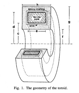
Figure 1 of this paper shows the toroid geometry
A illustration of a toroidal coil from Capabilities of a Toroid-Amplifier System for Magnetic Measurement of Current in Biological Tissue, by Gielen et al. (IEEE Trans Biomed Eng, 33:910-921, 1986)
When I was measuring biomagnetic fields back in graduate school, I wanted to relate the magnetic field in the toroid to the current passing through it. For simplicity, assume the current is in a wire passing through the toroid center. The magnetic field B a distance r from a wire carrying current i is (Eq. 8.7 in IPMB)
An equation giving the magnetic field produced by a current-carrying wire.
where μ is the magnetic permeability. The question is, what value should I use for r? Should I use the inner radius, the outer radius, the width, or some combination of these? The answer can be found by solving this new homework problem.
Section 8.2
Problem 11 1/2. Suppose a toroid having inner radius c, outer radius d, and width e is used to detect current i in a wire threading the toroids center. The voltage induced in the toroid is proportional to the magnetic flux through its cross section.
(a) Integrate the magnetic field produced by the current in the wire across the cross section of the ferrite core to obtain the magnetic flux.
(b) Calculate the average magnetic field in the toroid, which is equal to the flux divided by the toroid cross-sectional area.
(c) Define the “effective radius” of the toroid, reff, as the radius needed in Eq. 8.7 to relate the current in the wire to the average magnetic field. Derive an expression for reff in terms of the parameters of the toroid.
(d) If c = 1 mm, d = 2 mm, e = 1 mm, and μ=104μo, calculate reff.
The solution to this homework problem, the effective radius, appears on page 915 of our paper.

Finally, and just for fun, below I reproduce the short bios published with the paper, which appeared 30 years ago.
A brief bio of Frans Gielen, published in IEEE Trans Biomed Eng.

A brief bio of Brad Roth, published in IEEE Trans Biomed Eng.

A brief bio of John Wikswo, published in IEEE Trans Biomed Eng.

Friday, December 2, 2016

The Millikan Oil Drop Experiment

Selected Papers of Great American Physicists, superimposed on Intermediate Physics for Medicine and Biology.
Selected Papers of
Great American Physicists.
When I was in college, I was given a book published by the American Institute of Physics titled Selected Papers of Great American Physicists. Of the seven articles reprinted in this book, my favorite was “On the Elementary Electrical Charge and the Avogadro Constant” by Robert Millikan. Maybe I enjoyed it so much because I had performed the Millikan oil drop experiment as an undergraduate physics major at the University of Kansas. (I have discussed Millikan and his experiment previously in this blog.)

The charge of an electron is encountered often in Intermediate Physics for Medicine and Biology. Its one of those constants thats so fundamental to both physics and biology that its worth knowing how it was first measured. Below is a new homework problem requiring the student to analyze data like that obtained by Millikan. I have designated it for Chapter 6, right after the analysis of the force on a charge in an electric field and the relationship between the electric field and the voltage. I like this problem because it reinforces several concepts already discussed in IPMB (Stoke's law, density, viscosity, electrostatics), it forces the student to analyze data like that obtained experimentally, and it provides a mini history lesson.
Section 6.4

Problem 11 ½. Assume you can measure the time required for a small, charged oil drop to move through air (perhaps by watching it through a microscope with a stop watch in your hand). First, record the time for the drop to fall under the force of gravity. Then record the time for the drop to rise in an electric field. The drop will occasionally gain or lose a few electrons. Assume the drop’s charge is constant over a few minutes, but varies over the hour or two needed to perform the entire experiment, which consists of turning the electric field on and off so one drop goes up and down.

(a) When the drop falls with a constant velocity v1 it is acted on by two forces: gravity and friction given by Stokes’ law. When the drop rises at a constant velocity v2 it is acted on by three forces: gravity, friction, and an electrical force. Derive an expression for the charge q on your drop in terms of v1 and v2. Assume you know the density of the oil ρ, the viscosity of air η, the acceleration of gravity g, and the voltage V you apply across two plates separated by distance L to produce the electric field. These drops, however, are so tiny that you cannot measure their radius a. Therefore, your expression for q should depend on v1, v2, ρ, η, g, V, and L, but not a.
(b) You perform this experiment, and find that whenever the voltage is off the time required for the drop to fall 10 mm is always 12.32 s. Each time you turn the voltage on the drop rises, but the time to rise 10 mm varies because the number of electrons on the drop changes. Successive experiments give rise times of 162.07, 42.31, 83.33, 33.95, 18.96, and 24.33 s. Calculate the charge on the drop in each case. Assume η = 0.000018 Pa s, ρ = 920 kg m-3, V = 5000 V, L = 15 mm, and g = 9.8 m s-2.

(c) Analyze your data to find the greatest common divisor for the charge on the drop, which you can take as the charge of a single electron. Hint: it may be easiest to look at changes in the drops charge over time.
What impressed me most about Millikan’s paper was his careful analysis of sources of systematic error. He went to great pains to determine accurately the viscosity of air, and he accounted for small effects like the mean free path of the air molecules and the drop's buoyancy (effects you can neglect in the problem above). He worried about tiny sources of error such as distortions of the shape of the drop caused by the electric field. When I was a young graduate student, Millikans article provided my model for how you should conduct an experiment.

Friday, November 25, 2016

Intermediate Physicist for Medicine and Biology

In my more contemplative moments, I sometimes ponder: who am I? Or perhaps better: what am I? In my personal life I am many things: son, husband, father, brother, dog-lover, die-hard Cubs fan, Asimov aficionado, Dickens devotee, and mid-twentieth-century-Broadway-musical-theatre admirer.

What I am professionally is not as clear. By training I’m a physicist. Each month I read Physics Today and my favorite publication is the American Journal of Physics. But in many ways I don’t fit well in physics. I don’t understand much of what’s said at our weekly physics colloquium, and I have little or no interest in topics such as high energy physics. Quantum mechanics frightens me.

The term biophysicist doesn’t apply to me, because I don’t work at the microscopic level. I don’t care about protein structures or DNA replication mechanisms. I’m a macroscopic guy.

My work overlaps that of biomedical engineers, and indeed I publish frequently in biomedical engineering journals. But my work is not applied enough for engineering. In the 1990s, when searching desperately for a job, I considered positions in biomedical engineering departments, but I was never sure what I would teach. I have no idea what’s taught in engineering schools. Ultimately I decided that I fit better in a physics department.

Mathematical biologist is a better definition of me. I build mathematical models of biological systems for a living. But I’m at heart neither a mathematician nor a biologist. I find math papers—full of  theorem-proof-theorem-proof—to be tedious. Biologists celebrate life’s diversity, which is exactly the part of biology I like to sweep under the rug.

I’m not a medical physicist. Nothing I have worked on has healed anyone. Besides, medical physicists work in nuclear medicine and radiation therapy departments at hospitals, and they get paid a lot more that I do. No, I’m definitely not a medical physicist. Perhaps one of the most appropriate labels is biological physicist—whatever that means.

Another question is: at what level do I work? I’m not a popularizer of science or a science writer (except when writing this blog, which is more of a hobby; my “Hobbie hobby”). I write research papers and publish them in professional journals. Yet, in these papers I build toy models that are as simple as possible (but no simpler!). Reviewers of my manuscripts write things like “the topic is interesting and the paper is well-written, but the model is too simple; it fails to capture the underlying complexity of the system.” When my simple models grow too complicated, I change direction and work on something else. So my research is neither at an introductory level nor an advanced level.

I guess the best label for me is: Intermediate Physicist for Medicine and Biology.

Friday, November 18, 2016

Molybdenum-99 for Medical Imaging

Molybdenum-99 for Medical Imaging, published by the National Academies Press.
Molybdenum-99 for Medical Imaging,
published by the National Academies Press.
Between 2007 and 2011, I wrote several blog posts about a shortage of the radioisotope technetium-99m (here, here, here, and here). Chapter 17 of Intermediate Physics for Medicine and Biology discusses the many uses of 99mTc in nuclear medicine. It is produced from the decay of molybdenum-99, and shortages arise because of dwindling sources of 99Mo.

Recently, the Committee on State of Molybdenum-99 Production and Utilization and Progress Toward Eliminating Use of Highly Enriched Uranium addressed this issue in their report Molybdenum-99 for Medical Imaging, published by the National Academies Press. Below I reproduce excerpts from the executive summary.
This Academies study was mandated by the American Medical Isotopes Production Act of 2012. Key results for each of the five study charges are summarized below…

Study charge 1: Provide a list of facilities that produce molybdenum-99 (Mo-99) for medical use including an indication of whether these facilities utilize highly enriched uranium (HEU)… About 95 percent of the global supply of Mo-99 for medical use is produced in seven research reactors and supplied from five target processing facilities located in Australia, Canada, Europe, and South Africa. About 5 percent of the global supply is produced in other locations for regional use. About 75 percent of the global supply of Mo-99 for medical use is produced using HEU targets; the remaining 25 percent is produced with low enriched uranium targets….

Study charge 2: Review international production of Mo-99 over the previous 5 years … New Mo-99 suppliers have entered the global supply market since 2009 and further expansions are planned. An organization in Australia (Australian Nuclear Science and Technology Organisation) has become a global supplier and is currently expanding its available supply capacity; existing global suppliers in Europe (Mallinckrodt) and South Africa (NTP Radioisotopes) are also expanding ... A reactor in France (OSIRIS) that produced Mo-99 shut down permanently in December 2015. The reactor in Canada (NRU) will stop the routine production of Mo-99 after October 2016 and permanently shut down at the end of March 2018.

Study charge 3: Assess progress made in the previous 5 years toward establishing domestic production of Mo-99 and associated medical isotopes iodine-131 (I-131) and xenon-133 (Xe-133) … The American Medical Isotopes Production Act of 2012 and financial support from the Department of Energy’s National Nuclear Security Administration … have stimulated private-sector efforts to establish domestic production of Mo-99 and associated medical isotopes. Four NNSA-supported projects and several other private-sector efforts are under way to establish domestic capabilities to produce Mo-99; each project is intended to supply half or more of U.S. needs…. it is unlikely that substantial domestic supplies of Mo-99 will be available before 2018. Neither I-131 nor Xe-133 is currently produced in the United States, but one U.S. organization (University of Missouri Research Reactor Center) is developing the capability to supply I-131; some potential domestic Mo-99 suppliers also have plans to supply I-131 and/or Xe-133 in the future.

Study charge 4: Assess the adequacy of Mo-99 supplies to meet future domestic medical needs, particularly in 2016 and beyond …The United States currently consumes about half of the global supply of Mo-99/technetium-99m (Tc-99m) for medical use; global supplies of Mo-99 are adequate at present to meet domestic needs. Domestic demand for Mo-99/Tc-99m has been declining for at least a decade and has declined by about 25 percent between 2009-2010 and 2014-2015; domestic medical use of Mo-99/Tc-99m is unlikely to increase significantly over the next 5 years. The committee judges that there is a substantial ... likelihood of severe Mo-99/Tc-99m supply shortages after October 2016, when Canada stops supplying Mo-99, lasting at least until current global Mo-99 suppliers complete their planned capacity expansions (planned for 2017) and substantial new domestic Mo-99 supplies enter the market (not likely until 2018 and beyond)….

Study charge 5: Assess progress made by the DOE and others to eliminate worldwide use of HEU in reactor targets and medical isotope production facilities and identify key remaining obstacles for eliminating HEU use… The American Medical Isotopes Production Act of 2012 is accelerating the elimination of worldwide use of HEU for medical isotope production [to reduce the amount of HEU available for production of weapons of mass destruction by terrorist groups]. Current global Mo-99 suppliers have committed to eliminating HEU use in reactor targets and medical isotope production facilities and are making uneven progress toward this goal. Progress is … being impeded by the continued availability of Mo-99 produced with HEU targets … Even after HEU is eliminated from Mo-99 production, large quantities of HEU-bearing wastes from past production will continue to exist at multiple locations throughout the world…
News articles associated with the release of the report can be found here, here, here, and here. The message I get from this report is that the long-term prognosis for 99Mo supplies is promising, but the short-term outlook is worrisome. Let us hope I’m too pessimistic.

Friday, November 11, 2016

Mathematical Physiology

Mathematical Physiology, by James Keener and James Sneyd, with Intermediate Physics for Medicine and Biology.
Mathematical Physiology,
by James Keener and James Sneyd.
In a comment to the blog last week, Frankie mentioned the two-volume textbook Mathematical Physiology (MP), by James Keener and James Sneyd. Russ Hobbie and I cite Keener and Sneyd in Chapter 10 (Feedback and Control) of Intermediate Physics for Medicine and Biology. The Preface to the first edition of MP begins:
It can be argued that of all the biological sciences, physiology is the one in which mathematics has played the greatest role. From the work of Helmholtz and Frank in the last century through to that of Hodgkin, Huxley, and many others in this century [the first edition of MP was published in 1998], physiologists have repeatedly used mathematical methods and models to help their understanding of physiological processes. It might thus be expected that a close connection between applied mathematics and physiology would have developed naturally, but unfortunately, until recently, such has not been the case.

There are always barriers to communication between disciplines. Despite the quantitative nature of their subject, many physiologists seek only verbal descriptions, naming and learning the functions of an incredibly complicated array of components; often the complexity of the problem appears to preclude a mathematical description. Others want to become physicians, and so have little time for mathematics other than to learn about drug dosages, office accounting practices, and malpractice liability. Still others choose to study physiology precisely because thereby they hope not to study more mathematics, and that in itself is a significant benefit. On the other hand, many applied mathematicians are concerned with theoretical results, proving theorems and such, and prefer not to pay attention to real data or the applications of their results. Others hesitate to jump into a new discipline, with all its required background reading and its own history of modeling that must be learned.

But times are changing, and it is rapidly becoming apparent that applied mathematics and physiology have a great deal to offer one another. It is our view that teaching physiology without a mathematical description of the underlying dynamical processes is like teaching planetary motion to physicists without mentioning or using Kepler’s laws; you can observe that there is a full moon every 28 days, but without Kepler’s laws you cannot determine when the next total lunar or solar eclipse will be nor when Halley’s comet will return. Your head will be full of interesting and important facts, but it is difficult to organize those facts unless they are given a quantitative description. Similarly, if applied mathematicians were to ignore physiology, they would be losing the opportunity to study an extremely rich and interesting field of science.

To explain the goals of this book, it is most convenient to begin by emphasizing what this book is not; it is not a physiology book, and neither is it a mathematics book. Any reader who is seriously interested in learning physiology would be well advised to consult an introductory physiology book such as Guyton and Hall (1996) or Berne and Levy (1993), as, indeed, we ourselves have done many times. We give only a brief background for each physiological problem we discuss, certainly not enough to satisfy a real physiologist. Neither is this a book for learning mathematics. Of course, a great deal of mathematics is used throughout, but any reader who is not already familiar with the basic techniques would again be well advised to learn the material elsewhere.

Instead, this book describes work that lies on the border between mathematics and physiology; it describes ways in which mathematics may be used to give insight into physiological questions, and how physiological questions can, in turn, lead to new mathematical problems. In this sense, it is truly an interdisciplinary text, which, we hope, will be appreciated by physiologists interested in theoretical approaches to their subject as well as by mathematicians interested in learning new areas of application.
If you substitute the words “physics” for “mathematics,” “physical” for “mathematical,” and “physicist” for “mathematician,” you would almost think that this preface had been written by Russ Hobbie for an early edition of IPMB.

Many of the topics in MP overlap those in IPMB: diffusion, bioelectricity, osmosis, ion channels, blood flow, and the heart. MP covers additional topics not in IPMB, such as biochemical reactions, calcium dynamics, bursting pancreatic beta cells, and the regulation of gene expression. What IPMB has that MP doesn’t is clinical medical physics: ultrasound, x-rays, tomography, nuclear medicine, and MRI. Both books assume a knowledge of calculus, both average many equations per page, and both have generous collections of homework problems.

Which book should you use? Mathematical Physiology won an award, but Intermediate Physics for Medicine and Biology has an award-winning blog. I’ll take the book with the blog. I bet I know what Frankie will say: “I’ll take both!”