Friday, June 3, 2022

Roger Bacon, Biological Physicist

The Story of Civilization, by Will and Ariel Durant, surrounding a copy of Intermediate Physics for Medicine and Biology.
The Story of Civilization,
by Will and Ariel Durant.
About a year ago I began reading the eleven-volume series The Story of Civilization by Will and Ariel Durant. I just finished Volume 4, The Age of Faith. A History of Medieval Civilization—Christian, Islamic, and Judaic—from Constantine to Dante: A.D. 325–1300. Of course, I’m always on the lookout for how a book overlaps with Intermediate Physics for Medicine and Biology. In The Age of Faith I found a scholar from the Middle Ages who might qualify as a biological physicist: Roger Bacon. Durant writes (citations removed)
VII. ROGER BACON: c. 1214–92

The most famous of medieval scientists was born in Somerset about 1214. We know that he lived till 1292, and that in 1267 he called himself an old man. He studied at Oxford under Grosseteste, and caught from the great polymath a fascination for science; already in that circle of Oxford Franciscans the English spirit of empiricism and utilitarianism was taking form. He went to Paris about 1240, but did not find there the stimulation that Oxford had given him…
Bacon is known for his support of the role of experiment in science. So much of medieval thought was based on religion and mysticism, and an emphasis on science and experiment is refreshing.
We must not think of him [Bacon] as a lone originator, a scientific voice crying out in the scholastic wilderness. In every field he was indebted to his predecessors, and his originality was the forceful summation of a long development. Alexander Neckham, Bartholomew the Englishman, Robert Grosseteste, and Adam Marsh had established a scientific tradition at Oxford; Bacon inherited it, and proclaimed it to the world. He acknowledged his indebtedness, and gave his predecessors unmeasured praise. He recognized also his debt—and the debt of Christendom—to Islamic science and philosophy, and through these to the Greeks

Like Russ Hobbie and I, Bacon appreciated the role of math in science. Durant summarized Bacon’s view as “though science must use experiment as its method, it does not become fully scientific until it can reduce its conclusions to mathematical form.”

Bacon’s work on optics and vision overlaps with topics in IPMB. Durant notes that “one result of these studies in optics [performed by Bacon and others] was the invention of spectacles.” I can hardly think of a better example of physics interacting with physiology than eyeglasses. Durant concludes:

Experimenting with lenses and mirrors, Bacon sought to formulate the laws of refraction, reflection, magnification, and microscopy. Recalling the power of a convex lens to concentrate many rays of the sun at one burning point, and to spread the rays beyond that point to form a magnified image, he wrote:
We can so shape transparent bodies [lenses], and arrange them in such a way with respect to our sight and the objects of vision, that the rays will be refracted and bent in any direction we desire; and under any angle we wish we shall see the object near or at a distance. Thus from an incredible distance we might read the smallest letters…
These are brilliant passages. Almost every element in their theory can be found before Bacon, and above all in al-Haitham [an Arab scientist also known as Alhazen]; but the material was brought together in a practical and revolutionary vision that in time transformed the world. It was these passages that led Leonard Digges (d. c. 1571) to formulate the theory of which the telescope was invented.

I enjoy reading the Durants’ books. They contain not only the usual political and military history of the world, but also the history of science, art history, music history, comparative religion, linguistics, the history of medicine, philosophy, and literature. While The Story of Civilization may not be the definitive source on any of these topics, it is the best integration of all of them into one work that I am aware of. Had the Durants lived longer, future volumes (which they tentatively titled The Age of Darwin and The Age of Einstein) might have focused even more on the role of science in civilization. 

I won’t finish The Story of Civilization anytime soon; I still have seven volumes to go. The series runs to over ten thousand pages, single-spaced, small font (I had to buy more powerful reading glasses for this project). I’ll continue to search for discussions of medical physics and biological physics throughout.

Now, on to The Renaissance

The Story of Civilization. 1. Our Oriental Heritage, 2. The Life of Greece, 3. Caesar and Christ, 4. The Age of Faith, 5. The Renaissance, 6. The Reformation, 7. The Age of Reason Begins, 8. The Age of Louis XIV, 9. The Age of Voltaire, 10. Rousseau and Revolution, and 11. The Age of Napoleon.


In Our Time: Season 19/Episode 30, Roger Bacon (April 20, 2017)

https://www.youtube.com/watch?v=i3riF-F7hGY


The Durants—Will & Ariel Durant: The Story of Civilization Documentary.

https://www.youtube.com/watch?v=Jc8aFTuCAu0

Friday, May 27, 2022

Radiology in Our Changing Climate: A Call to Action

Schoen et al., (2021) “Radiology in Our Changing Climate: A Call to Action” JACR, 18:1041–1043, superimposed on Intermediate Physics for Medicine and Biology.
Schoen et al. (2021)
“Radiology in Our Changing Climate:
A Call to Action,”
JACR, 18:1041–1043.
Lately I’ve been thinking more and more about the importance of combating climate change, which may be the most urgent technological challenge of our time. But you haven’t seen much about it in this blog, because climate change doesn’t have much to do with Intermediate Physics for Medicine and Biology. Or does it? Last year, Julia Schoen, Geraldine McGinty, and Cody Quirk published an opinion piece in the Journal of the American College of Radiology titled “Radiology in Our Changing Climate: A Call to Action” (Volume 18, Pages 1041–1043). It’s short, clear, and well worth reading. The introduction begins:
Just as early radiologists did not understand the dangers of high radiation doses, today we are naive to imaging’s carbon footprint and its implications for public health. The world’s temperature has already risen more than 1 °C from preindustrial levels. We see the effects of climate change across the world, from extreme wildfires and stronger storms to rising sea levels and ocean acidification. If we continue with “business as usual,” children born today will experience a planet that is 4 °C warmer than in preindustrial times and the associated health consequences. These consequences are disproportionately felt by children, the elderly, those with preexisting conditions, and outdoor workers. As our climate crisis worsens, radiologists must urgently consider our role in climate change.
According to Schoen et al., the health care system may be responsible for nearly ten percent of American’s greenhouse gas emissions. TEN PERCENT! Yikes. They suggest that radiology departments are “likely a major contributor to energy use within hospital systems.” They identify four ways to address the energy use in radiology.

Imaging Exams

Schoen et al. claim that “over a year, the energy use of one CT [computed tomography] scanner was comparable with that of 5 four-person households, and the energy use of one MR [magnetic resonance] scanner was close to that of 26 four-person households.” I always thought MRI was the ideal imaging method, but it turns out it’s an energy hog, contributing significantly to radiology’s carbon footprint. There are few easy ways to reduce energy use; perhaps use ultrasound more when appropriate and adopt new technologies that shorten imaging time.

Scanners in the Off State

Imaging systems use a lot of energy even in standby mode. You must keep the superconducting coil of a MRI scanner cold all the time, not just when it’s imaging. Solutions are not simple. Schoen et al. suggest using scanners 24 hours a day (patients won’t like that) and working with manufacturers to find ways of reducing energy use when a scanner is not operating.

Wasteful Habits

We have to cut the waste in radiology departments. Simple improvements would be to turn off computers and picture archiving and communication systems (PACSs) at night or when not in use, and reducing excess packaging. I support these easy changes, but wonder if they’ll have a major impact on our carbon footprint.

Energy Sources

Alternative energy sources—including ones like wind, solar, and nuclear—will reduce greenhouse gas emissions. This is something individual radiologists, or even radiology departments, have little control over, but if major health care systems demand cleaner energy sources they might be able to influence regional utilities and politicians.

Conclusion

Schoen, McGinty, and Quirk discuss an important issue, and I thank them for raising it. Their call to action must be addressed by radiologists in collaboration with hospital administrators, academic researchers, and medical device companies. All of us—including the past, present, and future patients needing radiological services—must advocate for reducing our impact on the climate.

I’ll give Schoen et al. the last word by quoting the eloquent final paragraph of their publication.

Radiology faces many challenges, from improving diversity to changes in reimbursement in a budget-neutral system. Addressing climate change is an opportunity to protect vulnerable populations and increase our value in the health care system. Initiatives to address climate change align with the ACR’s [American College of Radiology’s] core purpose of serving both patients and society. Our field has made great strides in patient safety by decreasing radiation doses. Similarly, through our technological expertise and awareness, we can decrease our carbon footprint, with the ultimate goal of mitigating climate change and preventing a looming public health crisis.


Listen to a podcast of Julia Schoen discussing sustainability and radiology.

https://www.youtube.com/watch?v=_66M2PgnTxA


The group “Radiologists for a Sustainable Future” is on Twitter. Follow them at @Rads4SF.

Friday, May 20, 2022

Using the Mechanical Bidomain Model to Analyze the Biomechanical Behavior of Cardiomyocytes

During the decade of 2010–2020, my research shifted from bioelectricity and biomagnetism to biomechanics and mechanotransduction. I took the bidomain model of cardiac electrophysiology—described in Chapter 7 of Intermediate Physics for Medicine and Biology— and adapted it to describe growth and remodeling in response to mechanical forces. In other words, I traded resistors for springs. This effort was not entirely successful, but I think it provided some useful insights.

In 2015 I described the mechanical bidomain model in a chapter of Cardiomyocytes: Methods and Protocols. This book was part of the series Methods in Molecular Biology, and each chapter had a unusual format. The research was outlined, with the details relegated to an extensive collection of endnotes. A second edition of the book was proposed, and I dutifully submitted an updated chapter. However, the new edition never come to pass. Rather than see my chapter go to waste, I offer it to you, dear reader. You can download a draft of my chapter for the second edition here. For those of you who have time only for a summary, below is the abstract.

The mechanical bidomain model provides a macroscopic description of cardiac tissue biomechanics, and also predicts the microscopic coupling between the extracellular matrix and the intracellular cytoskeleton of cardiomyocytes. The goal of this chapter is to introduce the mechanical bidomain model, to describe the mathematical methods required for solving the model equations, to predict where the membrane forces acting on integrin proteins coupling the intracellular and extracellular spaces are large, and to suggest experiments to test the model predictions.

The main difference between the chapter in the first edition and the one submitted for the second was a new section called “Experiments to Test the Mechanical Bidomain Model.” There I describe how the model can reproduce data obtained when studying colonies of embryonic stem cells, sheets of engineered heart tissue, and border zones between normal and ischemic regions in the heart. The chapter ends with this observation:

The most important contribution of mathematical modeling in biology is to make predictions that can be tested experimentally. The mechanical bidomain model makes many predictions, in diverse areas such as development, tissue engineering, and hypertrophy.
I particularly like a new figure in the second edition. It’s a revision of a figure created by Xavier Trepat and Jeffrey Fredberg that compares mechanobiology to a game of tug-of-war. I added the elastic properties of the extracellular space (the green arrows), saying “It is as if the game of tug-of-war is played on a flexible surface, such as a flat elastic sheet.” In other words, tug-of-war on a trampoline

Enjoy!

The “tug-of-war” model of tissue biomechanics, adapted from an illustration by Trepat and Fredberg.
The “tug-of-war” model of tissue biomechanics, adapted from an illustrationby Trepat and Fredberg. Top: the intracellular (yellow), extracellular (green) and integrin (blue) forces acting on a monolayer of cells. Middle: The analogous forces among the players of a game of tug-of-war. This figure is extended beyond that of Trepat and Fredberg by allowing both the intracellular and extracellular spaces to move. Bottom: Representation of the mechanical bidomain model by a ladder of springs.

Friday, May 13, 2022

Aquaporins and Peter Agre

In Chapter 5 of Intermediate Physics for Medicine and Biology, Russ Hobbie and I mention aquaporins, a type of membrane channel. In a footnote, we write
Some aquaporins are permeable only to water, and not to any other small molecules or ions, even hydrogen ions (Preston et al. 1992). Aquaporins are formed by proteins that span the cell membrane. Their structure has been determined by x-ray crystallography (Murara et al. 2000). Their selectivity arises from a narrowing of the channel to about 0.3 nm, about the size of a single water molecule. Aquaporins allow water to cross cell membranes at a much higher rate than it could diffuse through. Genetically defective aquaporins may be responsible for some clinical diseases, such as nephrogenic diabetes insipidus and congenital cataracts (Agre et al. 2002).

In 2003, Peter Agre was awarded the Nobel Prize in Chemistry for the discovery of aquaporins. My goal in this post is to provide a bit more detail about aquaporins and Agre

To learn more about these water channels, let’s begin with this simple, fun Claymation video.

Claymation video about aquaporins by Sophia Dudte.
https://www.youtube.com/watch?v=7EGPtMqZ7pY

Next is a more rigorous simulation of an aquaporin

A simulation of a water channel in a cell membrane, performed by The Theoretical and Biophysics Group at the NIH Center for Macromolecular Modeling and Bioinformatics.
https://www.youtube.com/watch?v=GSi5-y6NHjY

Russ and I cite the paper by Murara et al. (2000). The full citation is

Murata K, Mitsuoka K, Hirai T, Walz T, Agre P, Heymann JB, Engel A, Fujiyoshi Y (2000) Structural determinants of water permeation through aquaporin-1. Nature, Volume 407, Pages 599–605.
The abstract is listed below.
Human red cell AQP1 is the first functionally defined member of the aquaporin family of membrane water channels. Here we describe an atomic model of AQP1 at 3.8 Å resolution from electron crystallographic data. Multiple highly conserved amino-acid residues stabilize the novel fold of AQP1. The aqueous pathway is lined with conserved hydrophobic residues that permit rapid water transport, whereas the water selectivity is due to a constriction of the pore diameter to about 3 Å over a span of one residue. The atomic model provides a possible molecular explanation to a longstanding puzzle in physiology—how membranes can be freely permeable to water but impermeable to protons.
Below is a illustration of the aquaporin molecule. The view is perpendicular to the membrane, and the little hole in the middle is the pore. 


Illustration of an aquaporin molecule. Drawn by David Goodsell.

Next is the introduction to the Wikipedia article about Agre (references removed).

Peter Agre /ˈɑːɡriː/ (born January 30, 1949) is an American physician, Nobel Laureate, and molecular biologist, Bloomberg Distinguished Professor at the Johns Hopkins Bloomberg School of Public Health and Johns Hopkins School of Medicine, and director of the Johns Hopkins Malaria Research Institute. In 2003, Agre and Roderick MacKinnon shared the 2003 Nobel Prize in Chemistry for "discoveries concerning channels in cell membranes." Agre was recognized for his discovery of aquaporin water channels. Aquaporins are water-channel proteins that move water molecules through the cell membrane. In 2009, Agre was elected president of the American Association for the Advancement of Science (AAAS) and became active in science diplomacy.
You can learn more about Agre in the videos below.

Peter Agre talking about aquaporin channels at the National Institutes of Health. https://www.youtube.com/watch?v=L1TyWo86w4Q 


Peter Agre answering questions about his life and research 


Peter Agre giving a TED Talk (2011)
https://www.youtube.com/watch?v=-eq5tfU1kZY

Friday, May 6, 2022

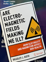
Are Electromagnetic Fields Making Me Ill? How Electricity and Magnetism Affect Our Health

Are Electromagnetic Fields Making Me Ill? by Brad Roth superimposed on Intermediate Physics for Medicine and Biology.
Are Electromagnetic Fields Making Me Ill?
How Electricity and Magnetism Affect Our Health
,
by Brad Roth
Big News! This week Springer published my new book: Are Electromagnetic Fields Making Me Ill? How Electricity and Magnetism Affect Our Health. This book is different from Intermediate Physics for Medicine and Biology: it’s short (122 pages), uses no math, and is aimed at a general audience. Readers of this blog may find parts of the book familiar; over the last couple years I’ve written posts that served as first drafts of some sections. Below is an excerpt from the Introduction.
This book is about electric and magnetic fields, and their effect on your body. We will examine the use of magnets for pain relief, the risk of power line magnetic fields, the safety of cell phones, and the possibility that microwave weapons are responsible for the Havana syndrome. Many medical treatments are based on electromagnetism, including well established ones like heart pacemakers and neural prostheses, and more questionable ones such as bone healing, transcutaneous electrical nerve stimulation, and transcranial direct current stimulation. Innumerable books and articles have been written about each of these topics; my goal in this book is to examine them together, to get the big picture.

This book is not a research monograph. It presents no original discoveries and makes no attempt to be comprehensive. Moreover, it omits numerous details and technicalities that experts often argue about. It does, however, try to offer an overall view of the field that is accurate.

My target readers are nonscientists: journalists, politicians, teachers, students, and anyone who has heard about electric and magnetic fields interacting with biological tissue and wants to learn more. I use no mathematics, avoid jargon, and employ abbreviations only when repeating the same mouthful of words over and over again becomes tedious. I tried my best to make the book understandable to a wide audience….

Sometimes the effect of electric and magnetic fields is controversial. For any debate, I have tried to present both sides. Nevertheless, readers will soon catch on that I’m a skeptic. Each chapter title is a question, of which my answer is usually “probably not” or “no.”
Here is the Table of Contents.
  1. Introduction 
  2. Can Magnets Cure All Your Ills? 
  3. Can a 9-Volt Battery Make You Smarter? 
  4. Do Power Lines Cause Cancer? 
  5. Will Electrical Stimulation Help Your Aching Back? 
  6. Is Your Cell Phone Killing You? 
  7. Did 5G Cell Phone Radiation Cause Covid-19? 
  8. Did Cuba Attack America with Microwaves? 
  9. Is That Airport Security Scanner Dangerous? 
  10. Conclusion

Although Russ Hobbie is not a coauthor on my new book, readers familiar with IPMB will see his influence on each page. In one of our last email exchanges before he passed away, I sent Russ an early draft of the book and he claimed to love it (that may have been Russ being kind, as he always was).

Enjoy!

Listen to me read the final chapter of Are Electromagnetic Fields Making Me Ill?

https://www.youtube.com/watch?v=5jJLkBsU4V0

Friday, April 29, 2022

How the Attenuation of Light Depends on Wavelength

Air and Water,
by Mark Denny.
In Chapter 14 of Intermediate Physics for Medicine and Biology, Russ Hobbie and I discuss the absorption of light by water. Our Figure 14.14 shows that the absorption coefficient of water increases with the wavelength of light over the range from 600 to 750 nanometers, which is the red end of the visible spectrum.

To examine this behavior in more detail, let’s turn to Mark Denny’s book Air and Water: The Biology and Physics of Life’s Media. Denny has an entire section on the consequences of the attenuation of light. Below I present a modified version of his Figure 11.13B, plotting the attenuation coefficient as a function of wavelength. The most important point is that the attenuation of air is much less than that of water. The difference doesn’t look too striking in this figure, because the attenuation is plotted on a logarithmic scale, but the attenuation of water is at least a hundred times greater than the attenuation of air, and for large wavelengths (red light) the difference is far greater. On the right I added a scale for the penetration depth, which is just the reciprocal of the attenuation coefficient. For air, the penetration depth is at least ten kilometers, and often much more. This is good, because we definitely want sunlight to pass through the atmosphere and reach the earth’s surface. 


For water, the attenuation coefficient has a minimum around 470 nm, which is in the blue part of the visible spectrum. It then rises as the wavelength increases into the green, yellow, and red parts of the spectrum. Again, don’t let the logarithmic plot fool you. Between blue and red the attenuation coefficient increases by a factor of a hundred. Red light can only penetrate a few meters into water, but blue light reaches depths of hundreds of meters. Except very near the surface, aquatic animals live in a blue world. No sunlight reaches the bottom of the ocean, ten kilometers down.

I’ll let Denny describe more ramifications of the strong dependence of attenuation on color.

The attenuation coefficient of water varies with wavelength… Attenuation is high in the UV [ultraviolet] and IR [infrared], and is minimal for light at visible wavelengths. Given that life initially evolved in an aqueous medium, it may not be a coincidence that “visible” light corresponds to those wavelengths for which water is most transparent. The same argument can be applied to the pigments used by plants to capture light for photosynthesis. All of the major photosynthetic pigments (chlorophylls, carotenoids, and phycobilins) absorb light in the range of 400 to 700 nm, the range at which water is least attenuating…

Even within the visible range, the attenuation coefficient of water varies substantially (fig. 11.13B); red light is attenuated much more strongly than blue light. Again this effect is well known to SCUBA divers, who note that the apparent color of objects changes rapidly with depth. For instance, a camera case that is bright red at the surface appears gray at a depth of only a few meters because all of the available red light has been absorbed by water above… The rapid absorption of red light has had evolutionary consequences for plants. Because chlorophyll a (the most common photosynthetic pigment) absorbs strongly at a wavelength of 680 nm (red light), it is a relatively ineffective means for gathering light at depth. However, plants which live deep beneath the water’s surface have accessory pigments (carotenoids and phycobilins) that absorb at shorter wavelengths.

Friday, April 22, 2022

So Simple a Beginning

So Simple a Beginning, by Raghuveer Parthasarathy, superimposed on Intermediate Physics for Medicine and Biology.
So Simple a Beginning,
by Raghuveer Parthasarathy.
My friend Raghuveer (Raghu) Parthasarathy (author of the blog The Eighteenth Elephant) recently published a biological physics book titled So Simple a Beginning: How Four Physical Principles Shape Our Living World. Here is an excerpt from his introduction.
I’ve already hinted at the view of nature… this book expands upon, which I identify as biophysical. The term implies a unification of biology and physics. It encapsulates the notion that the substances, shapes, and actions that constitute life are governed and constrained by the universal laws of physics, and that illuminating the connections between physical rules and biological manifestations reveals a framework upon which the dazzling variety of life is built. The notion of universality is central to the utility of physics, and to its appeal… Biophysics extends to the living world the quest for unity that lies at the heart of physics.
So, what are these four principles that Raghu says shape our living world?
  • Self-Assembly: “the idea that the instructions for building with biological components—whether molecules, cells, or tissues—are encoded in the physical characteristics of the components themselves.”
  • Regulatory Circuits: “The wet, squishy building blocks of life assemble into machines that can sense their environment, perform calculations, and make logical decisions.”
  • Predictable Randomness: “The physical processes underlying the machinery of life are fundamentally random but, paradoxically, their average outcomes are reliably predictable.”
  • Scaling: “Physical forces depend on the size and shape in ways that determine the forms accessible to living, growing, and evolving organisms.”
So Simple a Beginning is a very different book than Intermediate Physics for Medicine and Biology. SSaB is an introductory book for the general public; IPMB is an intermediate textbook for upper-level undergraduates in the sciences. SSaB examines life from the molecular scale to organs and organisms; IPMB focuses more on tissue-scale physiology and up, with only passing mention of molecular biology and biochemistry. SSaB has no math; IPMB has equations on nearly every page. SSaB has no end-of-chapter homework problems; one of IPMB’s strengths is its large collection of exercises for the reader. SSaB is elegantly and beautifully written; IPMB’s prose is workmanlike, nothing too graceful but adequate for the job. Finally, SSaB contains dozens of Raghu’s charming drawings and paintings; IPMB’s figures tend to be competent but not artistic. I’m gonna send out a strongly worded letter to whoever’s in charge of distributing talent. No one should have the ability to write well and draw skillfully. Raghu does both. That’s cheating.

I’ll end with the final paragraph of Raghu’s introduction. He quotes Darwin’s famous last paragraph of On the Origin of Species. He probably wanted to title his book This View of Life but Stephen Jay Gould already claimed that phrase for his series of essays about evolution. Instead, Raghu took So Simple a Beginning. Raghu’s writing reminds me of Gould, one of my favorite authors. He writes like Gould would have written had Gould been a physicist.
As interesting as these topics and examples may be, their cumulative effect is greater than the sum of their parts. Biophysics transforms the way we look at the world. At the end of On the Origin of Species, Darwin writes:
There is grandeur in this view of life, with its several powers, having been originally breathed into a few forms or into one; and that, whilst this planet has gone cycling on according to the fixed law of gravity, from so simple a beginning endless forms most beautiful and most wonderful have been, and are being, evolved.
I hope to convince you that Nature has a grandeur even deeper than what Darwin discerned. Rather than a contrast between the fixed, clockwork laws of physics and the generation of endless and beautiful forms, the two are inextricably linked. We can identify the crucial “simple beginning” not as the origin of life, nor the formation of our planet, but as the primeval emergence of the physical laws that characterize our universe. The influence of these laws on life didn’t end billions of years ago, but rather shaped and continue to shape all the wonderful forms around us and within us. To discern simplicity amid complexity and to draw connections between life’s diverse phenomena and universal physical concepts gives us a deeper appreciation of ourselves, our fellow living creatures, and the natural world that we inhabit. I hope you’ll agree.

I agree. Read So Simple a Beginning. You’ll love it.


Raghuveer Parthasarathy describes So Simple a Beginning.

https://www.youtube.com/watch?v=fxnqq9Dv18o

Friday, April 15, 2022

Louis Harold Gray: A Founding Father of Radiobiology

Louis Harold Gray: A Founding Father of Radiobiology, by Sinclair Wynchank, superimposed on Intermediate Physics for Medicine and Biology.
Louis Harold Gray:
A Founding Father of Radiobiology
,
by Sinclair Wynchank.
Only great scientists have units named after them: the newton, the joule, the watt. An important unit in medical physics is the gray. Chapter 15 of Intermediate Physics for Medicine and Biology states
The absorbed dose is the expectation value of the energy imparted per unit mass:

D = dE/dm .            (15.68)

It is measured in joules per kilogram or gray (Gy).
Is the gray named after a scientist or does it have something to do with the color? And if a scientist, then just who is Dr. Gray? The answer can be found in Sinclair Wynchank’s scientific biography Louis Harold Gray: A Founding Father of Radiobiology (Springer, 2007). My summary of Gray’s life is taken from Wynchank’s excellent book.

Gray—known to his friends as “Hal”—was an English radiobiologist born in London in 1905. His parents were poor, and he attended high school at Christ’s Hospital, a British public boarding school established to help boys who could not afford other institutions. The school was noted for its excellent teaching of science. Gray thrived and performed well enough in the sciences that in 1924 he was awarded a scholarship to Trinity College, part of the University of Cambridge.

Cambridge was famous for being the home of the Cavendish Laboratory, headed by Ernest Rutherford. When Gray graduated with a bachelors degree in physics he joined the Cavendish as a graduate student. His thesis advisor was James Chadwick, who discovered the neutron and was Rutherford’s right-hand-man. As a graduate student, Gray derived the Bragg-Gray relationship (see Equation 16.35 in IPMB). Wynchank writes
Hal’s first scientific publication was in 1929 and it provided a method of calculating the dose of X-rays that were used to irradiate someone (the cavity ionisation principle). This was a most important piece of work, for it allows someone to have an X-ray picture taken and then for it to be known what X-ray dosage had been given. Excessive use of X-rays is very dangerous and in the early days of X-ray applications, some doctors and patients died because they had received excessive doses of this radiation… every reader of these words, who has had a chest, or any other X-ray, has benefited from this work of Hal. It is now known as the Bragg-Gray principle, since both Hal and Professor W. H. Bragg, a friend of Rutherford, a Nobelist, former exhibitioner of Trinity College and professor of physics at Leeds, had both independently described the principle. But Bragg had not realised its importance and its long range implications.

After obtaining his PhD in 1931, Gray remained at Cambridge but changed his research direction to study the interaction of radiation with biological materials. After Chadwick discovered the neutron in 1932, Gray became interested how neutrons interacted with tissue. In that same year, Gray married Frieda (Freye) Marjorie Picot, an English Literature major at Girton College, Cambridge.

Wynchank continues:

Hal’s work at Cambridge ended in 1933 when he took up a post of hospital physicist at the Mount Vernon Hospital in Northwood, on the northern edge of London... Hal’s principal reason for the move was to be able to do full time research in his newly chosen field, the study of ionising radiation to aid cancer treatment... [In February 1938, Hal built] the world’s first accelerator neutron source for biological research... Hal’s insight with regard to this crucial function of energy deposition in tissue irradiation led finally to the unit of absorbed dose of radiation being re-defined in terms of energy and being adopted internationally in 1953. Later the unit was posthumously named after him, being officially termed the “Gray”... In 2 years of slog and improvising most creatively, [Hal and his collaborator] built the neutron generator and then studied the relative effectiveness of various radiations: neutrons, alphas, X-rays and gammas, when they cause cellular damage... Hal found that his neutrons when irradiating mouse tumours were 17 times more effective than gammas.
Gray and Freye had two sons, born in 1939 and 1943. Gray was a firm pacifist. During World War II his work was considered so important that he was exempted from military duty.
At the start of 1946, Hal was appointed senior physicist in the Radiotherapy Research Unit (RRU) of the Medical Research Council (MRC), located at the large and very prestigious postgraduate Hammersmith Hospital in West London... Hal was the first to explain the oxygen enhancement effect, although others had previously suggested that some parts of a tumour might lack oxygen and so be able to resist destruction by ionising radiation... Radioactive atoms (radioisotopes) were also investigated at the RRU. Their valuable clinical applications resulted from collaborative studies at the Hammersmith Hospital and elsewhere. This was the beginning of a new medical speciality, nuclear medicine.
In 1953, after a dispute with his supervisor about the relative priority of clinical versus basic research, Gray abruptly left the RRU and accepted a new position in a London laboratory funded by the charity known as the British Empire Cancer Campaign (BECC).
Hal’s life work after Cambridge can be summed up as relating radiobiology to radiotherapy, so that more effective treatment of cancer would result. Almost single-handedly he was the initiator of the relevance of oxygen, stressing its potential importance to the treatment of patients. This oxygen effect (that is its presence) increased radiation’s destructive power. He made many radiotherapists appreciate the importance, where it was appropriate, of experimental results to the better understanding of how to manage their patients… Free radicals’ effects on the DNA molecule, a crucial component of life, were also studied by Hal and his colleagues… Ways of improving the action of radiation were studied and the findings allowed more effective treatment. One successful such approach was to find pharmaceuticals which, if located in the region to be irradiated, cause the radiation to kill more cancer cells. These pharmaceutical products are radiosensitisers and Hal was one of the first to investigate them.
Gray received many prestigious awards, including election as a Fellow of the Royal Society of London. He died in July 1965, at the age of 59, from a stroke. Had Gray not died so young, he might have eventually been awarded a Nobel Prize.

I’ll end with a tribute to Gray that readers of IPMB will appreciate. Gray was
“the first—and quite possibly the last—scientist to have had a thorough appreciation in all four sectors of radiation research: physics, chemistry, biology and medicine.”

 ____________________________________________

A personal note: Academically speaking I am descended from James Chadwick, so Louis Harold Gray is my academic great-great-great uncle. It’s good to know you better, Uncle Hal!

Friday, April 8, 2022

Intergovernmental Panel on Climate Change Sixth Assessment Report

IPCC Sixth Assessment Report
This week, the Intergovernmental Panel on Climate Change (IPCC) released the full version of its Sixth Assessment Report. A news article posted by the Union of Concerned Scientists begins with the headline
New IPCC Report Finds Sharp Cuts in Fossil Fuels and Emissions Urgently Needed, Policymakers’ Failures Putting Climate Goals at Risk
What, you may ask, does the IPCC report have to do with physics applied to medicine and biology? Everything. The medical and biological consequences of ignoring the physics of climate change will be catastrophic. The advances in medical physics and biological physics that Russ Hobbie and I outline in Intermediate Physics for Medicine and Biology are insignificant compared to the disastrous health risks that could result from unchecked global warming.

Fighting the climate crisis is not new. Below are excerpts from a 1999 position statement published in the American Journal of Physics (my favorite journal).
Climate Change and Greenhouse Gases

The following position statement was released on 28 January 1999 by the American Geophysical Union. The Executive Board of the American Association of Physics Teachers endorsed this statement at its meeting on 20 March 1999. 
Atmospheric concentrations of carbon dioxide and other greenhouse gases have substantially increased as a consequence of fossil fuel combustion and other human activities. These elevated concentrations of greenhouse gases are predicted to persist in the atmosphere for times ranging to thousands of years. Increasing concentrations of carbon dioxide and other greenhouse gases affect the Earth-atmosphere energy balance, enhancing the natural greenhouse effect and thereby exerting a warming influence at the Earth’s surface…

The world may already be committed to some degree of human-caused climate change, and further buildup of greenhouse gas concentrations may be expected to cause further change. Some of these changes may be beneficial and others damaging for different parts of the world. However, the rapidity and uneven geographic distribution of these changes could be very disruptive. AGU recommends the development and evaluation of strategies such as emissions reduction, carbon sequestration, and adaptation to the impacts of climate change. AGU believes that the present level of scientific uncertainty does not justify inaction in the mitigation of human-induced climate change and/or the adaptation to it.

Alas, this statement was followed by two decades of inaction. The IPCC Sixth Assessment Report describes the danger we now face. We know the science; now we must act. I urge readers of IPMB to study the IPCC Report and to consider the issue of climate change when deciding who to vote for in future elections. 

Act Now on Climate Change

Friday, April 1, 2022

Diffusion with a Buffer

The Mathematics of Diffusion, by John Crank, superimposed on Intermediate Physics for Medicine and Biology.
The Mathematics of Diffusion,
by John Crank.
Homework Problem 27 in Chapter 4 of Intermediate Physics for Medicine and Biology examines diffusion in the presence of a buffer. The problem shows that the buffer slows diffusion and introduces the idea of an effective diffusion constant. I like this problem but I admit it’s rather long-winded. Recently, when thumbing through John Crank’s book The Mathematics of Diffusion (doesn’t everyone thumb through The Mathematics of Diffusion on occasion?), I found an easier way to present the same basic idea. Below is a simplified version of Problem 27.

Section 4.8

Problem 27½. Calcium ions with concentration C diffuse inside cells. Assume that this free calcium is in instantaneous local equilibrium with calcium of concentration S that is bound to an immobile buffer, such that

S = RC ,          (1)

where R is a dimensionless constant. Calcium released from the buffer acts as a source term in the diffusion equation

   C/∂t = D2C/∂x2 − ∂S/∂t .        (2)

(a) Explain in words why ∂S/∂t is the correct source term. Be sure to address why there is a minus sign.

(b) Substitute Eq. (1) into Eq. (2), derive an equation of the form

C/∂t = Deff2C/∂x2 ,         (3)

and obtain an expression for the effective diffusion constant Deff.

(c) If R is much greater than one, describe the physical affect the buffer has on diffusion.

(d) Show that this problem corresponds to the case of Problem 27 when [B] is much greater than [CaB]. Explain physically what this means.

To do part (d), you will need to look at the problem in IPMB

The bottom line: an immobile buffer hinders diffusion. The stronger the buffer (the larger the value of R), the slower the calcium diffuses. The beauty of the homework problem is that it illustrates this property with only a little mathematics.

Enjoy!