Friday, July 29, 2022

Essential Concepts in MRI

Essential Concepts in MRI, by Yang Xia, superimposed on Intermediate Physics for Medicine and Biology.
Essential Concepts in MRI,
by Yang Xia.
Suppose you’ve read Chapter 18 of Intermediate Physics for Medicine and Biology covering magnetic resonance imaging and you want to learn more. What do you read next? I suggest the new textbook by Yang Xia, Essential Concepts in MRI: Physics, Instrumentation, Spectroscopy, and Imaging. Xia writes in the Preface
In the fall of 1994, I became a new assistant professor of physics at Oakland University, in the specialization of medical physics. After receiving my assignment to teach a graduate-level one-semester course in magnetic resonance imaging (MRI) for the next semester, I sat in my nearly empty office and wondered what and how to teach my students…

As I went over [the MRI books available at the time] for a possible adaptation for my course, I could not find any single book that contained what I had in mind as the four essential and inseparable components of MRI—theory, instrumentation, spectroscopy, and imaging… I eventually realized, painfully, that I would have to put together the materials myself… My lecture notes, evolved and revised substantially during the last 26 years, became the basis for this book…

The book is grouped into five parts. Part I introduces the essential comcepts in magnetic resonance, including the use of the classical description and a brief introduction of the quantum mechanical description. It also includes the description for a number of nuclear interactions that are fundamental to magnetic resonance. Part II covers the essential concepts in experimental magnetic resonance, which are common for both NMR spectroscopy and MRI. Part III describes the essential concepts in NMR spectroscopy, which should also be beneficial for MRI researchers. Part IV introduces the essential concepts in MRI. The final part is concerned with the quantitative and creative nature of MRI research…

IPMB covers some of the material in Essential Concepts, particularly that dealing with physics and imaging. Nuclear magnetic resonance spectroscopy is entirely absent in IPMB. I had not seen the material in Essential Concepts about spectroscopy since taking an organic chemistry course while an undergraduate at the University of Kansas, and even then I didn’t understand much of it. IPMB has little to say about instrumentation and I found these sections of Essential Concepts to be among the most useful for me.

Essential Concepts is full of excellent images and illustrations. Some images, such as a high-resolution picture of a pickle, I had seen before on the door to Xia’s laboratory at Oakland University. We both were members of the physics department at OU for over twenty years. In fact, if you look at the acknowledgment section of Essential Concepts, you’ll find my name—along with many others—listed as reading and commenting on a draft of the book. Of course, this was done virtually, as Xia sat in his house and I in mine during the COVID-19 pandemic. This book is one of the few good things that arose from that plague.

The Early Development of Q-Space NMR Microscopy — Yang Xia

Friday, July 22, 2022

FLASH

In radiotherapy, a dose of radiation is usually not given all at once, but instead is applied in fractions. In Intermediate Physics for Medicine and Biology, Russ Hobbie and I discuss fractionation this way.
The central problem of radiation oncology is how much dose to give a patient, over what length of time, in order to have the greatest probability of killing the tumor while doing the least possible damage to surrounding normal tissue. While the dose is sometimes given all at once (over several minutes), it is usually given in fractions five days a week for four to six weeks.
Radiobiology for the Radiologist,
by Hall and Giaccia.
We can gain insight into why fractionation works from Eric Hall and Amato Giaccia’s book Radiobiology for the Radiologist.
The basis of fractionation in radiotherapy can be understood in simple terms. Dividing a dose into several fractions spares normal tissues because of repair of sublethal damage between dose fractions and repopulation of cells if the overall time is sufficiently long. At the same time, dividing a dose into several fractions increases damage to the tumor because of reoxygenation and reassortment of cells into radiosensitive phases of the cycle between dose fractions.
True Tales of Medical Physics,
by Jacob Van Dyk.
Despite the apparent advantages of delivering radiation in many fractions, recently a new technique has been proposed in which the radiation is applied very quickly all at once. In True Tales of Medical Physics (2022), Dr. Radhe Mohan writes
At the time of writing of this chapter, ultra-high dose rate radiotherapy, called FLASH radiotherapy, has become the rage. In contrast with the conventional low dose rate protracted radiotherapy, which requires a fractionated course of up to 40 (sometimes even more) treatments, with each daily fraction taking between 15 and 60 min, in FLASH radiotherapy, the entire treatment can be delivered in a fraction of a second. The question is whether FLASH is something real or just a flash in the pan. Around 2015, a medical physicist, Dr. Alejandro Mazal of Institut Curie, in Paris, France, presented results of a study conducted by Favaudon, et al. (https://www.ncbi.nlm.nih.gov/pubmed/25031268) at his institution showing sparing of normal tissues at ultrahigh dose rates. I was skeptical. Naively, I thought why should the dose rate matter? It is the dose deposited that determines the biological damage. Since Favaudon’s work, many experiments have been carried out all over the world confirming the normal tissue sparing effect of FLASH and, equally importantly, showing that the response of tumours to FLASH and conventional low dose rates is about the same. The number of researchers involved in FLASH as well as the number of publications is increasing exponentially. The underlying mechanisms are not yet understood; however, multiple hypotheses are being offered. It turns out that the sparing effect of ultra-high dose rates was discovered in the 1960s and 70s for electron beams. Research activities remained on the back burner until Favaudon’s efforts. The rekindling of interest in FLASH radiotherapy is being thought of as akin to “sleeping beauty awakened.”
What is the mechanism by which FLASH preferentially kills tumor cells while sparing normal cells? Mohan offers some speculation.
The more we learn about FLASH, the more questions arise. Our team is contributing to understanding the basic mechanisms, to designing and conducting experiments to acquire in vivo and in vitro data, and to interpreting the results. The current dominant hypothesis for FLASH seems to be that, at extremely high dose rates, oxygen is depleted, making normal tissues hypoxic (i.e., low in oxygen content) and, therefore, resistant to radiationdamage. Tumours are not spared, possibly because they are already low in oxygen. I have a different hypothesis: FLASH also spares cells of the immune system (T-lymphocytes) that infiltrate the tumour and kill tumour cells. Another hypothesis, that seems to be appropriate at least for radiationtherapy with carbon ions, is that FLASH may actually generate oxygen within the tumour, which sensitizes tumours. The FLASH effect overall may be a combination of all of these factors.

So, should we dribble radiation out in fractions over many weeks, or give it all in one big burst? I don’t know. I do know that if FLASH pans out, we textbook writers need to update our textbooks. I’m rooting for FLASH, because it will certainly be easier on the patient to have only one treatment instead of daily hospital visits over a month. But beware: you'd better aim your radiation beam accurately, because you only have one chance. Don’t throw away your shot!


The Emerging Story of FLASH Radiotherapy, presented by Marie-Catherine Vozenin.

https://www.youtube.com/watch?v=bP6Eve6OUJk


My Shot, from Hamilton, by Lin-Manuel Miranda.

Friday, July 15, 2022

Medical Physics for World Benefit

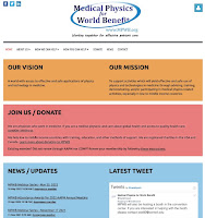
Screenshot of the website Medical Physics for World Benefit, www.mpwb.org.
Screenshot of the website
Medical Physics for World Benefit,
www.mpwb.org.
In the last two blog posts (here and here), I discussed the book True Tales of Medical Physics, edited by Jacob Van Dyk. Today, I want to bring to your attention something else I learned about when reading True Tales: the group Medical Physics for World Benefit.

[Jacob (Jake) Van Dyk] was the main founder of Medical Physics for World Benefit (www.MPWB.org), an organization devoted to supporting medical physics activities, largely by training and mentoring, especially for lower income settings.

The vision of this organization is to create

A world with access to effective and safe applications of physics and technology in medicine 

and its mission is

To support activities which will yield effective and safe use of physics and technologies in medicine through advising, training, demonstrating, and/or participating in medical physics-related activities, especially in low to middle income countries.

What’s there not to like? 

You can join or donate to the organization on its website.

We are physicists who work in medicine. If you are a medical physicist and care about global health and access to quality health care, consider joining us.

We help low to middle income countries with training, education, and other methods of support. We are registered charities in the USA and Canada. Learn more about donating to MPWB.

Too cheap to give? At least follow Medical Physics for World Benefit on Facebook or Twitter (@medphyswb).

Finally, I’m gonna give ya a tip. Many academic libraries subscribe to a package from Springer Publishing that lets institutional library members download pdf’s of Springer books for free. So, you can download Intermediate Physics for Medicine and Biology, True Tales of Medical Physics, and my recently published Are Electromagnetic Fields Making Me Ill? all at no charge. Then, take the money you saved and donate it to Medical Physics for World Benefit. 

Deal or no deal?

Friday, July 8, 2022

The Physics of Radiology

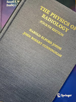
The Physics of Radiology, by Johns and Cunningham, superimposed on Intermediate Physics for Medicine and Biology.
The Physics of Radiology,
by Johns and Cunningham.
In last week’s blog post, I reviewed the recently published book True Tales of Medical Physics. A point I didn’t mention in my review was the central role of one textbook in the education of many of the authors who described their life story in True Tales. The Physics of Radiology was written by Harold Johns and John Cunningham. The first edition was published in 1953, but I have access through the Oakland University library to the fourth edition from 1983. This iconic book defined the field of medical physics and radiology in the second half of the twentieth century. Chapters 15–17 in Intermediate Physics for Medicine and Biology summarize material covered in more detail in The Physics of Radiology.

In True Tales, Jacob Van Dyk wrote
In 1971, I was hired by Professor Harold Elford Johns to work as a medical physicist at the Princess Margaret Hospital (PMH) in Toronto. Professor Jack Cunningham was my immediate boss. Professor Johns was considered the guru of Medical Physics with a world-renowned reputation for being a great scientist, a feared graduate student supervisor, and humanitarian. Over the years, he received multiple awards including five honorary doctorate degrees, and Officer of the Order of Canada. He was the first medical physicist to be inducted into the Canadian Medical Hall of Fame. Jack Cunningham also received the Officer of the Order of Canada along with multiple other awards, largely for his work on software development for computerized radiation treatment planning systems. Johns and Cunningham were the authors of The Physics of Radiology, the textbook which gave me my medical physics grounding as it did for all other young, aspiring medical physicists at that time….

[When Van Dyk was studying for his medical physics certification exams], Professors Johns and Cunningham were working on a draft of the fourth edition of their book, The Physics of Radiology. In January, I asked Jack Cunningham if I could review the available draft of this fourth edition. Considering that they would be contributors to the certification examination questions, my guess was that some, if not all, of the questions could be answered if I knew everything in this new edition. So, I went through this draft of the book from cover to cover and I solved (at least I worked on) every problem that was posed at the end of each chapter. As part of this process, I provided Jack with some comments on some questionable things that I found in the draft. As a result, my name was listed, along with others, in the acknowledgments when the book was published in 1983.
In his chapter of True Tales, Terry Peters wrote
During my early years at McGill, I became involved in the activities of the newly formed Canadian College of Physicists in Medicine (CCPM), which had begun the process of credentialling Medical Physicists in Canada. While I had Engineering, rather than Physics, training, I felt that my background had prepared me well for the roles I was playing in Diagnostic Radiology at the Neuro. Nevertheless, I felt it would do no harm to formally study radiation physics and its practical implementation in medicine, so in 1983 I embarked on a mission to devour The Physics of Radiology, by Johns and Cunningham, in preparation for the CCPM Fellowship exams in 1984. The examination process had evolved into an oral session, and a closed book examination—where three questions were selected from a previously published catalogue of questions covering all aspects of Medical Physics. Every Friday afternoon for almost a year I studied “Johns and Cunningham” with Gino Fallone, then a physicist at the Montreal General Hospital, who had also decided to take the certification examination. A gruelling process, but finally successful—we both became CCPM fellows that year.
Martin Yaffe's contribution to True Tales described a bit about Johns background and career.
Dr. Johns had been born in Chengdu, China to Canadian church missionary parents and he had spent his early years there, roaming about small communities in the mountains of Szechuan province with his father, a no-nonsense disciplinarian who believed strongly in devotion to duty and hard work. He learned to be focussed and driven to succeed at whatever was his mission. When the family eventually returned to Canada, he brought that to his graduate work in physics and later to the University of Saskatchewan where he built a strong medical physics research group, concentrating on developing and refining radiation therapy. There he developed the first (or possibly the second—there is some debate as his unit and a competitor, built by Eldorado Mining and Refining Ltd., were used to treat patients within a week or two of each other) cobalt-60 radiation treatment system and carried out pioneering work on radiation dosimetry and treatment planning. Dr. Johns and his work in Saskatchewan were actually mentioned in the film First Man, about the astronaut Neil Armstrong whose daughter had suffered from a brain tumour. Later, he began work on The Physics of Radiology, a textbook which he referred to jokingly (I think) as “The Bible”. This book truly became a guide to those working in radiation oncology all over the world and was published in multiple languages. While the book and its various editions consumed many of his evenings after a hard day at the lab, Johns reverse bragged that he earned about two cents per hour on his textbook writing efforts.

I once estimated that I make about ten cents an hour for my work on IPMB. I guess the difference represents inflation.

Yaffe also reminisced on Johns’ personality and mentoring technique.

Johns had an abrupt nature, not hesitating to poke you emphatically when he felt that you needed to think harder. Often, he would read your carefully written document, hold it up between you and slowly rip it to shreds before filing it in the trash bin. If it was late in the day, he would tell you to meet him the next morning at eight to re-write.

What I learned in those sessions was how to sharply focus your thinking on a problem and how to persist until you had a workable solution. Dr. Johns had two more senior students at the time—Aaron Fenster… and Don Plewes. These two and the lab technician, Dan Ostler, more or less adopted me and provided mentoring to prepare me for my sessions with Johns. Also, Johns used to invite me into his office when he was working on a paper with Aaron or Don and let me watch. While he was more respectful toward them, it was not uncommon for him to fix one of them with his laser-like glare which he held on them for what seemed like minutes and then say something like: “Plews” (he never pronounced the “e” that made it rhyme with Lewis; instead he made it rhyme with “news”), “If you sent that to a journal, they’d crap all over you”. Or, as he slowly ripped up a piece of writing that Aaron had proudly submitted, he’d say at a similar slow pace, “well (rrrrip) Fenster (rrrrip), your (rrrrip) writing (rrrrip) is improving.” So, rather than feel discriminated against, I simply realized that the standards were high, and I’d have to present my best game at all times.
The Physics of Radiology is one of those landmark textbooks (like Jackson’s Classical Electrodynamics in physics) that is a rite of passage for a student in that a field of study. As a coauthor on IPMB, I know what an honor it would be for your book to make that sort of impact. Johns and Cunningham was cited in the second edition of IPMB, but not in earlier or later editions.

The fourth edition is the last that Johns and Cunningham published. However, just last year an updated fifth edition was prepared by a team of five authors.

Friday, July 1, 2022

True Tales of Medical Physics: Insights into a Life-Saving Specialty

True Tales of Medical Physics: Insights into a Live-Saving Speciality, by Jacob Van Dyk, superimposed on Intermediate Physics for Medicine and Biology.
True Tales of Medical Physics:
Insights into a Live-Saving Specialty
,
by Jacob Van Dyk.
I’ve found the perfect book for readers of Intermediate Physics for Medicine and Biology who are fascinated by medical physics but who don’t want a lot of technical details and math. Jacob Van Dyk recently published True Tales of Medical Physics: Insights into a Life-Saving Specialty. The book consists of 22 chapters written by leading medical physicists, in which they each discuss their career, focusing on interesting anecdotes and life lessons. If I were a young student pondering what career to pursue, this is a book I’d want to read.

Van Dyk’s instructions to the contributors were
to communicate what medical physics is and what medical physicists do to a broad audience including science students, graduate students and residents, experienced medical physicists and their family members, and the general public who are wondering about medical physics. The book will consist of a series of short stories written by award-winning medical physicists—stories that are of personal interest as it relates to their careers. Each story will be unique to the author and could serve any one or more of the following purposes:
  1. Be an inspiration to young people searching for career directions, as well as more experienced physicists who are seeking direction on leadership development. 
  2. Provide an overview of what medical physicists do with a level of description that is understandable by the non-medical physicist.
  3. Provide lessons on life’s experiences from high-profile medical physicists who have significant experience and who are clearly at the top of the field as shown by the awards that they have won. 
  4. Be entertaining for those working in the field as well as others.
You can look at this book as a Plutarchian collection of comparative biographies, or you can focus on cross-cutting lessons that appear again and again in the various chapters. Here are some of the lessons I noticed.
  • The critical role of mentoring. All the authors stressed the importance of having supportive, inspirational mentors early in their career, and the satisfaction of mentoring their own students.
  • Crucial advances grow out of discussions at scientific meetings. Clearly the opportunity to travel and attend meetings is a part of a scientist’s career that’s highly valued, and often leads to new research directions and collaborations.
  • The division of their duties into three parts: research, teaching, and clinical work. The variety that arises from having these three different tasks keeps a medical physicist’s job from ever becoming dull or routine.
  • Leading scientific societies. The American Association of Physicists in Medicine (AAPM), the American Society for Radiation Oncology (ASTRO), the International Organization for Medical Physics (IOMP), and others professional groups are mentioned over and over by these authors.
  • The challenge of the American Board of Radiology (ABR) exams. Today, these certification exams act as a gateway to a career in clinical medical physics.
  • The interdisciplinary nature of medical physics. Many of these authors brought expertise from one field (say, computer programming) and integrated that knowledge with other fields (say, anatomy or medical imaging). 
  • Failures are opportunities. These scientists had their share of setbacks, but managed to overcome them and use them as springboards to success. They persisted.
  • The role of industry in medical physics research. Many authors tell stories of interacting with for-profit companies making imaging or therapy devices. Working with industry can be complicated and aggravating, but when successful the resulting products can have a huge impact on medical practice.
  • Science is an international activity. Many authors had collaborators and students from all over the world, leading to lifelong friendships.
True Tales of Medical Physics illustrates what a career in medical physics is like better than any textbook can (even IPMB!). Some of the authors worked on old, now obsolete devices—colbalt-60 radiation therapy, the betatron, film/screen cassettes—but they also helped develop today’s cutting edge technologies: computed tomography (CT), intensity-modulated radiation therapy (IMRT), magnetic resonance imaging (MRI), and medical ultrasound (US). These authors worked at leading medical institutions, such as the Memorial Sloan Kettering Cancer Center and the MD Anderson Cancer Center. Many were included in the IOMP’s 50th anniversary list of 50 medical physicists who have made an outstanding contribution to the advancement of medical physics over the last 50 years (I can’t help but compare this to the list of the 50 greatest basketball players prepared by the National Basketball Association on their 50th anniversary).

While I enjoyed all the chapters in True Tales, my favorite was by Marcel van Herk, a Steve Wozniak-like Dutch electronics guru. He started out as a 12-year-old hobbyist who built his own computer. Van Herk writes “One of the first things I did was to design and build a completely functional relay-based full adder (a circuit that can add two 4-bit binary numbers), soldered together while listening to Black Sabbath’s Iron Man in the living room, not totally to my mother’s liking due to the music and the spilled solder on the carpet. The parts I used were small electromechanical relays from 1950 punched card sorting machines, acquired cheaply.” He ended up developing software for the Elekta cone-beam CT imaging guidance system integrated with a medical linear accelerator. While his story is fascinating, it’s not uncommon; many of these scientists traveled individual, meandering paths into medical physics, taking them from a clueless neophyte to a giant in their field. A key lesson for students is that there’s no single route to success in science, and certainly not in medical physics.

If you are considering possible careers, I urge you to read True Tale of Medical Physics. It may change your life. You may change medicine.