Friday, November 9, 2018

Marie Curie and her X-ray Vehicles’ Contribution to World War I Battlefield Medicine

Sunday is Veterans Day. This year the holiday is particularly significant because it marks the 100th anniversary of the armistice ending World War I.

Marie Curie in a Mobile Military Hospital X-Ray Unit
Marie Curie in a Mobile Military Hospital X-Ray Unit.
Readers of Intermediate Physics for Medicine and Biology might wonder how the Great War influenced the application of physics to medicine. Timothy Jorgensen published a fascinating article on the website The Conversation discussing Nobel Prize-winner Marie Curie’s use of medical x-rays on the battlefield. Below are some annotated excerpts.
[In addition to her discovery of radium and polonium, Curie (1867-1934)] was also a major hero of World War I. In fact, a visitor to her Paris laboratory 100 years ago would not have found either her or her radium on the premises. Her radium was in hiding and she was at war.

The Guns of August,by Barbara Tuchman.
For Curie, the war started in early 1914, as German troops headed toward her hometown of Paris [“early” 1914? Germany declared war against France on August 3; see Guns of August by Barbara Tuchman]…[Curie] gathered her entire stock of radium, put it in a lead-lined container, transported it by train to Bordeaux…and left it in a safety deposit box at a local bank…

With the subject of her life’s work hidden far away…she decided to redirect her scientific skills toward the war effort; not to make weapons, but to save lives.

X-rays...had been discovered in 1895 by Curie’s fellow Nobel laureate, Wilhelm Roentgen.… Almost immediately after their discovery, physicians began using X-rays to image patients’ bones and find foreign objects – like bullets. But at the start of the war, X-ray machines were still found only in city hospitals, far from the battlefields where wounded troops were being treated. Curie’s solution was to invent the first “radiological car” – a vehicle containing an X-ray machine and photographic darkroom equipment – which could be driven right up to the battlefield where army surgeons could use X-rays to guide their surgeries....

[As the war progressed,] more radiological cars were needed. So Curie exploited her scientific clout to ask wealthy Parisian women to donate vehicles. Soon she had 20, which she outfitted with X-ray equipment. But the cars were useless without trained X-ray operators, so Curie started to train women volunteers. She recruited 20 women for the first training course, which she taught along with her daughter Irene [1897-1956, making her a teenager during much of the war], a future Nobel Prize winner herself….

Not content just to send out her trainees to the battlefront, Curie herself had her own “little Curie” [Petites Curies] – as the radiological cars were nicknamed – that she took to the front. This required her to learn to drive, change flat tires and even master some rudimentary auto mechanics, like cleaning carburetors. And she also had to deal with car accidents. When her driver careened into a ditch and overturned the vehicle, they righted the car, fixed the damaged equipment as best they could and got back to work [don’t you just love her?]....

Curie survived the war but was concerned that her intense X-ray work would ultimately cause her demise. Years later, she did contract aplastic anemia, a blood disorder sometimes produced by high radiation exposure. Many assumed that her illness was the result of her decades of radium work – it’s well-established that internalized radium is lethal [see The Radium Girls by Kate Moore]. But Curie was dismissive of that idea. She had always protected herself from ingesting any radium. Rather, she attributed her illness to the high X-ray exposures she had received during the war. (We will likely never know whether the wartime X-rays contributed to her death in 1934, but a sampling of her remains in 1995 showed her body was indeed free of radium.)
To learn more about Marie Curie, I recommend Jorgensen’s fine book Strange Glow: The Story of Radiation, or the article about Marie and her husband Pierre Curie and the discovery of polonium and radium, published by the Nobel Prize website. If you are a child at heart and enjoy Animated Hero Classic videos, watch this tearjerker.


To learn more about x-ray imaging, see Intermediate Physics for Medicine and Biology:
Chapter 16 describes the use of x rays for medical diagnosis and treatment. It moves from production to detection, to the diagnostic radiograph. We discuss image quality and noise, followed by angiography, mammography, fluoroscopy, and computed tomography. After briefly reviewing radiobiology, we discuss therapy and dose measurement. The chapter closes with a section on the risks from radiation.
To learn more about the First World War, visit the National World War I Museum and Memorial in Kansas City (know locally as Liberty Memorial).

National World War I Museum and Memorial in Kansas City
National World War I Museum and Memorial in Kansas City.
Happy Veterans Day all who have defended our country in the military (including my dad and my brother-in-law). Thank you for your service.

Friday, November 2, 2018

Roderick MacKinnon's Nobel Lecture

In Chapter 9 of Intermediate Physics for Medicine and Biology, Russ Hobbie and I write
Roderick MacKinnon and his colleagues determined the three-dimensional structure of a potassium channel using X-ray diffraction (Doyle et al. 1998; Jiang et al. 2003). MacKinnon received the 2003 Nobel Prize in Chemistry for his work on the potassium channel.
When I teach my graduate class on bioelectricity, we read the Doyle et al. article (“The Structure of the Potassium Channel: Molecular Basis of K+ Conduction and Selectivity,” Science, Volume 280, Pages 69–77, 1998). In my class, usually either my students and I discuss a paper or I explain some aspect of it. However, I’ve not found a better way to describe potassium channels than to watch MacKinnon’s brilliant Nobel lecture. I suggest you watch it too, using the embedded Youtube link below. It's 45 minutes long, but well worth the time.

If you have no time to spare, listen to the much shorter (less than two minute) interview where MacKinnon explains how being a scientist is like being an explorer.

Enjoy!

 
Roderick MacKinnon’s Nobel lecture.


“Being a scientist is like being an explorer.
 

Friday, October 26, 2018

Earl Bakken (1924-2018)

Earl Bakken (1924-2018)
Earl Bakken. From the Bakken Museum, via wikipedia.
Earl Bakken, cofounder of the medical device company Medtronic, died Sunday at the age of 94. In 1957 he developed the first external, battery-operated artificial pacemaker. Russ Hobbie and I don’t mention Bakken by name in Intermediate Physics for Medicine and Biology, but we do discuss pacemakers.
Cardiac pacemakers are a useful treatment for certain heart diseases. The most frequent are an abnormally slow pulse rate (bradycardia) associated with symptoms such as dizziness, fainting (syncope), or heart failure. These may arise from a problem with the SA node (sick sinus syndrome) or with the conduction system (heart block). One of the first uses of pacemakers was to treat complete or third degree heart block. The SA node and the atria fire at a normal rate but the wave front cannot pass through the conduction system. The AV node or some other part of the conduction system then begins firing and driving the ventricles at its own, pathologically slower rate…. A pacemaker stimulating the ventricles can be used to restore a normal ventricular rate.
In the 1950s, famed cardiac surgeon Dr. Walt Lillehei was at the University of Minnesota, where he met Bakken. Kirk Jeffrey tells the story of their collaboration in Machines In Our Hearts.
Machines In Our Hearts, by Kirk Jeffrey
The myocardial pacing wire was the first electrical device ever to be implanted in the human body and left there for a period of time. Surgeons at Minnesota were now able to pace children for days or weeks after heart surgery. By October 1957, they had used the technique with 18 patients. But Lillehei now grew uneasy about the Grass stimulator because it was bulky and plugged into the electrical system. The surgeon wanted to get his heart patients out of bed and moving around, but the stimulator had to accompany them on a wheeled cart. The electrical cord was a further nuisance. ‘‘Many of these [patients] were kids. They wanted to wander around and get active. Well, they were active. They couldn’t go any further than the cord. We had to string wires down the hall. . . . And then, if they needed an X ray or something that couldn’t be done in the room, you couldn’t get on the elevator so you had to string them down the stairwells. It seemed that almost everything you wanted was on a different floor. We needed something battery-operated.’’

From Machines In Our Hearts.
The plug-in stimulator was more than an inconvenience, for by introducing the myocardial pacing wire, Lillehei and his associates had connected the hearts of their surgical patients to the 110-volt electrical system of the hospital. Everyone in the program knew that an electrical surge might send patients into ventricular fibrillation or that a power outage could leave them without pacemaker support. On October 31, 1957, an equipment failure at a large Twin Cities power plant caused an outage lasting nearly three hours in Minneapolis. The University hospital had auxiliary power in its surgical suites and recovery area, but not in patients’ rooms. None of his heart patients died—but Lillehei viewed the event as a warning. Lillehei…turned to Earl Bakken…a young engineer who owned a small medical electronics business called Medtronic and repaired and serviced equipment for the Department of Surgery.

Bakken…realized that he could simply build a stimulator that used transistors and small batteries. ‘‘It was kind of an interesting point in history,’’ he recalled—‘‘a joining of several technologies.’’ In constructing the external pulse generator, Bakken borrowed a circuit design for a metronome that he had noticed a few months earlier in an electronics magazine for hobbyists. It included two transistors. Invented a decade earlier, the transistor was just beginning to spread into general use in the mid-1950s. Hardly anyone had explored its applications in medical devices. Bakken used a nine-volt battery, housed the assemblage in an aluminum circuit box, and provided an on-off switch and control knobs for stimulus rate and amplitude.

At the electronics repair shop that he had founded with his brother-in-law in 1949, Bakken had customized many instruments for researchers at the University of Minnesota Medical School…When Bakken delivered the battery-powered external pulse generator to Walt Lillehei in January 1958, it seemed to the inventor another special order, nothing more. The pulse generator was hardly an aesthetic triumph, but it was small enough to hold in the hand and severed all connection between the patient’s heart and the hospital power system. Bakken’s business had no animal-testing facility, so he assumed that the surgeons would test the device by pacing laboratory dogs. They did ‘‘a few dogs,’’ then Lillehei put the pacemaker into clinical use. When Bakken next visited the university, he was surprised to find that his crude prototype was managing the heartbeat of a child recovering from open-heart surgery.
The Bakken Museum. From wikipedia.
Russ Hobbie is retired from the University of Minnesota, and still lives in the area, so he is particularly familiar with Earl Bakken. He served on the board of the Bakken Museum, which is devoted to bioelectricity (my kind of museum). Russ say he ‘‘was impressed by Bakken's vision, energy, and desire to help people. The Bakken Museum has an extensive outreach program which does a lot of good things.’’ They recently posted a statement honoring their founder.

Medtronic is one of the largest medical device companies. I had a job interview there years ago, but I didn’t get the position. I came away impressed by the company, and wish I had bought stock.

Bakken is a member of the dwindling greatest generation; he was an airborne radar maintenance instructor during World War II. He had a long, full life, and we will miss him.

Below are a couple of videos about Earl Bakken. Enjoy!


Friday, October 19, 2018

The Bond Number

In graduate school, I did experiments on nerves submerged in saline. When dissecting or manipulating the tissue, I was amazed by the effect of surface tension compared to gravity; bubbles would stick to my forceps, and when I raised my scalpel out of the saline all kinds of stuff came along with it.

A dimensionless number describes this effect: the Bond number (sometimes called the Eötvös number). Several dimensionless numbers—such as the Reynolds number, the Peclet number, and the Lewis number—are mentioned in Intermediate Physics for Medicine and Biology. They indicate the relative importance of two physical mechanisms. Because they are dimensionless, their value does not depend on the units used. I find them useful.

The Bond number, Bo, is the ratio of the gravitational force mg (where m is the mass and g is the acceleration of gravity) to the force due to surface tension γL (where γ is the surface tension and L is some characteristic length such as the radius for a sphere), so Bo = mg/γL. If the Bond number is much greater than one, gravity dominates and surface tension is relatively insignificant. If the Bond number is much less than one, surface tension dominates and gravity hardly matters (like during my nerve experiments). When it’s close to one, both forces are similar.

The Bond number can help answer questions such as: Can animals stand on water? Insects can but people can’t. Why? Consider how the Bond number scales with size. An animal’s mass is equal to its density ρ times its volume, which varies as L3. In that case, the Bond number has an L3 in the numerator and an L in the denominator, so it scales as L2.

The Bond number
If an animal is large enough, gravity wins and it sinks.

Let's estimate the size of an animal when the Bond number is one.
The distance is equal to the square root of the surface tension divided by the density and the acceleration of gravity
Take g = 9.8 m/s2, ρ = 1000 kg/m3, and γ = 0.07 N/m (for water on earth). We find that L = 3 mm. For an insect, the good news is that it can stand on water. The bad news is that surface tension holds it in place so it can hardly move. As Steven Vogel says in Life in Moving Fluids: “Staying up is easy, but getting around is awkward.”

The Bond number is also useful for studying capillary action. Water will climb up the walls of a tube because of cohesion between the wall and the fluid. Technically, this effect depends on the nature of the surface as measured by its contact angle; you can coat a surface with a substance that repels water so that water climbs down the tube, but we will ignore all that and assume the contact angle is so small that the water “wets” the surface and cohesion is the same as surface tension. The gravitational force of the climbing water is gρπR2H, where R is the tube radius and H is the height that the water climbs. The surface tension force is γ times the circumference of the tube, 2πR. The Bond number becomes gρRH/2γ. We expect water to rise until gravity and surface tension are in balance, or until about Bo = 1. The height is then H = 2γ/gρR. For a 1 mm radius tube, the height is 14 mm, or roughly half an inch.

Can capillary action explain the ascent of sap in trees? The tubes in a tree's xylem have a radius of about 20 microns. In that case, water will rise about 0.6 meters. That’s about two feet, which doesn’t explain how water gets up a 100-m giant redwood. Suffice to say, the trunk of a redwood tree does not act as a giant wick, sucking water up its trunk by capillary action.

Wilfrid Noel Bond
Wilfrid Noel Bond, from Wikipedia.
The Bond number is named after the English physicist Wilfrid Noel Bond (1897-1937). He studied fluid dynamics, including viscosity and surface tension. He died tragically early at age 39.

If you want to learn more about the Bond number and other dimensionless numbers, see Vogel’s 1998 article in Physics Today about “Exposing Life’s Limits with Dimensionless Numbers.”

Friday, October 12, 2018

A Trick to Generate Exam Problems

Intermediate Physics for Medicine and Biology.
When teaching a class based on Intermediate Physics for Medicine and Biology, instructors need to write problems for their exams. My goal in this post is to explain a trick for creating good exam problems. 

One of my favorite homework problems in IPMB is from Chapter 4.
Problem 37. The goal of this problem is to estimate how large a cell living in an oxygenated medium can be before it is limited by oxygen transport. Assume the extracellular space is well stirred with uniform oxygen concentration C0. The cell is a sphere of radius R. Inside the cell oxygen is consumed at a rate Q molecule m−3 s−1. The diffusion constant for oxygen in the cell is D.
(a) Calculate the concentration of oxygen in the cell in the steady state.
(b) Assume that if the cell is to survive the oxygen concentration at the center of the cell cannot become negative. Use this constraint to estimate the maximum size of the cell.
(c) Calculate the maximum size of a cell for C0 = 8 mol m−3, D = 2 x 10⁻⁹ m2 s−1, Q = 0.1 mol m−3 s−1. (This value of Q is typical of protozoa; the value of C0 is for air and is roughly the same as the oxygen concentration in blood.)
Homework problems for Chapter 4 in Intermediate Physics for Medicine and Biology.
Homework problems for Chapter 4 in
Intermediate Physics for Medicine and Biology.
I usually work this problem in class. Not only do students practice solving the steady-state diffusion equation, but also they estimate the maximum size of a cell from some basic properties of oxygen. In the Solution Manual—available to instructors only (email us)—we explain the purpose of each problem in a preamble. Here is what the solution manual says about Problem 37:
This important “toy model” considers the maximum size of a spherical cell before its core dies from lack of oxygen. One goal of biological physics is to show how physics constrains evolution. In this case, the physics of diffusion limits how large an animal can be before needing a circulatory system to move oxygen around.
How do you create an exam problem on this subject? Here's the trick: Do Problem 37 in class and then put a question on the exam identical to Problem 37 except “sphere” is replaced by “cylinder”. The problem is only slightly changed; just enough to determine if the student is solving the problem from first principles or merely memorizing. In addition, nerve and muscle fibers are cylindrical, so the revised problem may provide an even better model for those cells. Depending on the mathematical abilities of your students, you may need to provide students with the Laplacian in cylindrical coordinates. (If the exam is open book then they can find the Laplacian in Appendix L).

Here’s a second example: Chapter 1 considers viscous flow in a tube; Poiseuille flow. On the exam, ask the student to analyze viscous flow between two stationary plates.
Section 1.17
Problem 36 ½. Consider fluid flow between two stationary plates driven by a pressure gradient. The pressure varies in the x direction with constant gradient dp/dx, the plates are located at y = +L and y = -L, and the system is uniform in the z direction with width H. The fluid has viscosity η.

(a) Draw a picture the geometry.
(b) Consider a rectangular box of fluid centered at the origin and derive a differential equation like Eq. 1.35 governing the velocity vx(y).

(c) Solve this differential equation to determine vx(y), analogous to Eq. 1.37. Assume a no-slip boundary condition at the surface of each plate. Plot vx(y) versus y.

(d) Integrate the volume fluence and find the total flow i. How does i depend on the plate separation, 2L? How does this compare with the case of flow in a tube?
An interesting feature of this example is that i depends on the third power of L, whereas for a tube it depends on the fourth power of the radius. Encourage the student to wonder why.

Third example: A problem in Chapter 7 compares three different functions describing the strength-duration curve for electrical stimulation. On your exam, have the students analyze a fourth case.
Section 7.10

Problem 46 ½. Problem 46 analyzes three possible functions that could describe the strength-duration curve, relating the threshold current strength required for neural excitation, i, to the stimulus pulse duration, t. Consider the function i = A/tan-1(t/B). Derive expressions for the rheobase iR and chronaxie tC in terms of A and B. Write the function in the form used in Problem 46. Plot i versus t.
And still more: Problem 32 in Chapter 8 examines magnetic stimulation of a nerve axon using an applied electric field Ei(x) = E0 a2/(x2 + a2). Give a similar problem on your exam but use a different electric field, such as Ei(x) = E0 exp(-x2/a2).

And yet another: Chapter 10 examines the onset of cardiac fibrillation and chaos. The action potential duration APD is related to the diastolic interval (time from the end of the previous action potential to the start of the following one) DI by the restitution curve. Have the student repeat Problem 41 but using a different restitution curve: APDi+1 = 300 DIi/(DIi + 100).

Final example: Problem 36 in Chapter 9 asks the student to calculate the electrical potential inside and outside a spherical cell in the presence of a uniform electric field (Figure 9.19). On your exam, make the sphere into a cylinder.

I think you get the point. On an exam, repeat one the of homework problems in IPMB, but with a twist. Change the problem slightly, using a new function or a modified geometry. You will be able to test the knowledge and understanding of the student without springing any big surprises on the exam. Many problems in IPMB that could be modified in this way.

Warning: This trick doesn’t always work. For instance, in Chapter 1 if you try to analyze fluid flow perpendicular to a stationary object, you run into difficulties when you change the sphere of Problem 46 into a cylinder. The cylindrical version of this problem has no solution! The lack of a solution for low Reynolds number flow around a cylinder is known as Stokes’ Paradox. In that case, you’re just going to have to think up your own exam question.

Friday, October 5, 2018

A Bidomain Model for the Extracellular Potential and Magnetic Field of Cardiac Tissue

My research notebooks from graduate school.
In graduate school, I worked with John Wikswo measuring the magnetic field of a nerve axon. We isolated a crayfish axon and threaded it through a wire-wound ferrite-core toroid immersed in saline. As the action currents propagated by, they produced a changing magnetic field that induced a signal in the toroid by Faraday induction. Ampere’s law tells us that the signal is proportional to the net current through the toroid, which is the sum of the intracellular current and the fraction of the current in the saline that passes through the toroid, called the return current.

For my PhD dissertation, Wikswo had me make similar measurements on strands of cardiac tissue, such as a papillary muscle. The instrumentation was the same as for the nerve, but the interpretation was different. Now the signal had three sources: the intracellular current, the return current in the saline, and extracellular current passing through the interstitial space within the muscle called the “interstitial return current.” Initially neither Wikswo nor I knew how to calculate the interstitial return current, so we were not sure how to interpret our results. As I planned these experiments, I recorded my thoughts in my research notebook. The July 26, 1984 entry stressed that “Understanding this point [the role of interstitial return currents] will be central to my research and deserves much thought.”

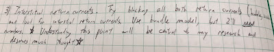
Excerpt from the July 26, 1984 entry in my Notebook 8, page 64.
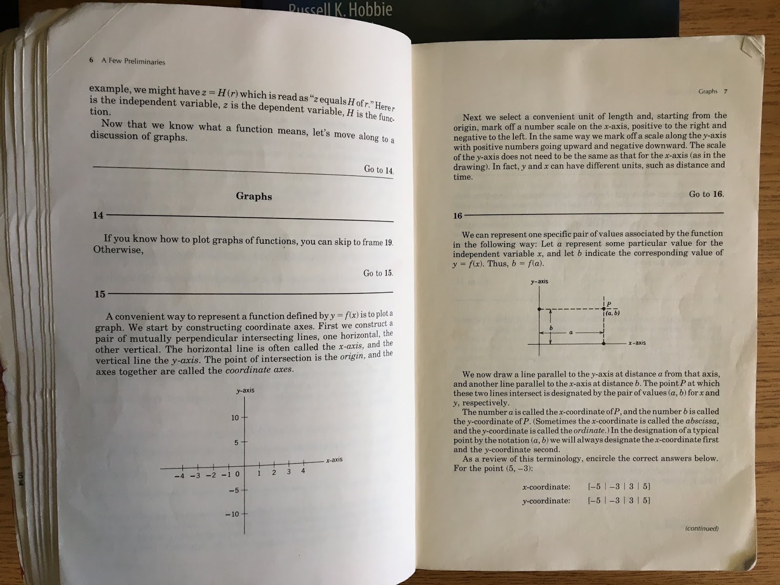
When working on nerves, I had studied articles by Robert Plonsey and John Clark, in which they calculated the extracellular potential in the saline from the measured voltage across the axon’s membrane: the transmembrane potential. I was impressed by this calculation, which involved Fourier transforms and Bessel functions (see Chapter 7, Problem 30, in Intermediate Physics for Medicine and Biology). I had used their result to calculate the magnetic field around an axon (see Chapter 8, Problem 16 in IPMB), so I set out to extend their analysis again to include interstitial return currents.

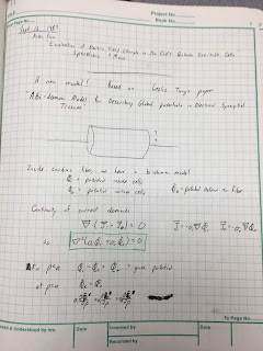
Page 1 of Notebook 9, from Sept 13, 1984.
The key was to use the then-new bidomain model, which accounts for currents in both the intracellular and interstitial spaces. The crucial advance came in September 1984 after I read a copy of Les Tung’s PhD dissertation that Wikswo had loaned me. After four days of intense work, I had solved the problem. My results looked a lot like those of Clark and Plonsey, except for a few strategically placed additional factors and extra terms.
Excerpt from Notebook 9, page 13, the Sept 16, 1984 entry.
First page of Roth and Wikswo (1986) IEEE Trans. Biomed. Eng., 33:467–469.
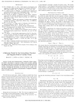
First page of Roth and Wikswo (1986).
Wikswo and I published these results as a brief communication in the IEEE Transactions on Biomedical Engineering.
Roth, B. J. and J. P. Wikswo, Jr., 1986, A bidomain model for the extracellular potential and magnetic field of cardiac tissue. IEEE Trans. Biomed. Eng., 33:467-469.

Abstract—In this brief communication, a bidomain volume conductor model is developed to represent cardiac muscle strands, enabling the magnetic field and extracellular potential to be calculated from the cardiac transmembrane potential. The model accounts for all action currents, including the interstitial current between the cardiac cells, and thereby allows quantitative interpretation of magnetic measurements of cardiac muscle.
Rather than explain the calculation in all its gory detail, I will ask you to solve it in a new homework problem.
Section 7.9
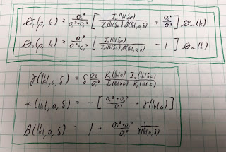
Problem 31½. A cylindrical strand of cardiac tissue, of radius a, is immersed in a saline bath. Cardiac tissue is a bidomain with anisotropic intracellular and interstitial conductivities σir, σiz, σor, and σoz, and saline is a monodomain volume conductor with isotropic conductivity σe. The intracellular and interstitial potentials are Vi and Vo, and the saline potential is Ve.
a) Write the bidomain equations, Eqs. 7.44a and 7.44b, for Vi and Vo in cylindrical coordinates (r,z). Add the two equations.
b) Assume Vi = σiz/(σiz+σoz) [A I0(kλr) + (σoz/σiz) Vm] sin(kz) and Vo = σiz/(σiz+σoz) [A I0(kλr) - Vm] sin(kz) where λ2 = (σiz+σoz)/(σir+σor). Verify that Vi - Vo equals the transmembrane potential Vm sin(kz). Show that Vi and Vo obey the equation derived in part a). I0(x) is the modified Bessel function of the first kind of order zero, which obeys the modified Bessel equation d2y/dx2 + (1/x) dy/dx = y. Assume Vm is independent of r.
c) Write Laplace’s equation (Eq. 7.38) in cylindrical coordinates. Assume Ve = B K0(kr) sin(kz). Show that Ve satisfies Laplace’s equation. K0(x) is the modified Bessel function of the second kind of order zero, which obeys the modified Bessel equation.
d) At r = a, the boundary conditions are Vo = Ve and σirVi/∂r + σorVo/∂r = σeVe/∂r. Determine A and B. You may need the Bessel function identities dI0/dx = I1 and dK0/dx = - K1, where I1(x) and K1(x) are modified Bessel functions of order one.
In the problem above, I assumed the potentials vary sinusoidally with z, but any waveform can be expressed as a superposition of sines and cosines (Fourier analysis) so this is not as restrictive as it seems.

In part b), I assumed the transmembrane potential was not a function of r. There is little data supporting this assumption, but I was stuck without it. Another assumption I could have used was equal anisotropy ratios, but I didn’t want to do that (and initially I didn’t realize it provided an alternative path to the solution).

The calculation of the magnetic field is not included in the new problem; it requires differentiating Vi, Vo, and Ve to find the current density, and then integrating the current to find the magnetic field via Ampere’s law. You can find the details in our IEEE TBME communication.

Some of you might be thinking “this is a nice homework problem, but how did you get those weird expressions for the intracellular and interstitial potentials used in part b)”? Our article gives some insight, and my notebook provides more. I started with Clark and Plonsey’s result, used ideas from Tung’s dissertation, and then played with the math (trial and error) until I had a solution that obeyed the bidomain equations. Some might call that a strange way to do science, but it worked for me.

I was very proud of this calculation (and still am). It played a role in the development of the bidomain model, which is now considered the state-of-the-art model for simulating the heart during defibrillation.

Wikswo and I carried out experiments on guinea pig papillary muscles to test the calculation, but the cardiac data was not as clean and definitive as our nerve data. We published it as a chapter in Cell Interactions and Gap Junctions (1989). The cardiac work made up most of my PhD dissertation. Vanderbilt let me include our three-page IEEE TBME communication as an appendix. It’s the most important three pages in the dissertation.


Coda: While browsing through my old research notebooks, I found this gem passed from Prof. Wikswo to his earnest but naive graduate student, who dutifully wrote it down in his research notebook for posterity.

Excerpt from Notebook 10, Page 62, January 3, 1985.
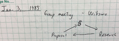
Excerpt from Notebook 10, Page 62.

Friday, September 28, 2018

Steven Strogatz Lectures on Youtube

Nonlinear Dynaics and Chaos, by Steven Strogatz
Nonlinear Dynamics and Chaos.
Previously (here, here, and here), I’ve written about Steven Strogatz, Professor of Applied Mathematics at Cornell University. Strogatz wrote one of my favorite textbooks: Nonlinear Dynamics and Chaos. Russ Hobbie and I cite it in Chapter 10 of Intermediate Physics for Medicine and Biology.

Nowadays students rarely read textbooks; they prefer watching videos. Well, I have good news. Strogatz taught a course based on his book, and his lectures are posted on YouTube. You can learn chaos straight from the horse’s mouth. You better get started: there are over 24 hours of video (all embedded below).

The second edition of Nonlinear Dynaics and Chaos, by Steven Strogatz
The 2nd Edition of Nonlinear Dynamics and Chaos.
Strogatz is an enormously successful mathematician. According to Google Scholar, his 1998 article with Duncan Watts—“Collective Dynamics of ‘Small-World’ Networks” (Nature, Volume 393, Pages 440-442)—has been cited over 35,000 times! His textbook has over ten thousand citations. (To put this in perspective, IPMB has 392.) He won the Lewis Thomas Prize for Writing about Science, tweets at @stevenstrogatz, and is buddies with M*A*S*H star and science communicator Alan Alda. In IPMB, Russ and I cite the first edition of Nonlinear Dynamics and Chaos, but Strogatz published a second edition in 2015.

Enjoy!

 
1. Introduction and Overview

2. One Dimensional Systems

3. Overdamped Bead on a Rotating Hoop

4. Model of Insect Outbreak

5. Two Dimensional Nonlinear Systems

6. Two Dimensional Nonlinear Systems Fixed Points

7. Conservative Systems

8. Index Theory and Introduction to Limit Cycles

9. Testing for Closed Orbits

10. Van der Pol Oscillator

11. Averaging Theory for Weakly Nonlinear Oscillators

12. Bifurcations in Two Dimensional Systems

13. Hopf Bifurcations in Aeroelastic Instabilities and Chemical Oscillators

14. Global Bifurcations of Cycles

 15. Chaotic Waterwheel

16. Waterwheel Equations and Lorenz Equations

17. Chaos in the Lorenz Equations

18. Strange Attractor for the Lorenz Equations

19. One Dimensional Maps

20. Universal Aspects of Period Doubling

21. Feigenbaum’s Renormalization Analysis of Period Doubling

22. Renormalization: Function Space and a Hands-on Calculation

23. Fractals and the Geometry of Strange Attractors

24. Henon Map

25. Using Chaos to Send Secret Messages

Friday, September 21, 2018

Quick Calculus

In Intermediate Physics for Medicine and Biology, Russ Hobbie and I assume the reader knows calculus. Some readers, however, have weak or rusty math skills. Is there an easy way to learn what is needed?

Quick Calculus is a self-teaching guide written by Daniel Klepner and Norman Ramsey
Quick Calculus.
Yes! Quick Calculus is a self-teaching guide written by Daniel Klepner and Norman Ramsey. Their preface states:
Before you plunge into Quick Calculus, perhaps we ought to tell you what it is supposed to do. Quick Calculus should teach you the elementary techniques of differential and integral calculus with a minimum of wasted effort on your part; it is designed for you to study by yourself. Since the best way for anyone to learn calculus is to work problems, we have included many problems in this book. You will always see the solution to your problem as soon as you have finished it, and what you do next will depend on your answer. A correct answer generally sends you to new material, while an incorrect answer sends you to further explanations and perhaps another problem.
The book covers nearly all the calculus needed in IPMB.
  • Chapter One reviews functions and graphs, emphasizing trigonometry, exponentials, and logarithms.
  • Chapter Two discusses differentiation—including the product rule and the chain rule—and maximum/minimum problems.
  • Chapter Three analyzes integration, both definite and indefinite, and covers techniques such as change of variable, integration by parts, and multiple integrals.
  • Chapter Four summarizes all the results in a few pages.
Math Book useful for Intermediate Physics for Medicine and Biology
Math Books Useful for IPMB.
The only calculus in IPMB that Quick Calculus doesn’t teach is vector calculus; for that you should consult Div, Grad, Curl and All That. Used Math covers more ground than Quick Calculus, but it’s a handbook rather than a self-teaching guide.

Quick Calculus has several virtues. It is clearly written, it emphasizes understanding math visually with lots of plots, and it focuses on utilitarian techniques without distracting rigor. If you want to understand math at a fundamental level, you should take a real calculus class. If you want to brush up on what's needed to get through IPMB, use Quick Calculus.

One disadvantage is that Quick Calculus is old. The second edition—the most recent one I am aware of—was published in 1985. It might be difficult to purchase, although Amazon seems to have copies for sale. The authors make quaint comments about “readers who have an electronic calculator,” as opposed to slide rules I suppose. I also found several typos, which might frustrate readers using the book for self-study.

A sample from Quick Calculus
A sample from Quick Calculus.
The format is unusual. The text is divided into approximately half-page “frames,” and the reader is guided from one frame to the next. Someone should put this book online, because it would lend itself to an interactive online format. Rather than explain how the book is organized, I’ve taken Section 1.17 of IPMB and rewritten it in the style of Quick Calculus (see below). In my opinion, if all of Intermediate Physics for Medicine and Biology were organized like this it would be tedious. What do you think?


Friday, September 14, 2018

Gulliver was a Bad Biologist

Gulliver's Travels
Gulliver’s Travels by Jonathan Swift.
Most of my reading is nonfiction, but recently I read Jonathan Swift’s Gulliver’s Travels. The story describes Englishman Lemuel Gulliver’s journeys to exotic lands, including Lilliput inhabited by tiny people, and Brobdingnag where giants live. Swift was a delightful and funny writer, but Florence Moog claims “Gulliver was a Bad Biologist” (Scientific American, Volume 179, November 1948, Pages 52–55). The problem is scaling, which Russ Hobbie and I discuss in Chapter 2 of Intermediate Physics for Medicine and Biology. The properties of animals change as they get bigger or smaller; you can’t just scale people up or down and expect them to function correctly. As Moog writes “for a student of comparative biology Gulliver’s book may serve as an unpremeditated textbook on biological absurdities.”

Gulliver was a Bad Biologist
“Gulliver was a Bad Biologist,” by Florence Moog.
Moog’s first example was the 60-foot tall Brobdingnagians. She notes that because their mass increases as the cube of their height, supporting their body would “necessitate a truly ponderous skeleton” (A point I’ve discussed before in this blog when contemplating elephants). The giants would need thick stubby legs and fat bones.

Gulliver's Travels Title Page
Title Page of Gulliver’s Travels.
Moog then considers the six-inch-tall Lilliputians. “If the Brobdingnagians were too big to exist, the mouse-sized Lilliputians were too small to be human.” She explains that smaller animals have a higher specific metabolic rate (that is, rate per unit mass) than larger animals. “Gulliver … failed to realize that the creatures of his invention would have spent the larger part of their time stuffing themselves with food.”

Why was I reading Gulliver’s Travels? Blame Neil deGrasse Tyson. The Public Broadcasting System is sponsoring the Great American Read this summer, where we vote for our favorite of one hundred famous books. In their Launch Special, various celebrities select their personal favorite, and Tyson—one of the few scientists featured on the special—chose Gulliver. Apparently he hasn’t studied Chapter 2 of IPMB. Regular readers of this blog know that I am a fan of Isaac Asimov, and I have been voting for his Foundation Series twice a day (once using the Firefox browser, and once using Safari) all summer.

Neil deGrasse Tyson likes Gulliver's Travels
Neil deGrasse Tyson discussing Gulliver’s Travels.
Maybe Tyson has a point. Moog concludes that “after all, we must not be too hard on Gulliver for failing to understand the biological conditions that made him a man—and an implausible liar. His talents … were in the psychological realm.” His satirical story provides great insight into human behavior.

Friday, September 7, 2018

Microwave Weapons are Prime Suspect in Ills of U.S. Embassy Workers

Last Saturday, The New York Times published an article by Pulitzer Prize-winning science writer William Broad with the headline “Microwave Weapons are Prime Suspect in Ills of U.S. Embassy Workers.”
Doctors and scientists say microwave strikes may have caused sonic delusions and very real brain damage among embassy staff and family members.
The article has made quite a splash; I even heard about it on the news.


This topic is relevant to Intermediate Physics for Medicine and Biology, so Ill address it in this post. I hesitate, however, because the science is uncertain and the topic of electromagnetic effects on health is fraught with conspiracy theories and voodoo science. Yet, the issue has more than academic importance; U.S.-Cuban relations suffered because of these unexplained health effects. So, reluctantly, I wade in.
I begin with a report from last March in the prestigious Journal of the American Medical Association (JAMA) by Swanson et al. about “Neurological Manifestations Among US Government Personnel Reporting Directional Audible and Sensory Phenomena in Havana, Cuba” (Volume 319, Pages 1125–1133).
  • Question: Are there neurological manifestations associated with reports of audible and sensory phenomena among US government personnel in Havana, Cuba? 
  • Findings: In this case series of 21 individuals exposed to directional audible and sensory phenomena, a constellation of acute and persistent signs and symptoms were identified, in the absence of an associated history of blunt head trauma. Following exposure, patients experienced cognitive, vestibular, and oculomotor dysfunction, along with auditory symptoms, sleep abnormalities, and headache. 
  • Meaning: The unique circumstances of these patients and the consistency of the clinical manifestations raised concern for a novel mechanism of a possible acquired brain injury from a directional exposure of undetermined etiology.
The articles claim of cognitive dysfunction has been hotly debated. A post in the blog Neuroskeptic was….er….skeptical. It concludes
Overall … the JAMA paper is pretty weak. Clearly, something has happened to make these 21 people experience so many unpleasant symptoms, but at present I don’t think we can rule out the possibility that the cause is psychological in nature.
Last weeks New York Times article was triggered by the recently proposed hypothesis that microwaves are responsible for these health issues. Russ Hobbie and I discuss the biological effects of electric and magnetic fields in Section 9.10 of IPMB. We focus on the potential of microwaves to induce tumors, and conclude that nonthermal mechanisms are implausible. In other words, radiofrequency fields can heat tissue—just like in your microwave oven—but they don’t cause cancer. The hypothesis touted in the Times article, however, is a thermal mechanism: a thermoelastic pressure wave sensed as sound by part of the inner ear called the cochlea.

Hearing induced by microwaves has been studied for years, and is known as the “Frey effect” after Allen Frey, who first reported it. A 2007 article in the journal Health Physics by James Lin and Zhangwei Wang (Volume 92, Pages 621-628) describes this phenomenon.
Hearing of Microwave Pulses by Humans and Animals: Effects, Mechanism, and Thresholds

The hearing of microwave pulses is a unique exception to the airborne or bone-conducted sound energy normally encountered in human auditory perception. The hearing apparatus commonly responds to airborne or bone-conducted acoustic or sound pressure waves in the audible frequency range. But the hearing of microwave pulses involves electromagnetic waves whose frequency ranges from hundreds of MHz to tens of GHz. Since electromagnetic waves (e.g., light) are seen but not heard, the report of auditory perception of microwave pulses was at once astonishing and intriguing. Moreover, it stood in sharp contrast to the responses associated with continuous-wave microwave radiation. Experimental and theoretical studies have shown that the microwave auditory phenomenon does not arise from an interaction of microwave pulses directly with the auditory nerves or neurons along the auditory neurophysiological pathways of the central nervous system. Instead, the microwave pulse, upon absorption by soft tissues in the head, launches a thermoelastic wave of acoustic pressure that travels by bone conduction to the inner ear. There, it activates the cochlear receptors via the same process involved for normal hearing. Aside from tissue heating, microwave auditory effect is the most widely accepted biological effect of microwave radiation with a known mechanism of interaction: the thermoelastic theory. The phenomenon, mechanism, power requirement, pressure amplitude, and auditory thresholds of microwave hearing are discussed in this paper. A specific emphasis is placed on human exposures to wireless communication fields and magnetic resonance imaging (MRI) coils.
Their introduction gives some useful numbers.
The microwave auditory phenomenon or microwave hearing effect pertains to the hearing of short-pulse, modulated microwave energy at high peak power by humans and laboratory animals (Frey 1961, 1962; Guy et al.1975a, b; Lin 1978, 1980, 2004). The effect can arise, for example, at an incident energy density threshold of 400 mJ m-2 for a single, 10-µs-wide pulse of 2,450 MHz microwave energy, incident on the head of a human subject (Guy et al. 1975a, b; Lin 1978). It has been shown to occur at a specific absorption rate (SAR) threshold of 1.6 kW kg-1 for a single 10-µs-wide pulse of 2,450 MHz microwave energy. A single microwave pulse can be perceived as an acoustic click or knocking sound, and a train of microwave pulses to the head can be sensed as an audible tune, with a pitch corresponding to the pulse repetition rate (Lin 1978).
The temperature increase caused by such a microwave pulse is rapid (microseconds) and tiny (microdegrees Celsius), and the associated pressure is small (tenths of a Pascal, or equivalently millionths of an atmosphere). People can hear these sounds because the cochlea is so sensitive.

One reason that microwaves might be a more plausible mechanism than sound waves for the apparent embassy attacks is acoustic impedance, discussed in Chapter 13 of IPMB. Air and water have very different impedances. When a sound wave impinges on a person, most of the acoustic energy is lost by reflection, and little (perhaps one part in a thousand) enters the fluid-filled body. Animals have evolved elaborate structures in the middle ear to mitigate this acoustic mismatch. However, a pressure wave caused by microwave heating originates inside the ear. No energy is lost by sound reflecting from the air-tissue interface.

I am no expert on thermoelastic effects, but it seems plausible that they could be responsible for the perception of sound by embassy workers in Cuba. By modifying the shape and frequency of the microwave pulses, you might even induce sounds more distinct than vague clicks. However, I don’t know how you get from little noises to brain damage and cognitive dysfunction. My brain isn’t damaged by listening to clicky sounds. Either there is more to this that I don’t understand, or—as neuroskeptic speculates—the rest of the cause is “psychological in nature.”

Right now, our country could use a hard-nosed scientist or engineer expert in the bioeffects of microwave radiation to look into this problem. Where have you gone John Moulder and Ken Foster? We need you!