The
200th anniversary of the birth of
Charles Dickens occurs this week (he was born February 7, 1812). I am a big Dickens fan, so I had to fit him into this week’s blog entry somehow. It is not easy, since there is little overlap between Dickens’ novels and the 4th edition of
Intermediate Physics for Medicine and Biology. But let us try.
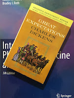 |
Great Expectations,
by Charles Dickens. |
Dickens’ life spanned an incredibly productive era of
Victorian science in England. He was born the same year as English Chemist
Humphry Davy published his
Elements of Chemical Philosophy. Dickens’ birth fell almost exactly halfway between the births of the two greatest of 19th century British physicists:
Michael Faraday (September 1791) and
James Maxwell (November 1831). Just as Maxwell was publishing his eponymous
Maxwell’s equations, Dickens was publishing
Great Expectations. The physician
John Snow was born one year after Dickens. It was Snow who famously traced the source of the
1854 cholera epidemic to the
Broad Street pump in London (read more about this story in
The Ghost Map by
Steven Johnson). Dickens was born just two years after the birth of
Charles Darwin, and
On The Origin of Species appeared almost simultaneously with Dickens’ masterpiece
A Tale of Two Cities.
William Thomson (Lord Kelvin) was 12 years younger than Dickens. He formulated his version of the
second law of thermodynamics in 1851, soon after Dickens published
David Copperfield.
 |
David Copperfield,
by Charles Dickens. |
A young Charles was working long hours at Warren’s Blacking Warehouse when the first issue of the British medical journal
The Lancet appeared in 1823. Dickens published his first story the year after Faraday proposed his vision of electric and magnetic fields, and he got married and published his first novel (
The Pickwick Papers) in 1836, the year Darwin returned from his
voyage on the Beagle.
Martin Chuzzlewit came out in 1843, the same year
James Joule determined the
mechanical equivalent of heat. Dickens traveled to France and Italy the year that the prominent English chemist
John Dalton died in England. His last complete novel,
Our Mutual Friend, appeared in 1865, the year before the
first transatlantic telegraph cable was laid (for more about this fascinating story, read
A Thread Across the Ocean by John Gordon). Kelvin developed the
cable equation to govern the transmission of signals over this telegraph line, and the same equation is used nowadays to describe nerve axons. An elderly Dickens came to the United States for a reading tour in 1867, the year English surgeon
Joseph Lister pioneered the use of
antiseptic to sterilize surgical instruments. Charles Dickens died of a stroke in 1870—leaving the
Mystery of Edwin Drood unfinished—just a few months before the birth of the greatest experimental physicist since Faraday,
Ernest Rutherford.
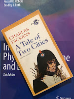 |
A Tale of Two Cities,
by Charles Dickens. |
The medical literature contains several studies of how medicine was portrayed in Dickens’ books. Howard Markel writes about “
Charles Dickens and the Art of Medicine” in the
Annals of Internal Medicine (Volume 101, Pages 408–411, 1984).
Charles Dickens, the novelist, humanist, and social reformer, was a keen observer of all the characteristics of the people in his novels. Dickens observed physicians and visited hospitals so that he could record various illnesses and diseases of people he met during his life. Dickens also worked for many public health reforms in Victorian England. The author used his observations of sick people in many of his novels and produced several accurate descriptions of disease, including Ménière's disorder and acute leukemia.
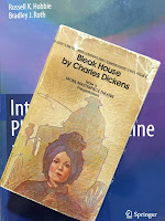 |
Bleak House,
by Charles Dickens. |
The article analyzes how, in
Bleak House, Phil Squod (who was always “shoulding his way along walls”) demonstrated symptoms consistent with “dysfunction of the vestibular nerve…most likely Meniere’s disorder.” In
Dombey and Son, the symptoms of young Paul Dombey “resemble those of a child with an acute form of leukemia.”
Kerrie Schoffer and John O’Sullivan focus on
movement disorders in their
study of Charles Dickens in the
Journal of Clinical Neuroscience (Volume 13, Pages 898–901, 2006)
Nineteenth-century Victorian novelists played an important role in developing our understanding of medicine and illness. With the eye of an expert clinician, Charles Dickens provided several detailed accounts of movement disorders in his literary works, many of which predated medical descriptions. His gift for eloquence, imagery, and precision attest not only to the importance of careful clinical observation, but also provide an insightful and entertaining perspective on movement disorders for modern students of neuroscience.
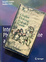 |
Pickwick Papers,
by Charles Dickens. |
So is Dickens a medical physicist? I guess not. But he was a great writer. My favorite Dickens book is
A Christmas Carol; I read it every Christmas. I reread
A Tale of Two Cities during my
Paris trip two years ago (“…It is a far, far better thing that I do, than I have ever done; it is a far, far better rest that I go to than I have ever known.”). I enjoyed
Bleak House a few years ago, although it took me a long time to plow through that 818 page tome. I love Dickens’ characters, like the
Artful Dodger in
Oliver Twist, and
Wilkins Micawber in
David Copperfield. What will be my next Dickens book? I haven’t read
Nicholas Nickleby yet; I think it will be next.


