Friday, May 31, 2019

Introduction to Radiological Physics and Radiation Dosimetry

Introduction to Radiological Physics and Radiation Dosimetry, by Frank Herbert Attix, superimposed on Intermediate Physics for Medicine and Biology.
Introduction to Radiological Physics
and Radiation Dosimetry
,
by Frank Herbert Attix.
In Chapters 15 and 16 of Intermediate Physics for Medicine and Biology, Russ Hobbie and I often cite Introduction to Radiological Physics and Radiation Dosimetry by Frank Herbert Attix. This book, published in 1986, is an oldie but goodie. It is one of a handful of textbooks that Steven Ratliff recommends you own if you plan a career in medical physics (“Resource Letter MPRT-1: Medical Physics in Radiation Therapy,” American Journal of Physics, Volume 77, Pages 774-782, 2009).

In his introduction, Attix discusses how small amounts of radiation can cause so much biological damage.
The reason why so much attention is paid to ionizing radiation, and that an extensive science dealing with these radiations and their interactions with matter has evolved, stems from the unique effects that such interactions have upon the irradiated material. Biological systems (e.g., humans) are particularly susceptible to damage by ionizing radiation, so that the expenditure of a relatively trivial amount of energy (~4 J/kg) throughout the body is likely to cause death, even though that amount of energy can only raise the gross temperature by about 0.001°C. Clearly the ability of ionizing radiations to impart their energy to individual atoms, molecules, and biological cells has a profound effect on the outcome. The resulting high local concentrations of absorbed energy can kill a cell...
In Section 15.3 of IPMB, Russ and I analyze the photoelectric cross section, but don’t derive it from first principles. Attix writes
Theoretical derivation of the interaction cross section for the photoelectric effect is more difficult than for the Compton effect, because of the complications arising from the binding of the electron. There is no simple equation for the differential photoelectric cross section that corresponds to the K-N [Klein-Nishina] formula [relating the Compton cross section to the photon energy, ]. However, satisfactory solutions have been reported by different authors for several photon energy regions…

The interaction cross section per atom for [the] photoelectric effect [τ], integrated over all angles of photoelectron emission, can be written as

τ = k Zn/()m     (cm2/atom)

where [Z is the atomic number,] k is a constant, n ~ 4 at = 0.1 MeV, gradually rising to about 4.6 at 3 MeV, and m ~ 3 at = 0.1 MeV, gradually decreasing to about 1 at 5 MeV.
In the energy region ~ 0.1 MeV and below, where the photoelectric effect becomes most important, it is convenient to remember that

τ ∝ Z4/()3     (cm2/atom)...
Figure 15.2 of Intermediate Physics for Medicine and Biology. The contribution of the photonuclear scattering cross section is circled in red.
Figure 15.2 of IPMB.
The contribution of the photonuclear
scattering cross section is circled in red.
Figure 15.2 of IPMB shows the cross section for the interaction of photons with carbon as a function of photon energy. The plot includes an odd little blip at 20-30 MeV, and all Russ and I say about it is “The photonuclear scattering cross section PHN is also shown.” Attix gives more detail.
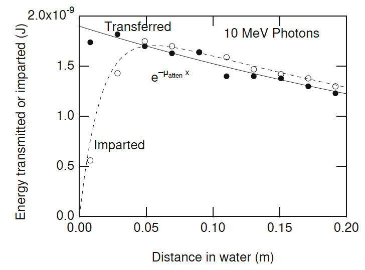
In a photonuclear interaction an energetic photon (exceeding a few MeV) enters and excites a nucleus, which then emits a proton or neutron. (γ, p) events contribute directly to the kerma, but the relative amount remains less than 5% of that due to pair production. Thus it has been commonly neglected in dosimetry considerations.
(γ, n) interactions have a greater practical importance because the neutrons thus produced may lead to problems in radiation protection…All of these consequences of (γ, n) interactions can be regarded as unwanted side effects of the use of higher-energy radiotherapy x-ray beams...
Attix provides insight into the difference between how photons interact with tissue and how charged particles interact.
Charged particles lose their energy in a manner that is distinctly different from that of uncharged radiations (x- or γ-rays and neutrons). An individual photon or neutron incident upon a slab of matter may pass through it with no interaction at all, and consequently no loss of energy. Or it may interact and thus lose its energy in one or a few “catastrophic” events.

By contrast, a charged particle, being surrounded by its Coulomb electric force field, interacts with one or more electrons or with the nucleus of practically every atom it passes. Most of these interactions individually transfer only minute fractions of the incident particle’s kinetic energy…A 1-MeV charged particle would typically undergo ~105 interactions before losing all of its kinetic energy.
Herb Attix was born in 1925 in Portland, Oregon, and served as a lieutenant in the navy during World War II. He was a professor at the University of Wisconsin, where he chaired the Department of Medical Physics before retiring in 1987. He has served on the board of directors for both the American Association of Physicists in Medicine and the Health Physics Society. In 1994 he received the William D. Coolidge Award, the highest award given by the AAPM.

The International Organization for Medical Physics marked its 50th anniversary by publishing short biographical sketches of 50 medical physicists who have made outstanding contributions to the advancement of medical physics over the last 50 years. You can read it here. Herb Attix was on the list. It reminds me of the NBA’s list of the top 50 players, but the IOMP honorees have contributed much more to mankind.

Friday, May 24, 2019

The Physics of Scuba Diving

In Section 1.15 of Intermediate Physics for Medicine and Biology, Russ Hobbie and I discuss scuba diving.
1.15 Diving

Air is easily compressible, so swimming at large depths can be dangerous as the volume of air in the lungs decreases. One can swim safely for depths of tens of meters (several atmospheres of pressure) using a self-contained underwater breathing apparatus (SCUBA). Compressed air tanks are used to supply air to the lungs, and the pressure of the air is adjusted to match the pressure of the surrounding water.

One physiological effect of breathing high-pressure air is that nitrogen dissolves into the blood, which can lead to a mental impairment known as nitrogen narcosis. Moreover, if the swimmer returns rapidly to the surface after a long deep dive, the lowered pressure allows the dissolved nitrogen to form bubbles in the blood that block blood flow and cause decompression sickness, often called “the bends” (Benedek and Villars 2000). To avoid the bends, swimmers must return to the surface slowly, or replace nitrogen by other gasses, such as helium, that are less soluble in blood.
The Physics of Scuba Diving, by Marlow Anderson, superimposed on Intermediate Physics for Medicine and Biology.
The Physics of Scuba Diving,
by Marlow Anderson.

To learn more, I recommend The Physics of Scuba Diving, by Marlow Anderson. This fascinating book explains many aspects of diving. For instance, what happens if you don’t adjust the air in your lungs to match the surrounding pressure? Anderson explains.
Suppose [at a depth of 30 meters]… the diver fills his lungs with air [at 4 atm], and begins to ascend, while holding his breath. As the ambient pressure decreases, the air in the lungs expands. Since the diver is holding his breath, the air has nowhere to go, and so the flexible lung must expand in volume. If he were to hold his breath until reaching the surface, the lungs would have to expand to 4 times their original volume. Of course, this does not happen—instead, the lungs rupture, which can be an exceedingly dangerous injury. This leads to the number one principle drilled into divers when they train:
The Number One Rule of Scuba Diving
Breathe continuously, and never, never hold your breath.
Any air cavity, not just the lungs, is at risk when diving. Another example is the ear. Anderson writes
When a scuba diver descends, pressure begins to build on the eardrum. In order to continue without pain or damage, this pressure must be counterbalanced by a corresponding pressure in the middle ear. However, ordinary respiration of pressurized air through the scuba regulator will not necessarily convey this needed pressure into the middle ear. Consequently, a typical diver must take action to push the pressurized air into these tiny spaces. Sometimes just a movement of the head and neck is enough to stretch the Eustachian tubes and bring this newly respirated (and pressurized) air into the middle ear. More often, however, the diver finds it necessary to perform a valsalva maneuver. This is an action many of us have taken to counteract the change of pressure experienced when flying in an airplane. Namely, you merely need to block the nostrils, and gently blow; the ears then “pop”. This is enough to move the air from the mouth and nostrils up the Eustachian tubes and into the middle ear. Scuba divers call this process equalization, for obvious reasons. You are taught in scuba training to equalize early, and often!
Anderson then discusses another air pocket that I never thought of.
There is one more air space that must be equalized—the space between the [diving] mask and the eyes…It is for this reason that a diver’s mask includes the nostrils. The diver need only exhale through the nostrils into the mask in order to introduce the denser air with the appropriate pressure she has been breathing from the regulator. If the diver does not equalize this air space, the increasing ambient pressure at depth will push the mask even tighter against the face, eventually causing bruising… and this pressure can even burst the tiny capillaries in the eyes, making them bloodshot. But after only a little experience, divers equalize this air space almost without thinking about it.
Gosh, scuba diving sounds complicated and even dangerous. I don’t think I’ll take up the sport.

The conduction and convection of heat also is important when diving. Russ and I discuss biological thermodynamics and heat transport in Chapters 3 and 4 of IPMB. In particular, we define the convection coefficient for heat loss in Homework Problem 51 of Chapter 3, when analyzing Newton’s law of cooling. The convection coefficient at the surface of our body is much larger in water than in air. Anderson notes
Convection can and does happen in the air too; this is especially true on a windy day, when the moving air continually replaces the air molecules that have been warmed by the body with cold ones: this is the origin of the concept of wind chill. However, the larger [thermal] conductivity of water makes this effect more powerful in water than in air. The practical consequence of this is that a diver in 80° [Fahrenheit] air can happily survive indefinitely. Our body continues to produce heat while turning food into energy. Some of this heat is lost to the surrounding air; indeed, we would quickly become overheated if we did not dissipate some of this energy. But in water of that same temperature more heat is lost than our body produces. The result is that the diver gets colder and colder. Indeed, a diver continually immersed even in warm tropical waters will eventually suffer from hypothermia. This is one of the major problems for divers lost at the surface.
Yikes! Have you ever watched the end of the film Titanic?

Jack’s death scene in Titanic.

Another issue that arises under water is localizing the origin of sounds. Homework Problem 20 of Chapter 13 in IPMB says
Problem 20. People use many cues to estimate the direction a sound came from. One is the time delay between sound arriving at the left and right ears. Estimate the maximum time delay. Ignore any diffraction effects caused by the head.
The solution’s simple: divide the distance between the ears by the speed of sound. Anderson explains
The four-fold increase in the speed of sound in water as compared to air means that our brain cannot as effectively detect the direction sounds come from! Hearing a boat motor overhead but being unable to determine the direction it is coming from is a common and frustrating experience for scuba divers.
As you might expect, The Physics of Scuba Diving contains a long section about nitrogen absorption and the bends. Interestingly, the differential equation governing the absorption of nitrogen by the body is the same as described in Section 2.8 of IPMB: decay plus input at a constant rate. It is also the same equation governing Newton’s law of cooling

dP/dt = (Pa - P)/τ ,

where P is the partial pressure of nitrogen in the blood, Pa is the partial pressure of nitrogen being breathed (a function of depth), t is time, and τ is the time constant. Unfortunately, the process is complicated because the body contains several compartments, each with its own time constant.
It was the eminent British physiologist John Scott Haldane who first made use of the model for nitrogen on-gassing and off-gassing that we have described… His goal in understanding these rates was to help divers avoid the serious health effects of overly rapid decompressions… Haldane observed that various tissues in the body should be expected to have different rates of nitrogen absorption. A liquid (like blood) should be expected to very rapidly absorb nitrogen under pressure brought into contact with it. But a solid tissue like bone might be expected to absorb extra nitrogen at a much slower rate. Haldane consequently built a mathematical model to describe this. He assumed that body tissues could be put into five categories he called compartments, with different rates of absorption.
Anderson then analyzes scuba dive tables using this model. He also gives some historical background about the bends, including an explanation about how the disease got its name.
The illness we call decompression sickness (or the bends) was first reported in the scientific literature in France in 1845, which a mining engineer named Charles-Jean Triger described the limb pain suffered by coal miners; he called their difficulties caisson disease… The mine had been filled with pressurized air to prevent ground water from entering the passages...

The construction (1869-1883) of the Brooklyn Bridge in New York was one of the engineering marvels of the nineteenth century. Massive caissons were constructed, including one as deep as 75 feet. Many workers suffered from caisson disease, including not only limb pain, but also paralysis and even death. The chief engineer for the project was stricken by the disease and remained paralyzed for the rest of his life [his wife took over the role of chief engineer, see David McCullough's wonderful book The Great Bridge]. It was during this period that the illness became popularly know as the bends, in reference to the exaggerated bending of the back workers attempted in a futile effort to avoid the pain. The name compared the posture of the bridge workers to the “Grecian Bend”, a posture adopted by fashionable women of the day.
My daughter Stephanie is taking scuba lessons, and plans on diving this summer at the Great Barrier Reef. Have fun, Stephanie, but be careful. Maybe she should read The Physics of Scuba Diving before she dives.

Friday, May 17, 2019

Defending Thermodynamics in a Diet Debate

Homework Problem 47 in Chapter 3 of Intermediate Physics for Medicine and Biology states
Problem 47. The “Calorie” we see listed on food labels is actually 1000 [calories] or 1 kcal. How many kcal do you expend each day if your average metabolism is 100 [watts]?
This problem is as close as Russ Hobbie and I get to diet advice in IPMB. But weight-loss blogs get a lot more page views than do blogs about physics applied to medicine and biology, so I better post about dieting to beef up my numbers.

Fortunately, Physics—the free online magazine from the American Physical Society—recently published an article titled Defending Thermodynamics in a Diet Debate, by Katherine Wright.
Experiments show that calories from different food types are equivalent and that the laws of thermodynamics apply to human metabolism, despite claims to the contrary.
The article begins
Not all calories are equivalent, say some nutrition experts, because the human body extracts energy differently from different types of food. A related concern in the field is that diet advice based on the first law of thermodynamics is inappropriate. However, these claims are countered by those studying human metabolism, who point to experiments that show that the calorie counts on food packaging correctly account for the differences between foods. Both camps agree that thermodynamics has earned a bad reputation in diet science, thanks to certain myths about weight loss, but they disagree on whether that reputation is deserved.
Wright continues
“A calorie is of course a calorie,” says Kevin Hall, who trained as a physicist and currently conducts experiments and develops mathematical models for metabolism and body weight regulation at the National Institutes of Health in Maryland. Hall agrees that different macronutrients—think fats versus carbohydrates—have very different effects on the body, but he strongly disagrees with Preece’s claim [that “a calorie may not be a calorie”]. If the question is just about the number of calories burned by the body, rather than stored as fat, it’s “practically the same” for two foods having the same calorie rating, regardless of their fat or carb content, he says.
Seriously, the laws of thermodynamics are in no danger. Einstein believed that classical thermodyanamics “is the only physical theory ... [that] will never be overthrown.” The question is if the laws are being applied appropriately.
At first glance, the laws of thermodynamics may seem inappropriate for modeling energy fluxes through the human body, as the body is not a closed, isolated system. “Living organisms are not in equilibrium,” so thermodynamics is not relevant, says Richard Feinman, a biochemist at the State University of New York Health and Science Center at Brooklyn, who also agrees with the “calorie is not a calorie” point-of-view. He argues that even if the oxidation pathways for different macronutrients use the same total energy, they still generate different amounts of work and heat and thus their calories are inequivalent. This line of argument is erroneous, says Dale Schoeller, who studies metabolism and nutrition at the University of Wisconsin in Madison. He notes that the human feeding experiments conducted to determine the calorie content of foods factor in variations in how the body handles different macronutrients. These numbers are the ones used to calculate the values that appear on the sides of cereal boxes, for example. “It’s not a perfect number; it varies a few percent between individuals due to differences in their metabolisms,” Schoeller says. But it’s close to being spot on.
The article concludes
Hall says that he and others have made headway in educating physicians and dieticians about the equivalence of calories from different macronutrients and also about the 3500 calorie rule. For example, they have developed tools that allow physicians to make more accurate predictions. Hall’s software, the NIH Body Weight Planner, encodes a simplified version of his energy flux model and can be used by patients to predict the calorie reduction necessary to reach a target weight. “The website has been used by millions of people, so the message is getting out there,” he says. But, he adds, diffusing dieting myths in the wider public is a whole different ball game.
I love a good argument. Although I'm not an expert in this field, I'm gonna go out on a limb and side with the physicist: A calorie is a calorie is a calorie.

Friday, May 10, 2019

Oh, My Aching Back

Oh, my aching back. I was unloading a dresser from a van, and I thought I could handle it myself. What a mistake. I think I strained a muscle in my lower back; probably the erector spinae. Then I aggravated the injury when mowing the lawn. Ouch.

Back pain is interesting. Mine is intense for a few specific movements, and otherwise hardly bothers me. For instance, if I lean over to tie my shoes, it hurts. I feel fine when walking my dog Harvest, except when I bend over to pick up her poop. I was able to paint the bathroom two days after the injury—which involves a lot of reaching up—with no discomfort. Rising from a chair, however, is painful. Driving is no problem; my car seat feels particularly comfortable. Here’s one that surprised me: my back hurts when I sneeze.

I know better than to lift a heavy load like that, using my back instead of my legs. Every time I teach Biological Physics (PHY 3250), the students and I solve Problem 10 in Chapter 1 of Intermediate Physics for Medicine and Biology, which begins “consider the forces on the spine when lifting…” The problem is intermediate in difficulty, and the figure associated with it is shown below.
The figure associated with Problem 10 of Chapter 1
in Intermediate Physics for Medicine and Biology.
My back pain feels lower than where the erector spinae muscle attaches to the spine (the insertion point). I suspect the injury was to the other end of the muscle (near its origin), where it attaches to the pelvis (or more correctly, the sacrum).

The homework problem is a simplification of the true geometry of the spine. It is a toy model, which is useful for gaining insight but should not be taken literally. For instance, the erector spinae is actually a muscle group consisting of the iliocostalis, the longissimus, and the spinalis, which all have slightly different origins and insertion points. The spine is neither stiff nor straight.

An interesting feature of this homework problem is that you can solve it using one of two natural coordinate systems: horizontal (x) and vertical (y), or along (x') and transverse to (y') the spine. In the solution manual for IPMB, Russ Hobbie and I use x' and y'. Students might benefit, however, from solving the problem both ways, so they can see that the choice of coordinate systems doesn’t matter.  

The solution manual has a short preamble for each problem, explaining its goal. The preamble for Problem 10 says
This problem helps students develop physical intuition about forces and torques, and is our first example of a mathematical model in which the students can examine limiting cases to build physical intuition.
The main message of this problem is that you should lift with your legs while keeping your back upright. If you lean over to lift—like I did—the forces must be huge to balance the torques acting on the spine. The solution manual says
The force on the spine by the pelvis is over seven times larger if the spine is horizontal than if it is vertical. You really should lift with your legs (θ = 90º), not with your back (θ = 0º)”!
I guess you could say I’ve developed a new laboratory experiment for IPMB: first lift with your legs and then with your back, and see which one hurts the most!

How am I treating my injury? Mostly with ibuprofen (for the pain and inflammation). Once the worst of the pain is gone (it’s healing rapidly), I’ll begin gently exercising my back, slowly building up strength. I’m not prone to these types of injuries, so I hope this is a one-time problem that will soon be resolved.

From now on, I’m going to take the lessons learned from Intermediate Physics for Medicine and Biology more seriously.

Lift with your legs, not with your back. 

Friday, May 3, 2019

If Only I Had a Few Negative X-rays, I’d be All Set

In Chapter 16 of Intermediate Physics for Medicine and Biology, Russ Hobbie and I discuss intensity-modulated radiation therapy. The goal is to fire x-rays from many different directions, each direction having a different distribution of intensity, to provide a large dose to the tumor while sparing the surrounding normal tissue.
In classical radiotherapy, the beam was either of uniform fluence across the field, or it was shaped by an attenuating wedge placed in the field. Intensity-modulated radiation therapy (IMRT) is achieved by stepping the collimator leaves during exposure so that the fluence varies from square to square in Fig. 16.45 (Goitein 2008; Khan 2010, Ch. 20)

It was originally hoped that CT [computed tomography] reconstruction techniques could be used to determine the collimator settings at different angles. This does not work because it is impossible to make the filtered radiation field negative, as the CT reconstruction would demand. IMRT with conventional treatment planning improves the dose pattern (Goitein 2008; Yu et al. 2008), providing better sparing of adjacent normal tissue and allowing a boost in dose to the tumor.
What’s up with this talk in IPMB about “negative” radiation? To gain insight, read what Steve Webb wrote in his review article “The Physical Basis of IMRT and Inverse Planning” (British Journal of Radiology, Volume 76, Pages 678-689, 2003).
With the glorious wisdom of hindsight some very early developments—we may call them pre-history—might be considered part of the development of IMRT. The mathematician George Birkhoff showed in 1940 that any drawing could be made up of lines of varying pencil thickness so long as negative pencils were allowed [20]. If we read ‘‘X-rays’’ for ‘‘pencils’’ and ‘‘dose distribution’’ for ‘‘picture’’ the analogy with IMRT is clear. Sadly there are no negative X-rays or uncomplicated tumour control would be 100% guaranteed...

[20]. Birkhoff GD (1940) “On Drawings Composed of Uniform Straight Lines,” Journal de Mathématiques Pures et Appliquées, Volume 19, Pages 221–236.
Oh well,
The best laid schemes o' mice an' men 
Gang aft a-gley
If only I had a few of those negative x-rays, I’d be all set.

An example of how a drawing can be made up of lines (in this case, of uniform thickness) based on a figure from “On Drawings Composed of Uniform Straight Lines” by George Birkhoff. An analogous problem is faced when designing radiation treatment plans using IMRT.
An example of how a drawing can be made up of lines (in this case, of uniform thickness), based on a figure from “On Drawings Composed of Uniform Straight Lines” by George Birkhoff. An analogous problem is faced when designing a treatment plan using intensity-modulated radiation therapy.

Friday, April 26, 2019

Neurological Control Systems

In Section 10.12 of Intermediate Physics for Medicine and Biology, Russ Hobbie and I provide examples of negative feedback. One of these examples analyzes how pupil size is controlled by the light impinging on the retina.
The pupil changes diameter in response to the amount of light entering the eye. This is one of the most easily studied feedback systems in the body, because it is possible to break the loop and to change the gain of the system…. [This feedback loop] has been studied extensively by Stark (see Stark 1957, 1968, 1984).
Neurological Control Systems, by Lawrence Stark, on top of Intermediate Physics for Medicine and Biology.
Neurological Control Systems,
by Lawrence Stark.
The 1968 reference is to Lawrence Stark’s book Neurological Control Systems: Studies in Bioengineering. In the introduction, Stark outlines his philosophy of science, which is similar to the approach in IPMB.
Science proceeds by alternate steps: formulation of an intuitive concept for a new phenomenon, incorporation of the new and other related phenomena into a mathematical structure, and finally, again, development of intuitive concepts of further phenomena utilizing the added perspective obtained with the formal elegance of the mathematical model or theory.
I like how he emphasizes the connection between mathematics and intuition.

In Neurological Control Systems, Stark describes his motivation for choosing the pupil of the eye as his model system.
The pupil was chosen for study from a host of possible examples of biological servosystems for several reasons... First, its motor mechanism, the iris, lies exposed behind the transparent cornea for possible measurement without prior dissection. This had previously been exploited for scientific and clinical researches by using high-speed motion picture cameras. Further, by employing invisible infrared photographic techniques, measurements can be made without disturbing the system, because its sensitivity is limited (by definition) to the visible spectrum. Second, the system can be disturbed or driven by changes in intensity of visible light, a form of energy fairly easy to control, and painless in its administration to the subject. The first two advantages lead to still a third: the possibility of performing experiments on awake, unanesthetized animals whose nervous system is fully intact and functional. In fact, all of the experiments to be discussed below have been performed on human subjects. Last, the system responds with a movement having only one degree of freedom, a change in pupil size, which simplifies the system equation analysis.
A photograph of Fig. 6 from Section II, Chapter 1
of Neurological Control Systems, by Lawrence Stark.
In Stark’s system you can break the feedback loop by restricting the light to a small dot at the center of the pupil, so changes in pupil area do not affect the amount of light impinging on the retina. Figure 10.34 in IPMB illustrates this point, as does Fig. 6 in Section II, Chapter 1 of Neurological Control Systems (shown at the right).

Stark goes on to analyze the transfer function (how the pupil area responds to an oscillating light intensity), oscillations induced by focusing the light on the border of the iris and pupil to increase the gain (in this case the light intensity is constant but the pupil area oscillates), random fluctuations of pupil noise (using many of the techniques discussed in Chapter 11 of IPMB), and nonlinearities in the pupil feedback loop.

After this exhaustive discussion of pupil area, Stark moves on to another feedback system: accommodation of the lens of the eye. But that’s another story.

Below, listen to Stark describe his life and work.

Listen to Lawrence Stark describe his research.

Friday, April 19, 2019

Me, Me, Me

Most of my blog posts are about the textbook Intermediate Physics for Medicine and Biology. This post, however, is all about me. IPMB makes a few appearances, but its mainly me, me, me.

OUTV Interview

I was recently featured in a Focus on Faculty interview filmed by the Oakland University TV station (OUTV). I uploaded a copy to Youtube, and you can view it here. I apologize for the hair; I was supposed to get a haircut before filming began, but I got busy. Watch for a cameo by IPMB.

OUTV interviews Brad Roth at Oakland University.
https://www.youtube.com/watch?v=IKjab7_unRA

Daughters Kathy and Stephanie with me, and with Auggie, Smokie, and Harvest.
(l-r) Daughters Kathy and Stephanie with me,
and with Auggie, Smokie, and Harvest.

Harvest

Long-time readers of this blog will remember Suki, my beloved Cocker Spaniel-Westie mix who helped explain concepts in IPMB. After her death about a year ago, my wife and I decided to get another dog. Let me introduce you to Harvest, our 65-pound Treeing Walker Coonhound. She is as lovable as Suki (though not quite as smart). We adopted her from the Making Miracles Animal Rescue. On the right is a picture of me with my daughters Kathy and Stephanie, along with Harvest and my two granddogs Auggie (the foxhound) and Smokie (the greyhound), about to start a 5k walk. We like to take them to a dog park, and as we enter yell "Release the Hounds!"

The photo of Harvest and me published in the October 2018 issue of Physics Today
The photo of Harvest and me published
in the October 2018 issue of Physics Today.
Harvest is already famous. She was featured in the October 2018 issue of Physics Today. The magazine had a selfie contest that Harvest and I entered. Unfortunately, in the magazine our location is listed incorrectly; we are actually in our home in Rochester Hills, Michigan. To the left is the selfie that appeared in Physics Today.

Harvest with the IPMB Ideal Bookshelf.

Live Action IPMB Ideal Bookshelf

The logo for the Intermediate Physics for Medicine and Biology Facebook page is a drawing of the IPMB Ideal Bookshelf. You know how Disney often makes live-action movies out of previous animated shows? (Dumbo is the most recent example.) I’ve done the same thing. Below is a photograph of the “live-action” version of IPMB's My Ideal Bookshelf. Harvest helped me with the filming, so on the right I include a photo of her on the set.

The IPMB Ideal Bookshelf, consisting of books cited in Intermediate Physics for Medicine and Biology.

How to Get Published

Below is a video (divided into two parts) of Michael Sevilla (Distinguished Professor of Chemistry at Oakland University) and me talking to a group of graduate students about how to publish their research. Enjoy!

Part 1 of a discussion about Academic Publishing: How to Get Published in a Peer Review Journal, held Nov. 15, 2013 at Oakland University and hosted by the graduate student group Grad Connection. The host is then-graduate student George Corser, and the guests are Brad Roth and Michael Sevilla.

Part 2.

Friday, April 12, 2019

Kerma

Radiation Therapy Physics,
by Hendee, Ibbott, and Hendee.
In Section 15.15 of Intermediate Physics for Medicine and Biology, Russ Hobbie and I define the kerma. It’s measured in the same units as absorbed dose: J/kg, or gray. What’s the difference between the two? Kerma indicates the energy transferred to charged particles, while dose indicates the energy imparted to (or absorbed by) the tissue. Kerma is more closely related to the number of photons in the tissue, but absorbed dose is more closely related to biological damage. In Radiation Therapy Physics, Hendee, Ibbott and Hendee distinguish between kerma and dose.
The kerma (an acronym for kinetic energy released in matter) is the sum of the initial kinetic energies of all IP [ion pairs] liberated in a volume element of matter, divided by the mass of the matter in the volume element. The absorbed dose is the energy actually absorbed per unit mass in the volume element. If ion pairs escape the volume element without depositing all of their energy, and if they are not compensated by ion pairs originating outside the volume element but depositing energy within it (electron equilibrium), the kerma exceeds the absorbed dose. The kerma also is greater than the absorbed dose when energy is radiated from the volume element as bremsstrahlung or characteristic radiation. Under conditions in which electron equilibrium is achieved and the radiative energy loss is negligible, the kerma and absorbed dose are identical. The output of x-ray tubes is sometimes described in terms of air kerma expressed as the energy released per unit mass of air.
Figure 15.32 of IPMB plots the energy transferred and the energy imparted in 2-cm-thick slices versus depth when a 10 MeV photon beam is incident on water, calculated using Russ’s program MacDose.

Figure 15.32 of Intermediate Physics for Medicine and Biology. A plot of energy transferred and energy imparted for a simulation using 40,000 photons of energy 10 MeV.
Figure 15.32 of IPMB. A plot of energy transferred and energy imparted
for a simulation using 40,000 photons of energy 10 MeV.
If we divide both energies by the mass of the slice and average over many simulations, we get plots of the absorbed dose (dashed curve) and the kerma (solid curve). Hendee et al. provide a similar plot in their Figure 5-7.
The difference between kerma and absorbed dose is useful in explaining the skin-sparing effect of high-energy photons such as multi-MV x rays used in radiation therapy. As shown in Figure 5-7, the kerma is greatest at the surface of irradiated tissue because the photon intensity is highest at the surface and causes the greatest number of interactions with the medium. The photon intensity diminishes gradually as the photons interact on their way through the medium. The electrons set into motion during the photon interactions at the surface travel several millimeters in depth before their energy is completely dissipated… These electrons add to the ionization produced by photon interactions occurring at greater depths. Hence, the absorbed dose increases over the first few millimeters below the surface to reach the greatest dose at the depth of maximum dose (dmax) several millimeters below the surface [dmax is about 0.05 m in Fig. 15.32 of IPMB]. This buildup of absorbed dose over the first few millimeters below the skin is responsible for the clinically important skin-sparing effect of high-energy x and γ rays. Beyond dmax, the absorbed dose also decreases gradually as the photons are attenuated. At depths greater than dmax, the kerma curve falls below that for absorbed dose because kerma reflects the photon intensity at each depth, whereas absorbed dose reflects in part the photon intensity at shallower depths that sets electrons into motion that penetrate to the depth.
Medical Imaging Physics, by Hendee and Ritenour, superimposed on Intermediate Physics for Medicine and Biology.
Medical Imaging Physics,
by Hendee and Ritenour.
William Hendee, the lead author of Radiation Therapy Physics, is a giant in medical physics. He was the editor of the journal Medical Physics from 2005 to 2013. In addition to Radiation Therapy Physics, he wrote another textbook, with E. Russell Ritenour, about Medical Imaging Physics. These two books are at a level similar to IPMB, but with less mathematics and a narrower focus, overlapping our Chapters 13-18. A fourth edition of Radiation Therapy Physics is out, with a new title: Hendee‘s Radiation Therapy Physics.

Robert Lagemann's engraved copy of The Handbook of Chemistry and Physics, which I inherited when I become the Robert T. Lagemann Assistant Professor of Living State Physics at Vanderbilt
Robert Lagemann's engraved copy of
The Handbook of Chemistry and Physics,
which I inherited when I become the
Robert T. Lagemann Assistant Professor
of Living State Physics at Vanderbilt.
My connection to Hendee is that—according to an interview for the American Society for Radiation Oncology—he worked for a couple years as a graduate student at Vanderbilt University with Robert Lagemann. I knew Lagemann when I was a graduate student at Vanderbilt twenty years after Hendee was there, and I served as the Robert T. Lagemann Assistant Professor of Living State Physics at Vanderbilt from 1995 to 1998.

Listen to Hendee discuss medical physics in his own words.


Interview with William Hendee, in which he reflects on the history of the 
Radiological Society of North American, its influence on his career, 
radiology's progress, and improved patient care.
https://www.youtube.com/watch?v=Me06HwRQNvc

Two textbooks by William Hendee, along with
Intermediate Physics for Medicine and Biology.
 

Friday, April 5, 2019

Power Lines and Cancer FAQ

The Power Lines and Cancer FAQ,
by John Moulder.
In the 4th edition of Intermediate Physics for Medicine and Biology, Russ Hobbie and I wrote
An excellent discussion of the all aspects of the problem [whether radiofrequency and power-line electromagnetic fields cause cancer] is available at a frequently updated website, Powerlines and Cancer FAQ [Moulder (Web)].
Then we quoted from the website extensively
Moulder (Web, question 20A) says…
The reference was to
Moulder, J. E. (Web). Power Lines and Cancer: Frequently Asked Questions, www.mcw.edu/gcrc/cop/powerlines-cancer-FAQ/toc.html.
In the 5th edition of IPMB, the story became
John Moulder, the author of a web site about power lines and cancer that unfortunately no longer exists, said…
Yet, I wonder... Nothing disappears from the internet. After a few minutes of googling, I found the entire website saved as a pdf, available at large.stanford.edu/publications/crime/references/moulder/moulder.pdf or https://www.nrc.gov/docs/ML1126/ML112660019.pdf. You can also download it from the IPMB website, or just click here. It begins with a brief summary.
Questions and answers on the connection between power lines, electrical occupations and cancer; includes discussion of the biophysics of interactions, summaries of the laboratory and human studies, information on standards, and a bibliography.
The question-and-answer format includes cross-references to other questions (e.g., Q12” or Q27J). References are listed in the bibliography (e.g., B12). Below, I reproduce the first question.
1) Is there a concern about power lines and cancer?
The concern about power lines and cancer comes largely from studies of people living near power lines (see Q12) and people working in electrical occupations (see Q15). Some of these studies appear to show a weak association between exposure to power-frequency magnetic fields and the incidence of some cancers. However:
  • the more recent epidemiological studies show little evidence that either power lines or electrical occupations are associated with an increase in cancer (see Q19); 
  • laboratory studies have shown little evidence of a link between power-frequency fields and cancer (see Q16); 
  • an extensive series of studies have shown that life-time exposure of animals to power-frequency magnetic fields does not cause cancer (see Q16B); 
  • a connection between power line fields and cancer is physically implausible (see Q18).
The International Commission on Non-Ionizing Radiation Protection (2001):

“In the absence of evidence from cellular or animal studies, and given the methodological uncertainties and in many cases inconsistencies of the existing epidemiologic literature, there is no chronic disease for which an etiological [causal] relation to [power-frequency fields] can be regarded as established.” (See B12)
The International Agency for Research on Cancer (2001):
There is limited evidence in humans for the carcinogenicity of extremely low-frequency magnetic fields in relation to childhood leukaemia.... There is inadequate evidence in humans for the carcinogenicity of extremely low-frequency magnetic fields in relation to all other cancers [and] there is inadequate evidence in humans for the carcinogenicity of extremely low-frequency electric fields. (see Q27J)
The U.S. National Institutes of Health (2002):
The overall scientific evidence for human health risk from [exposure to power-frequency fields] is weak. No consistent pattern of biological effects from exposure to [power-frequency fields] has emerged from laboratory studies with animals or with cells. However, epidemiological studies... had shown a fairly consistent pattern that associated potential [exposure to power-frequency fields] with a small increased risk of leukemia in children and chronic lymphocytic leukemia in adults... For both childhood and adult leukemias interpretation of the epidemiological findings has been difficult due to the absence of supporting laboratory evidence or a scientific explanation linking [exposure to power-frequency fields] with leukemia.(see Q27G).
The U.K. National Radiological Protection Board (2004):
The epidemiological evidence indicates that exposure to power-frequency magnetic fields above 0.4 microT [4 milliG] is associated with a small absolute raised risk of leukaemia in children... However, the epidemiological evidence is not strong enough to justify a firm conclusion that [power-frequency magnetic] fields cause leukemia in children. There is little evidence to suggest... that cancer risks of other types, in children and adults, might arise from exposure to [power-frequency magnetic] fields... The results of epidemiological studies, taken individually or as collectively reviewed by expert groups, cannot be used as a basis for derivation of quantitative restrictions on exposure to [power-frequency magnetic] fields. (see Q27H)
Overall, most scientists consider that the evidence that power line fields cause or contribute to cancer is weak to nonexistent.
The document answers 35 questions, which together provide a detailed analysis of the controversy through 2006. How I wish the FAQ was up-to-date.

The final question is
35) Who wrote this FAQ?
This FAQ document originated in the early 1990's as a USENET FAQ in sci.med.physics. The USENET FAQ was maintained by Dr. John Moulder, Professor of Radiation Oncology, Radiology and Pharmacology/Toxicology at the Medical College of Wisconsin. Dr. Moulder has taught, lectured and written on the biological effects of non-ionizing radiation and electromagnetic fields since the late 1970’s.
The USENET FAQ was converted to html in 1997 by Bob Mueller and Dennis Taylor of the General Clinical Research Center at the Medical College of Wisconsin. The FAQ was expanded and updated to serve as a teaching aid at the Medical College of Wisconsin. The web server and web management was provided by the General Clinical Research Center at the Medical College of Wisconsin. The development and maintenance of this document was not supported by any person, agency, group or corporation outside the Medical College of Wisconsin.
In August 2005, Dr. Moulder became Director of the NIH-funded Medical College of Wisconsin Center for Medical Countermeasures Against Radiological Terrorism. This new job does not leave him the time required to keep these FAQs up-to-date. When the FAQs had become more than two years out-of-date they were discontinued. There is no version more up-to-date that this PDF version....
Parts of this FAQ were derived from the following peer-reviewed publications:

  • JE Moulder: Une approache biomédicale: le point de vue d'un chercheur en cancérologie. In: J Lambrozo, I Le Bis (Eds), Champs Électriques et Magnétique de Très Basse Fréquency: Electricité de France, 1998. 
  • JE Moulder: The controversy over powerlines and cancer, III Jornadas sobre Líneas Eléctricas y Medio Ambiente, Red Eléctrica de España, Madrid, 2000, pp. 159168.
Dr. Moulder maintained similar FAQ documents on Mobile (Cell) Phone Base Antennas and Human Health and Static EM Fields and Cancer.
I discovered a version of the static fields FAQ at https://stason.org/TULARC/health/static-fields-cancer/index.html. I have not found the cell phone FAQ; maybe things can disappear from the internet after all. If you find it, let me know (roth@oakland.edu).

I like the Power Line FAQ’s poetic closing lines.

Public controversy about electricity and health will continue
until:
future research shows conclusively that the fields are hazardous,
or
until the public learns that science cannot guarantee absolute safety,
or
until the public and media gets bored by the subject. 

Neither of the first two outcomes are particularly likely, 
But the third may happen.