Friday, April 19, 2019

Me, Me, Me

Most of my blog posts are about the textbook Intermediate Physics for Medicine and Biology. This post, however, is all about me. IPMB makes a few appearances, but its mainly me, me, me.

OUTV Interview

I was recently featured in a Focus on Faculty interview filmed by the Oakland University TV station (OUTV). I uploaded a copy to Youtube, and you can view it here. I apologize for the hair; I was supposed to get a haircut before filming began, but I got busy. Watch for a cameo by IPMB.

OUTV interviews Brad Roth at Oakland University.
https://www.youtube.com/watch?v=IKjab7_unRA

Daughters Kathy and Stephanie with me, and with Auggie, Smokie, and Harvest.
(l-r) Daughters Kathy and Stephanie with me,
and with Auggie, Smokie, and Harvest.

Harvest

Long-time readers of this blog will remember Suki, my beloved Cocker Spaniel-Westie mix who helped explain concepts in IPMB. After her death about a year ago, my wife and I decided to get another dog. Let me introduce you to Harvest, our 65-pound Treeing Walker Coonhound. She is as lovable as Suki (though not quite as smart). We adopted her from the Making Miracles Animal Rescue. On the right is a picture of me with my daughters Kathy and Stephanie, along with Harvest and my two granddogs Auggie (the foxhound) and Smokie (the greyhound), about to start a 5k walk. We like to take them to a dog park, and as we enter yell "Release the Hounds!"

The photo of Harvest and me published in the October 2018 issue of Physics Today
The photo of Harvest and me published
in the October 2018 issue of Physics Today.
Harvest is already famous. She was featured in the October 2018 issue of Physics Today. The magazine had a selfie contest that Harvest and I entered. Unfortunately, in the magazine our location is listed incorrectly; we are actually in our home in Rochester Hills, Michigan. To the left is the selfie that appeared in Physics Today.

Harvest with the IPMB Ideal Bookshelf.

Live Action IPMB Ideal Bookshelf

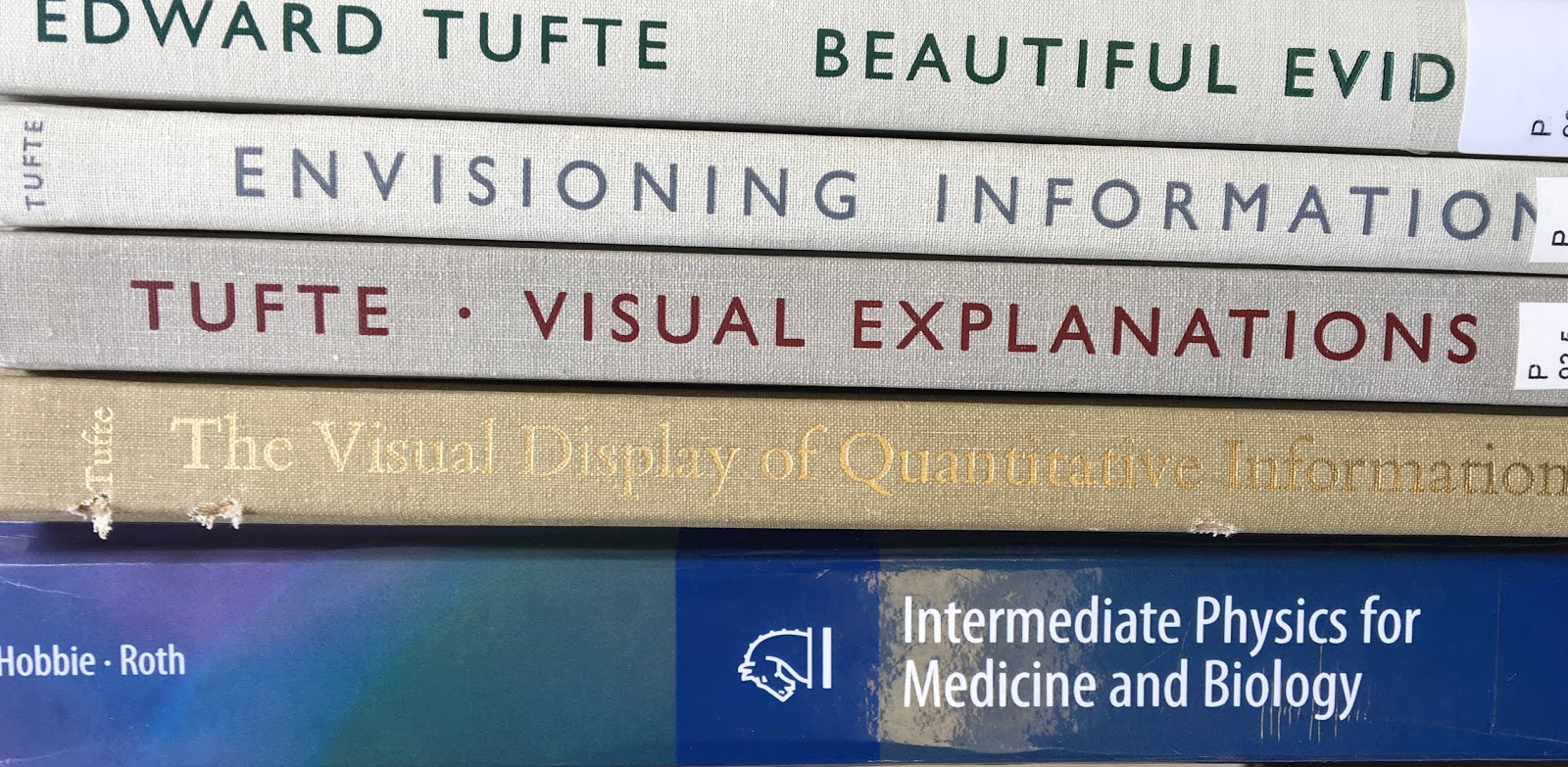
The logo for the Intermediate Physics for Medicine and Biology Facebook page is a drawing of the IPMB Ideal Bookshelf. You know how Disney often makes live-action movies out of previous animated shows? (Dumbo is the most recent example.) I’ve done the same thing. Below is a photograph of the “live-action” version of IPMB's My Ideal Bookshelf. Harvest helped me with the filming, so on the right I include a photo of her on the set.

The IPMB Ideal Bookshelf, consisting of books cited in Intermediate Physics for Medicine and Biology.

How to Get Published

Below is a video (divided into two parts) of Michael Sevilla (Distinguished Professor of Chemistry at Oakland University) and me talking to a group of graduate students about how to publish their research. Enjoy!

Part 1 of a discussion about Academic Publishing: How to Get Published in a Peer Review Journal, held Nov. 15, 2013 at Oakland University and hosted by the graduate student group Grad Connection. The host is then-graduate student George Corser, and the guests are Brad Roth and Michael Sevilla.

Part 2.

Friday, April 12, 2019

Kerma

Radiation Therapy Physics,
by Hendee, Ibbott, and Hendee.
In Section 15.15 of Intermediate Physics for Medicine and Biology, Russ Hobbie and I define the kerma. It’s measured in the same units as absorbed dose: J/kg, or gray. What’s the difference between the two? Kerma indicates the energy transferred to charged particles, while dose indicates the energy imparted to (or absorbed by) the tissue. Kerma is more closely related to the number of photons in the tissue, but absorbed dose is more closely related to biological damage. In Radiation Therapy Physics, Hendee, Ibbott and Hendee distinguish between kerma and dose.
The kerma (an acronym for kinetic energy released in matter) is the sum of the initial kinetic energies of all IP [ion pairs] liberated in a volume element of matter, divided by the mass of the matter in the volume element. The absorbed dose is the energy actually absorbed per unit mass in the volume element. If ion pairs escape the volume element without depositing all of their energy, and if they are not compensated by ion pairs originating outside the volume element but depositing energy within it (electron equilibrium), the kerma exceeds the absorbed dose. The kerma also is greater than the absorbed dose when energy is radiated from the volume element as bremsstrahlung or characteristic radiation. Under conditions in which electron equilibrium is achieved and the radiative energy loss is negligible, the kerma and absorbed dose are identical. The output of x-ray tubes is sometimes described in terms of air kerma expressed as the energy released per unit mass of air.
Figure 15.32 of IPMB plots the energy transferred and the energy imparted in 2-cm-thick slices versus depth when a 10 MeV photon beam is incident on water, calculated using Russ’s program MacDose.

Figure 15.32 of Intermediate Physics for Medicine and Biology. A plot of energy transferred and energy imparted for a simulation using 40,000 photons of energy 10 MeV.
Figure 15.32 of IPMB. A plot of energy transferred and energy imparted
for a simulation using 40,000 photons of energy 10 MeV.
If we divide both energies by the mass of the slice and average over many simulations, we get plots of the absorbed dose (dashed curve) and the kerma (solid curve). Hendee et al. provide a similar plot in their Figure 5-7.
The difference between kerma and absorbed dose is useful in explaining the skin-sparing effect of high-energy photons such as multi-MV x rays used in radiation therapy. As shown in Figure 5-7, the kerma is greatest at the surface of irradiated tissue because the photon intensity is highest at the surface and causes the greatest number of interactions with the medium. The photon intensity diminishes gradually as the photons interact on their way through the medium. The electrons set into motion during the photon interactions at the surface travel several millimeters in depth before their energy is completely dissipated… These electrons add to the ionization produced by photon interactions occurring at greater depths. Hence, the absorbed dose increases over the first few millimeters below the surface to reach the greatest dose at the depth of maximum dose (dmax) several millimeters below the surface [dmax is about 0.05 m in Fig. 15.32 of IPMB]. This buildup of absorbed dose over the first few millimeters below the skin is responsible for the clinically important skin-sparing effect of high-energy x and γ rays. Beyond dmax, the absorbed dose also decreases gradually as the photons are attenuated. At depths greater than dmax, the kerma curve falls below that for absorbed dose because kerma reflects the photon intensity at each depth, whereas absorbed dose reflects in part the photon intensity at shallower depths that sets electrons into motion that penetrate to the depth.
Medical Imaging Physics, by Hendee and Ritenour, superimposed on Intermediate Physics for Medicine and Biology.
Medical Imaging Physics,
by Hendee and Ritenour.
William Hendee, the lead author of Radiation Therapy Physics, is a giant in medical physics. He was the editor of the journal Medical Physics from 2005 to 2013. In addition to Radiation Therapy Physics, he wrote another textbook, with E. Russell Ritenour, about Medical Imaging Physics. These two books are at a level similar to IPMB, but with less mathematics and a narrower focus, overlapping our Chapters 13-18. A fourth edition of Radiation Therapy Physics is out, with a new title: Hendee‘s Radiation Therapy Physics.

Robert Lagemann's engraved copy of The Handbook of Chemistry and Physics, which I inherited when I become the Robert T. Lagemann Assistant Professor of Living State Physics at Vanderbilt
Robert Lagemann's engraved copy of
The Handbook of Chemistry and Physics,
which I inherited when I become the
Robert T. Lagemann Assistant Professor
of Living State Physics at Vanderbilt.
My connection to Hendee is that—according to an interview for the American Society for Radiation Oncology—he worked for a couple years as a graduate student at Vanderbilt University with Robert Lagemann. I knew Lagemann when I was a graduate student at Vanderbilt twenty years after Hendee was there, and I served as the Robert T. Lagemann Assistant Professor of Living State Physics at Vanderbilt from 1995 to 1998.

Listen to Hendee discuss medical physics in his own words.


Interview with William Hendee, in which he reflects on the history of the 
Radiological Society of North American, its influence on his career, 
radiology's progress, and improved patient care.
https://www.youtube.com/watch?v=Me06HwRQNvc

Two textbooks by William Hendee, along with
Intermediate Physics for Medicine and Biology.
 

Friday, April 5, 2019

Power Lines and Cancer FAQ

The Power Lines and Cancer FAQ,
by John Moulder.
In the 4th edition of Intermediate Physics for Medicine and Biology, Russ Hobbie and I wrote
An excellent discussion of the all aspects of the problem [whether radiofrequency and power-line electromagnetic fields cause cancer] is available at a frequently updated website, Powerlines and Cancer FAQ [Moulder (Web)].
Then we quoted from the website extensively
Moulder (Web, question 20A) says…
The reference was to
Moulder, J. E. (Web). Power Lines and Cancer: Frequently Asked Questions, www.mcw.edu/gcrc/cop/powerlines-cancer-FAQ/toc.html.
In the 5th edition of IPMB, the story became
John Moulder, the author of a web site about power lines and cancer that unfortunately no longer exists, said…
Yet, I wonder... Nothing disappears from the internet. After a few minutes of googling, I found the entire website saved as a pdf, available at large.stanford.edu/publications/crime/references/moulder/moulder.pdf or https://www.nrc.gov/docs/ML1126/ML112660019.pdf. You can also download it from the IPMB website, or just click here. It begins with a brief summary.
Questions and answers on the connection between power lines, electrical occupations and cancer; includes discussion of the biophysics of interactions, summaries of the laboratory and human studies, information on standards, and a bibliography.
The question-and-answer format includes cross-references to other questions (e.g., Q12” or Q27J). References are listed in the bibliography (e.g., B12). Below, I reproduce the first question.
1) Is there a concern about power lines and cancer?
The concern about power lines and cancer comes largely from studies of people living near power lines (see Q12) and people working in electrical occupations (see Q15). Some of these studies appear to show a weak association between exposure to power-frequency magnetic fields and the incidence of some cancers. However:
  • the more recent epidemiological studies show little evidence that either power lines or electrical occupations are associated with an increase in cancer (see Q19); 
  • laboratory studies have shown little evidence of a link between power-frequency fields and cancer (see Q16); 
  • an extensive series of studies have shown that life-time exposure of animals to power-frequency magnetic fields does not cause cancer (see Q16B); 
  • a connection between power line fields and cancer is physically implausible (see Q18).
The International Commission on Non-Ionizing Radiation Protection (2001):

“In the absence of evidence from cellular or animal studies, and given the methodological uncertainties and in many cases inconsistencies of the existing epidemiologic literature, there is no chronic disease for which an etiological [causal] relation to [power-frequency fields] can be regarded as established.” (See B12)
The International Agency for Research on Cancer (2001):
There is limited evidence in humans for the carcinogenicity of extremely low-frequency magnetic fields in relation to childhood leukaemia.... There is inadequate evidence in humans for the carcinogenicity of extremely low-frequency magnetic fields in relation to all other cancers [and] there is inadequate evidence in humans for the carcinogenicity of extremely low-frequency electric fields. (see Q27J)
The U.S. National Institutes of Health (2002):
The overall scientific evidence for human health risk from [exposure to power-frequency fields] is weak. No consistent pattern of biological effects from exposure to [power-frequency fields] has emerged from laboratory studies with animals or with cells. However, epidemiological studies... had shown a fairly consistent pattern that associated potential [exposure to power-frequency fields] with a small increased risk of leukemia in children and chronic lymphocytic leukemia in adults... For both childhood and adult leukemias interpretation of the epidemiological findings has been difficult due to the absence of supporting laboratory evidence or a scientific explanation linking [exposure to power-frequency fields] with leukemia.(see Q27G).
The U.K. National Radiological Protection Board (2004):
The epidemiological evidence indicates that exposure to power-frequency magnetic fields above 0.4 microT [4 milliG] is associated with a small absolute raised risk of leukaemia in children... However, the epidemiological evidence is not strong enough to justify a firm conclusion that [power-frequency magnetic] fields cause leukemia in children. There is little evidence to suggest... that cancer risks of other types, in children and adults, might arise from exposure to [power-frequency magnetic] fields... The results of epidemiological studies, taken individually or as collectively reviewed by expert groups, cannot be used as a basis for derivation of quantitative restrictions on exposure to [power-frequency magnetic] fields. (see Q27H)
Overall, most scientists consider that the evidence that power line fields cause or contribute to cancer is weak to nonexistent.
The document answers 35 questions, which together provide a detailed analysis of the controversy through 2006. How I wish the FAQ was up-to-date.

The final question is
35) Who wrote this FAQ?
This FAQ document originated in the early 1990's as a USENET FAQ in sci.med.physics. The USENET FAQ was maintained by Dr. John Moulder, Professor of Radiation Oncology, Radiology and Pharmacology/Toxicology at the Medical College of Wisconsin. Dr. Moulder has taught, lectured and written on the biological effects of non-ionizing radiation and electromagnetic fields since the late 1970’s.
The USENET FAQ was converted to html in 1997 by Bob Mueller and Dennis Taylor of the General Clinical Research Center at the Medical College of Wisconsin. The FAQ was expanded and updated to serve as a teaching aid at the Medical College of Wisconsin. The web server and web management was provided by the General Clinical Research Center at the Medical College of Wisconsin. The development and maintenance of this document was not supported by any person, agency, group or corporation outside the Medical College of Wisconsin.
In August 2005, Dr. Moulder became Director of the NIH-funded Medical College of Wisconsin Center for Medical Countermeasures Against Radiological Terrorism. This new job does not leave him the time required to keep these FAQs up-to-date. When the FAQs had become more than two years out-of-date they were discontinued. There is no version more up-to-date that this PDF version....
Parts of this FAQ were derived from the following peer-reviewed publications:

  • JE Moulder: Une approache biomédicale: le point de vue d'un chercheur en cancérologie. In: J Lambrozo, I Le Bis (Eds), Champs Électriques et Magnétique de Très Basse Fréquency: Electricité de France, 1998. 
  • JE Moulder: The controversy over powerlines and cancer, III Jornadas sobre Líneas Eléctricas y Medio Ambiente, Red Eléctrica de España, Madrid, 2000, pp. 159168.
Dr. Moulder maintained similar FAQ documents on Mobile (Cell) Phone Base Antennas and Human Health and Static EM Fields and Cancer.
I discovered a version of the static fields FAQ at https://stason.org/TULARC/health/static-fields-cancer/index.html. I have not found the cell phone FAQ; maybe things can disappear from the internet after all. If you find it, let me know (roth@oakland.edu).

I like the Power Line FAQ’s poetic closing lines.

Public controversy about electricity and health will continue
until:
future research shows conclusively that the fields are hazardous,
or
until the public learns that science cannot guarantee absolute safety,
or
until the public and media gets bored by the subject. 

Neither of the first two outcomes are particularly likely, 
But the third may happen.

Friday, March 29, 2019

Life Atomic

Life Atomic: A History of Radioisotopes in Science and Medicine, by Angela Creager, superimposed on Intermediate Physics for Medicine and Biology.
Life Atomic, by Angela Creager.
Chapter 17 of Intermediate Physics for Medicine and Biology discusses nuclear physics and nuclear medicine. To learn more about this topic, I recently read Life Atomic: A History of Radioisotopes in Science and Medicine, by Angela Creager. It begins (I removed the references in all excerpts)
At the close of World War II, the nuclear detonations over Hiroshima and Nagasaki demonstrated the devastating power of the atom. As Americans became aware of their country’s secret development of nuclear weapons, the US government swiftly turned attention to the peaceful benefits of nuclear knowledge. Foremost was harnessing the energy of atomic fission for electrical power and transportation, but these applications would require time and technology to realize. Another by-product of atomic energy, however, was ready immediately. Nuclear reactors could be used to generate radioactive isotopes—unstable variants of chemical elements that give off detectable radiation. Scientists began using radioisotopes in biomedical experiments two decades before the atomic age, but their availability remained small-scale until nuclear reactors were developed for the bomb project. In planning for postwar atomic energy, leaders of the Manhattan Project proposed converting a large reactor at Oak Ridge, part of the infrastructure for the bomb project, into a production site for radioisotopes for civilian scientists. The US Atomic Energy Commission (AEC) inherited this plan and oversaw an expansive program making isotopes available for research, therapy, and industry. This book is an account of the uses of radioisotopes as a way to shed new light on the consequences of the "physicists’ war" for postwar biology and medicine.
Creager develops an interesting analogy between radioisotopes as tracers for following biochemical pathways, and her use of radioisotopes to trace the history of twentieth-century science.
...Life Atomic uses radioisotopes as historical tracers, analyzing how they were introduced into systems of scientific research, how they circulated, and what new developments they enabled. I analyze the movement of radioisotopes through government facilities, laboratories, and clinics, both in the United States and around the world, as a way to make visible key transformations in the politics and epistemology of postwar biology and medicine.
My favorite chapter in Life Atomic was "Pathways." It considered two case studies: The Hershey-Chase experiment (which I discussed before in this blog) and the use of radiocarbon to sort out the chemical reactions during photosynthesis. In this post, I focus on the photosynthetic research.
“[Berkeley chemist Melvin] Calvin became an early purchaser of Oak Ridge [radioisotope] carbon-14... [that allowed] Calvin’s group to isolate the earliest, fleeting products of carbon dioxide fixation. [Calvin’s collaborator Andrew] Benson devised an ingenious piece of glassware, nicknamed the ‘lollipop,’ holding the culture suspension [of single-celled green algae Chlorella] into which 14C-labeled CO2 could be injected. A glass tube went into the top of the lollipop, enabling air to be bubbled in while the culture was exposed to light. This would allow photosynthesis to occur at an active rate. Then the bubbler would be removed, the remaining air would be flushed out with nitrogen, and…the flask would then be sealed and shaken in the light, as the radiolabeled carbon was taken up. At the end of a predetermined period of time, from seconds to minutes, the researcher would then drain the suspension into boiling ethanol to kill the cells...

The next step was analysis of the radioactive contents of the Chlorella. Assuming all of the assimilated carbon dioxide entered the photosynthetic pathway, every metabolic intermediate of the reduction from CO2 to sugar should be labeled. Limited progress was made in the first few years identifying the labeled intermediates…

[One] challenge was simply to identify the labeled compounds... Initial studies relied on traditional techniques of chemical extraction and analysis, but in 1948 collaborator William A. Stepka of the Department of Plant Nutrition introduced a newer separation method, paper chromatography, which could distinguish the groups of chemically similar radiolabeled compounds. Researchers separated the algae juices using two different eluting fluids in sequence, along two perpendicular sides of the paper. Different chemical compounds migrated in the two-dimensional space as discrete spots. Exposing the paper chromatogram to medical x-ray film enabled the researcher to pinpoint the radioactive compounds…

By comparing autoradiograms from short exposures of carbon dioxide to those of longer exposures, a researcher could follow the appearance of radioactivity in new compounds. The appearance of these new spots over time revealed the chemical transformations involving the labeled carbon as it proceeded down various metabolic pathways, including—and especially—that for photosynthesis…

By 1958 Calvin and his remaining coworkers had elucidated each step in the pathway and composed what became an eponymous schematic diagram of interlocking cycles.”
Radioactive tracers played a key role in mid-20th-century biology. Their use is not as common in biological research today, but they are still important in medicine. To learn more, listen to Creager talk about the history of radioisotopes in science and medicine.


Listen to Angela Creager talk about the history of radioisotopes in biology and medicine.
https://www.youtube.com/watch?v=w9F5VSTjsxE

Friday, March 22, 2019

Edward Tufte and Drawing Figures

Ten years ago, I wrote a blog post about Edward Tufte's book The Visual Display of Quantitative Information. Recently, we discussed this book in a graduate class I am teaching. One goal of the course is for students to learn how to write scientific papers, including drawing figures.

I like Tufte's advice (below) about friendly data graphics. I agree with everything he says, except for his distaste of sans serif fonts.

Advice about making friendly data graphics. From The Visual Display of Quantitative Information, By Edward Tufte.
Advice about making friendly data graphics,
from The Visual Display of Quantitative Information,
by Edward Tufte.
I wanted to give the students examples of how to improve illustrations but I didn't want to pick on a colleague, so I found a couple figures from my own papers that could be friendlier. The first one is from an article about magnetic stimulation that Peter Basser and I wrote. On the left is the original, and on the right is my revision.
Figure 3c from Roth and Basser (1990) A model for the stimulation of a nerve fiber by electromagnetic induction. IEEE Trans Biomed Eng 37:588-597.
Figure 3c from Roth and Basser (1990) A model for the stimulation of a nerve fiber
by electromagnetic induction
. IEEE Trans Biomed Eng 37:588-597.
The purpose of this figure was to show where magnetic stimulation occurs. That message was in the original figure, but you had to read the caption carefully to find it. In my revision, I clearly marked the locations where depolarization (that is, excitation) and hyperpolarization occur. The big square surrounding the original figure is what Tufte would call "chartjunk" and I deleted it. Instead, I tried to focus on the data. I also labeled the nerve and the coil, so you don't have to read the figure caption to determine what's what. Including units on one of the numerical values practically eliminated the need for a caption at all. I confess, the original figure was cropped from a mediocre scan of the article. Therefore, let's not focus on which figure is crisper, but rather on the overall design. Also, the 30-year-old original data is long lost so I had to retrace the contours in powerpoint using the polygon tool. If you look at the revised figure using high magnification, you may be able to see this. Nevertheless, in my opinion, the revised figure is better.

Another example is from a paper about the electrical stimulation of cardiac tissue.
Figure 3f from Sepulveda, Roth and Wikswo (1989) Current injection into a two-dimensional anisotropic bidomain. Biophys J 55:987-999.
Figure 3f from Sepulveda, Roth and Wikswo (1989) Current injection into a
two-dimensional anisotropic bidomain
. Biophys J 55:987-999.
The message of this figure is that adjacent regions of depolarization and hyperpolarization form when tissue is stimulated by a cathode. By shading the hyperpolarized region, I emphasized this message. I indicated the fiber direction, which is crucial to the main conclusion (tissue hyperpolarizes along the fiber direction). I eliminated the outer circle and subdued the coordinate axes to highlight the data, and inserted a black dot at the location of the cathode. You can decide if it's an improvement. A different version using color and containing all four quadrants is shown below.
A color, four-quadrant version of Figure 3f from Sepulveda, Roth and Wikswo (1989) Current injection into a two-dimensional anisotropic bidomain. Biophys J 55:987-999.
A color version of Fig. 3f from
Sepulveda, Roth and Wikswo (1989).
Another confession: each original illustration was one frame from a multipanel figure. They might have been drawn differently were they stand-alone figures (but I doubt it).

Five good books: The Visual Display of Quantitative Information, Visual Explanations, Evnisioning Information, and Beautiful Evidence (all by Edward Tufte) and Intermediate Physics for Medicine and Biology.
Five good books,
four by Edward Tufte.
Tufte has published several books on visualizing information, but my favorite remains The Visual Display of Quantitative Information. You decide if Russ Hobbie and I follow his advice in Intermediate Physics for Medicine and Biology. Sign up for his course on presenting data and information at his website.

Listen to Tufte talk about the future of data analysis in the video below.

Edward Tufte talking at the Machine Learning and Data Science Summit 2016 Keynote Session.

Friday, March 15, 2019

Ion Channels of Excitable Membranes

Ion Channels of Excitable Membranes, by Bertil Hille, superimposed on Intermediate Physics for Medicine and Biology.
Ion Channels of Excitable Membranes,
by Bertil Hille.

In Intermediate Physics for Medicine and Biology, Russ Hobbie and I claim
“The classic monograph on ion channels is the book by Hille (2001).”
Not only is Bertil Hille’s book Ion Channels of Excitable Membranes a classic, but also it's extraordinarily well written. To learn about ion channels, read this book.

The introduction begins eloquently
Ion channels are macromolecular pores in cell membranes. When they evolved and what role they may have played in the earliest forms of life we still do not know, but today ion channels are most obvious as the fundamental excitable elements in the membranes of excitable cells. Ion channels bear the same relation to electrical signaling in nerve, muscle, and synapse as enzymes bear to metabolism. Although their diversity is less broad than that of enzymes, there are still many types of channels working in concert, opening and closing to shape the signals and responses of the nervous system. Sensitive but potent amplifiers, they detect the sounds of chamber music and guide the artist's paintbrush, yet also generate the violent discharges of the electric eel or the electric ray. They tell the Paramecium to swim backward after a gentle collision, and they propagate the leaf-closing response of the Mimosa plant.
Hille appreciates the role of physics in electrophysiology.
More than in most areas of biology, we see in the study of ion channels how much can be learned by applying simple laws of physics. Much of what we know about ion channels is deduced from electrical measurements. Therefore it is essential to remember some rules of electricity…
Let's look at some topics covered by both IPMB and ICEM.

Toxins

Russ and I briefly mention toxins, saying “an example is tetrodotoxin (TTX), which binds to sodium channels and blocks them, making it a deadly poison.” Hille goes into more detail, explaining how toxins help separate currents and identify channels.
Pharmacological experiments with [channel blocking toxins] provided the first evidence needed to define channels as discrete entities. The magic bullet was tetrodotoxin (dubbed TTX by K.S. Cole), a paralytic poison of some puffer fish... In Japan this potent toxin attracted medical attention because puffer fish is prized there as a delicacy—with occasional fatal effects. Tetrodotoxin blocks action potential conduction in nerve and muscle. Toshio Narahashi brought a sample of TTX to John Moore’s laboratory in the United States. Their first voltage-clamp study with lobster giant axons revealed that TTX blocks INa [the sodium current] selectively, leaving IK and IL [the potassium and leak currents] untouched... Only nanomolar concentrations were needed.

Patch Clamping

Russ and I continue “the next big advance was patch-clamp recording…[which] revealed that the [ion channel] pores open and close randomly.” Hille expands on this idea.
Patch clamp ... forced a revision of the kinetic description of channel gating. At the single-channel level, the gating transitions are stochastic: they can be predicted only in terms of probabilities. Each trial with the same depolarizing step shows a new pattern of openings! Nevertheless, as Hodgkin and Huxley showed, gating does follow rules... Brief openings of Na channels are induced by repeated depolarizing steps... The openings appear after a short delay and cluster early in the sweep. When many records like this are averaged together, the ensemble average has a smoother transient time course of opening and closing, resembling the classical activation-inactivation sequence for macroscopic INa.
Hille's Figures 3.16 and 3.17—showing many individual patch clamp recordings averaged to reproduce the macroscopic Hodgkin and Huxley sodium and potassium currents—are my favorite illustrations in ICEM.

Calcium Channels

Russ and I have one paragraph about calcium channels. Hille has a whole chapter.
The biophysical properties of Ca channels might have been determined by classical voltage-clamp methods if the channels occurred in high density on a reliably clampable membrane. However, these channels are never found in high density, and many of the interesting ones occupy membranes that are difficult to clamp, such as dendrites, nerve terminals, and the complex infoldings of muscle cells. Even when Ca channels are on the surface membranes, as in the cell bodies of neurons, their small currents tend to be masked by those of many other channels, especially K [potassium] channels. The ambiguities caused by these problems delayed biophysical understanding of Ca channels.

Inward Rectification

Russ and I relegate inward rectification to a homework problem. Hille discusses why inward rectifiers are important.
Axons seem to be built for metabolic economy at rest. At the negative resting potential, all their channels tend to shut, minimizing the flow of antagonistic inward and outward currents and minimizing the metabolic costs of idling. Depolarization, on the other hand, tends to open channels and dissipate ion gradients, but the inactivation of Na channels and the delayed activation of K channels in axons keeps even this expenditure at a minimum.

Consider, however, the electrical activity of a tissue that cannot rest: the heart... Its cells spend almost half their time in the depolarized state... Furthermore, each depolarization lasts 100-600 ms. Metabolic economy in this busy but slow electrical activity is achieved in two ways. First, most ion channels are present at very low densities in heart cells, so even when activated, they pass [only small] currents…

The second economy, in non-pacemaker cells of the heart, is a type of K channel, the inward rectifier, that stops conducting during depolarization. The total membrane conductance is actually lower during the plateau phase of such action potentials than during the period between action potentials... Again antagonistic current flows are minimized. Heart muscle has a variety of K channels, many of which have the property of inward rectification or of rapid inactivation.
Other topics covered in both IPMB and ICEM are Roderick MacKinnon’s study of the structure of the potassium channel, and the Hodgkin and Huxley model for the action potential in a squid axon.

Having coauthored a textbook, I appreciate how much work must have gone into writing ICEM.
  • The figures are all clear, drawn in a uniform style, with a focus on the data. To have a consistent look, you can’t just cut and paste figures from an article into a book. They had to be lovingly reproduced and reformatted. 
  • The list of references contains about 1800 articles summarizing the literature up to 2001, the date of the most recent edition. 
  • The language is clear and readable. Young scientists looking for an example of effective scientific writing should read ICEM
  • Hille appreciates the history of his subject. Concepts are clearer when placed in historical context.
  • The book is authoritative because the author is a giant in his field. He received the Lasker award (America's Nobel) for his work on ion channels
  • I would compare ICEM to the robust hybrid offspring of a marriage between Solid State Physics (Ashcroft and Mermin) and Nerve, Muscle, and Synapse (Katz).
Intermediate Physics for Medicine and Biology is superior to Ion Channels of Excitable Membranes in one way: homework problems. ICEM has none and IPMB has hundreds.

Friday, March 8, 2019

The Atomic Energy Merit Badge

My Atomic Energy Merit Badge.
Chapter 17 of Intermediate Physics for Medicine and Biology discusses nuclear physics and nuclear medicine. I began studying nuclear physics fifty years ago. It all started in the Boy Scouts.

Boy Scout troop 96 in Morrison, Illinois.
Troop 96, Morrison, Illinois.
When growing up in Morrison, Illinois, I was a member of the Cub Scouts and then the Boy Scouts. I enjoyed the camping, hiking, and canoeing. Each summer I spent a week at scout camp, and loved it. In the winter, we would have a Klondike Derby, which involved pushing a large sled over the snow and then camping in the cold. I was a member of Morrison’s Troop 96 and Mr. Glenn Van Eaton was our Scoutmaster; behind his back we called him “General Glenn.” One of my fondest memories was being inducted into the Order of the Arrow. At a campfire ceremony, several of us were “tapped-out” for initiation, which involved spending a night in the woods alone.

My Scout Handbook, superimposed on the cover of Intermediate Physics for Medicine and Biology.
My Scout Handbook.
Between campouts, we earned merit badges. Some of them you'd expect, such as first-aid, rowing, swimming, and pioneering (knot tying). Others examined adult topics, such as atomic energy.

I found my old Scout Handbook—molding in a box in our basement—and looked up the requirements for the atomic energy merit badge. They are impressive. Completing this merit badge provides a good preparation for Chapter 17 of IPMB.
  1. Tell the meaning of the following: alpha particle, atom, background radiation, beta particle, curie, fallout, half-life, ionization, isotope, neutron activation, nuclear reactor, particle accelerator, radiation, radioactivity, roentgen, and X-ray
  2. Make three-dimensional models of the atoms of the three isotopes of hydrogen. Show neutrons, protons, and electrons. Use these models to explain the difference between atomic weight and number
  3. Make a drawing showing how nuclear fission happens. Label all details. Draw a second picture showing how a chain reaction could be started. Also show how it could be stopped. Show what is meant by “critical mass.”
  4. Tell who five of the following people were. Explain what each of the five discovered in the field of atomic energy: Henri Becquerel, Niels Bohr, Marie Curie, Albert Einstein, Enrico Fermi, Otto Hahn, Ernest Lawrence, Lise Meitner, William Rontgen, and Sir Ernest Rutherford. Explain how any one person’s discovery was related to one other person’s work. 
  5. Draw and color the radiation hazard symbol. Explain where it should be used and not used. Tell why and how people must use radiation or radioactive materials carefully.
  6. Do any THREE of the following:
    1. Build an electroscope. Show how it works. Put a radiation source inside it. Explain any difference seen. 
    2. Make a simple Geiger counter. Tell the parts. Tell which types of radiation the counter can spot. Tell how many counts per minute of what radiation you have found in your home. 
    3. Build a model of a reactor. Show the fuel, the control rods, the shielding, the moderator, and any cooling material. Explain how a reactor could be used to change nuclear into electrical energy or make things radioactive. 
    4. Use a Geiger counter and a radiation source. Show how the counts per minute change as the source gets closer. Put three different kinds of material between the source and the detector. Explain any differences in the counts per minute. Tell which is the best to shield people from radiation and why. 
    5. Use fast-speed film and a radiation source. Show the principles of autoradiography and radiography. Explain what happened to the films. Tell how someone could use this in medicine, research, or industry. 
    6. Using a Geiger counter (that you have built or borrowed), find a radiation source that has been hidden under a covering. Find it in at least three other places under the cover. Explain how someone could use this in medicine, research, agriculture, or industry. 
    7. Visit a place where X-rays are used. Draw a floor plan of the room in which it is used. Show where the unit is. Show where the unit, the person who runs it, and the patient would be when it is used. Describe the radiation dangers from X-rays. 
    8. Make a cloud chamber. Show how it can be used to see the tracks caused by radiation. Explain what is happening. 
    9. Visit a place where radioisotopes are being used. Explain by a drawing how and why they are used. 
    10. Get samples of irradiated seeds. Plant them. Plant a group of nonirradiated seeds of the same kind. Grow both groups. List any differences. Discuss what irradiation does to seeds.
Build a Geiger counter? Mom would have vetoed that!

Working on the atomic energy merit badge may have been my initial exposure to physics; the first step in a long journey. Now it is called the nuclear science merit badge. Some of the requirements are the same, but there is more emphasis on radiation hazards (for example, radon) and nuclear medicine. Probably it is even better at preparing you for Intermediate Physics for Medicine and Biology.

My dad made it to Eagle Scout when he was young, but I didn’t uphold the family tradition. I quit scouts with the rank of Life. Most boys enter high school and lose interest in scouting, but a few hang on and make it to Eagle. I was planning on being one of the few, but when we moved out of town after my sophomore year I didn't restart with a new troop. Besides, I attended high school in the post-Vietnam/Watergate era, when scouting went out of fashion. Over the years, I came to disagree with the Boy Scouts’ positions on homosexuality and religion, so I don’t regret dropping out. But when I was a kid in Morrison, those issues never came up. We just had fun.

My Boy Scout Order of the Arrow sash, superimposed on Intermediate Physics for Medicine and Biology.
My Order of the Arrow sash.
My merit badges: Stamp Collecting, First Aid, Music, Swimming, Cooking, Canoeing, Rowing, Camping, Reading, Citizenship in the Nation, Emergency Preparedness, Citizenship in the Community, Citizenship in the World, Atomic Energy, Scholarship, Fish and Wildlife Management, Pioneering, and Environmental Science.
My 18 merit badges (left to right, then top to bottom):
Stamp Collecting, First Aid, Music,
Swimming, Cooking, Canoeing,
Rowing, Camping, Reading,
Citizenship in the Nation, Emergency Preparedness, Citizenship in the Community,
Citizenship in the World, Atomic Energy, Scholarship,
Fish and Wildlife Management, Pioneering, and Environmental Science.
Those with a silver rim are required for Eagle.

Friday, March 1, 2019

Sunscreen

In Chapter 14 of Intermediate Physics for Medicine and Biology, Russ Hobbie and I discuss ultraviolet light, and specifically sunscreen.
Protection from the sun certainly reduces erythema [sunburn] and probably reduces skin cancer. Protection is most important in childhood years, both because children receive three times the annual sun exposure of adults and because the skin of children is more susceptible to cancer-causing changes. The simple sun protection factor (SPF) alone is not an adequate measure of effectiveness, because it is based on erythema, which is caused mainly by UVB [ultraviolet B light, with wavelengths from 280 to 315 nm]. Some sunscreens do not adequately protect against UVA radiation [315-400 nm]. Buka (2004) reviews both sunscreens and insect repellents for children. He finds several products that adequately block both UVA and UVB. Look for a sunscreen labeled “broad spectrum” or with at least three stars in a UVA rating system. An adequate amount must be used: for children he recommends 1 fluid ounce (30 ml) per application of a product with SPF of 15 or more. The desired application of sunscreen is 2 mg cm−2. Typical applications are about half this amount. It has been suggested that one make two applications (Teramura et al.2012) or use a sunscreen with a very high SPF (Hao et al.2012).
This week the Food and Drug Administration has issued a proposed update to sunscreen rules.
This significant action is aimed at bringing nonprescription, over-the-counter (OTC) sunscreens that are marketed without FDA-approved applications up to date with the latest science to better ensure consumers have access to safe and effective preventative sun care options. Among its provisions, the proposal addresses sunscreen active ingredient safety, dosage forms, and sun protection factor (SPF) and broad-spectrum requirements. It also proposes updates to how products are labeled to make it easier for consumers to identify key product information.

“Broad spectrum sunscreens with SPF values of at least 15 are critical to the arsenal of tools for preventing skin cancer and protecting the skin from damage caused by the sun’s rays, yet some of the essential requirements for these preventive tools haven’t been updated in decades. Since the initial evaluation of these products, we know much more about the effects of the sun and about sunscreen’s absorption through the skin. Sunscreen usage has changed, with more people using these products more frequently and in larger amounts. At the same time, sunscreen formulations have evolved as companies innovated. Today’s action is an important step in the FDA’s ongoing efforts to take into account modern science to ensure the safety and effectiveness of sunscreens,” said FDA Commissioner Scott Gottlieb, M.D. “The proposal we’ve put forward would improve quality, safety and efficacy of the sunscreens Americans use every day. We will continue to work with industry, consumers and public health stakeholders to ensure that we’re striking the right balance. To further advance these goals, we’re also working toward comprehensive OTC reform, which will help foster OTC product innovation as well as facilitate changes necessary for the FDA to keep pace with evolving science and new safety data.”

The agency is issuing this proposed rule to put into effect final monograph regulations for OTC sunscreen drug products as required by the Sunscreen Innovation Act. OTC monographs establish conditions under which the FDA permits certain OTC drugs to be marketed without approved new drug applications because they are generally recognized as safe and effective (GRASE) and not misbranded. Over the last twenty years, new scientific evidence has helped to shape the FDA’s perspective on the conditions, including active ingredients and dosage forms, under which sunscreens could be considered GRASE.
The feds then get specific.
Of the 16 currently marketed active ingredients, two ingredients – zinc oxide and titanium dioxide – are GRASE for use in sunscreens; two ingredients – PABA and trolamine salicylate – are not GRASE for use in sunscreens due to safety issues. There are 12 ingredients for which there are insufficient safety data to make a positive GRASE determination at this time.
Latha et al. (Sunscreening Agents: A Review, The Journal of Clinical and Aesthetic Dermatology, Volume 6, Pages 16-26, 2013) discuss the mechanism of sunscreen action (references removed).
Sunscreening agents contain titanium dioxide (TiO2), kaolin, talc, zinc oxide (ZnO), calcium carbonate, and magnesium oxide. Newer chemical compounds, such as bemotrizinol, avobenzone, bisoctizole [sic], benzophenone-3 (BZ-3, oxybenzone), and octocrylene, are broad-spectrum agents and are effective against a broad range of solar spectrum both in experimental models and outdoor settings. Ecamsule (terephthalylidene dicamphor sulphonic acid), dometrizole trisiloxane [sic], bemotrizinol, and bisoctrizole are considered organic UVA sunscreening agents... Commercial preparations available in the market include a combination of these agents to cover a wide range of UV rays.

Composition and mechanism of action of sunscreening agents vary from exerting their action through blocking, reflecting, and scattering sunlight. Chemical sunscreens absorb high-energy UV rays, and physical blockers reflect or scatter light. Multiple organic compounds are usually incorporated into chemical sunscreening agents to achieve protection against a range of the UV spectrum. Inorganic particulates may scatter the microparticles in the upper layers of skin, thereby increasing the optical pathway of photons, leading to absorption of more photons and enhancing the sun protection factor (SPF), resulting in high efficiency of the compound.
Researchers are postulating that the generation of sunlight-induced free radicals causes changes in skin; use of sunscreens reduces these free radicals on the skin, suggesting the antioxidant property. Broad-spectrum agents have been found to prevent UVA radiation-induced gene expression in vitro in reconstructed skin and in human skin in vivo.
Sunscreens may have unanticipated side effects. For instance, Key West banned sunscreens containing oxybenzone and octinoxate, two chemicals that damage coral reefs.

Do sunscreens cause cancer? Read what Harriet Hall, the SkepDoc, thinks (quick summary: no).

To learn more about sunscreens, here’s a video from one of my favorite explainers of physics: Dianna Cowern, The Physics Girl. She’s GRASE.

Happy physicsing!