Friday, January 25, 2013

Aliasing

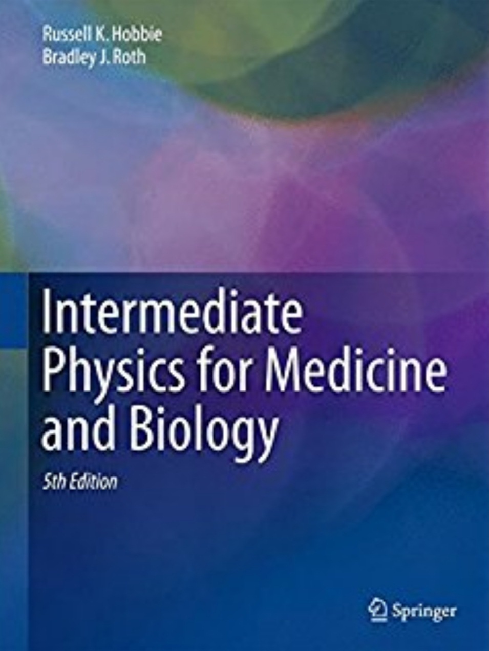
In Chapter 11 of the 4th edition of Intermediate Physics for Medicine and Biology, Russ Hobbie and I discuss aliasing.
If a component [in the Fourier spectrum] is present whose frequency is more than half the sampling frequency, it will appear in the analysis at a lower frequency. This is the familiar stroboscopic effect in which the wheels of the stagecoach appear to rotate backward because the samples (movie frames) are not made rapidly enough. In signal analysis, this is called aliasing. It can be seen in Fig. 11.15, which shows a sine wave sampled at regularly spaced intervals that are longer than half a period.
First of all, what is all this business about a stagecoach? Fifty years ago, when westerns were all the rage in movies and on TV, aliasing often occurred if the frame rate (typically 24 frames per second for old movies) was lower than the rotation rate of the wheel (if all the spokes of the wheel are equivalent, then you can take the “period of rotation” as the time it takes for one spoke to rotate to the position of the adjacent one, which may be much shorter than the time for the wheel to make one complete rotation). You can see an example of this in the John Wayne movie Winds of the Wasteland (1936), especially in the climactic scene of the stagecoach race. In this video of the movie, you can see aliasing of the stagecoach wheel briefly at time 55:40. For those of you who are more discriminating in your movie tastes, you can see another example of aliasing 14 minutes and 15 seconds into Stagecoach, a John Wayne classic from 1939 directed by John Ford. In my opinion, the greatest western is the John Ford masterpiece The Man Who Shot Liberty Valance. What more could you ask for than both John Wayne and Jimmy Stewart in the same production? You can see aliasing briefly when Stewart drives his buckboard out of Shinbone to practice his pistol shooting (without much success). Another time when you see a wheel rotate backwards in this movie does not involve aliasing; it is (Spoiler Alert!) after Stewart Wayne kills Valance (Lee Marvin), when Pompey (Woody Strode) takes the drunken Wayne to his ranch house where he backs up the buckboard (that was a joke….).

But I digress. Aliasing can happen in space as well as time, and can therefore affect images. If spatial frequencies in the structure of an object correspond to wavelengths smaller than the twice the pixel size, low spatial frequency artifacts, such as Moire patterns, can appear in the image, shown nicely in this figure. One can minimize aliasing by first filtering (anti-aliasing) before sampling. Some rather extreme cases of aliasing can been seen in Figs. 11.41 and 12.11 of Intermediate Physics for Medicine and Biology.

 Stagecoach, with John Wayne.

Friday, January 18, 2013

The Magic Angle

I recently found another error in the 4th edition of Intermediate Physics for Medicine and Biology. In Chapter 18 about magnetic resonance imaging, Homework Problem 18 reads
Problem 18  Suppose the two dipoles of the water molecule shown below point in the z direction while the line between them makes an angle θ with the x axis. Determine the angle θ for which the magnetic field of one dipole is perpendicular to the dipole moment of the other. For this angle the interaction energy is zero. This θ is called the “magic angle” and is used when studying anisotropic tissue such as cartilage [Xia (1998)].
Technically there is nothing wrong with this problem. However, if I were doing it over I would have the angle θ measured from the z axis, not the x axis. One reason is that this is the way θ is defined most often in the literature. Another is that in the solution manual we solve the problem as if θ were relative to the z axis, so the book and the solution manual are not consistent on the definition of θ. I should add, this problem was not present in the 3rd edition of Intermediate Physics for Medicine and Biology. It is a new problem I wrote for the 4th edition, so I can’t blame Russ Hobbie for this one (rats).

The citation in the homework problem is to the paper
Xia, Y. (1998) “Relaxation Anisotropy in Cartilage by NMR Microscopy (μMRI) at 14-μm Resolution,” Magnetic Resonance in Medicine, Volume 39, Pages 941–949.
The author, Yang Xia, is a good friend of mine, and a colleague here in the Department of Physics at Oakland University. He is well-known around OU because over the last decade he had the most grant money from the National Institutes of Health of anyone on campus. He uses a variety of techniques, including micro-magnetic resonance imaging (μMRI), to study cartilage and osteoarthritis. The abstract of his highly-cited paper reads
To study the structural anisotropy and the magic-angle effect in articular cartilage, T1 and T2 images were constructed at a series of orientations of cartilage specimens in the magnetic field by using NMR microscopy (μMRI). An isotropic T1, and a strong anisotropic T2 were observed across the cartilage tissue thickness. Three distinct regions in the microscopic MR images corresponded approximately to the superficial, transitional, and radial histological zones in the cartilage. The percentage decrease of T2 follows the pattern of the curve of (3cos2θ - 1)2 at the radial zone, where the collagen fibrils are perpendicular to the articular surface. In contrast, little orientational dependence of T2 was observed at the transitional zone, where the collagen fibrils are more randomly oriented. The result suggests that the interactions between water molecules and proteoglycans have a directional nature, which is somehow influenced by collagen fibril orientation. Hence, T2 anisotropy could serve as a sensitive and noninvasive marker for molecular-level orientations in articular cartilage.
Perhaps a better reference for our homework problem is another paper of Xia’s.
Xia, Y. (2000) “Magic Angle Effect in MRI of Articular Cartilage: A Review,” Investigative Radiology, Volume 35, Pages 602–621.
There in Fig. 3 of Xia’s review is a picture almost identical to the figure that immediately follows Homework Problem 18 in our book, except the angle θ is measured from the direction of the static magnetic field rather than perpendicular to it. Xia writes
T2 corresponds to the decay in phase coherence (dephasing) between the individual nuclear spins in a sample (protons in our case). Because each proton has a magnetic moment, it generates a small local dipolar magnetic field that impinges on its neighbor’s space (Fig. 3).43 This local field fluctuates constantly because the molecule is tumbling randomly. The T2 process can occur under the influence of this fluctuating magnetic field. At the end of signal excitation during an MRI experiment, the net magnetization (which produces the MRI signal) is coherent and points along a certain direction in space in the rotating frame of reference. This coherent magnetization vector soon becomes dephased because the local magnetic fields associated with the magnetic properties of neighboring nuclei cause the precessing nuclei to acquire a range of slightly different precessional frequencies. The time scale of this signal dephasing is reported as T2 and is characteristic of the molecular environment in the sample.43,44

For simple liquids or samples containing simple liquidlike molecules, the molecules tumble rapidly. The dipolar spin Hamiltonian (HD) that describes the dipolar interaction is averaged to zero, and its contribution to the spin relaxation vanishes. Relaxation characteristics exhibit a simple exponential decay that is well described by the Bloch equations.45 For samples containing molecules that are less mobile, HD is no longer averaged to zero and makes a significant contribution to the relaxation, resulting in a shorter T2. When HD is not zero, it is dominated by a geometric factor, (3cos2θ - 1), where θ is the angle between the position vector joining the two spins and the external magnetic field (see Fig. 3). A useful feature of this geometric factor is that it approaches zero as θ approaches 54.74° (Fig. 4). Therefore, even when HD is not zero, the contribution of HD to spin relaxation can be minimized if θ is set to 54.74°. This angle is called the magic angle in NMR.46
So, in the errata you will now find this:
Page 539: In Chapter 18, Homework Problem 18, “while the line between them makes an angle θ with the x axis” should be “while the line between them makes an angle θ with the z axis”. Also, in the accompanying figure following the homework problem, the angle θ should be measured from the z (vertical) axis, not the x (horizontal) axis. Corrected 1-18-13.
Is this the last error that we’ll find in our book? I doubt it; there are sure to be more we haven’t found yet. If you find any, please let us know.

Friday, January 11, 2013

5th Edition of Intermediate Physics for Medicine and Biology

Russ Hobbie and I are starting to talk about a 5th edition of Intermediate Physics for Medicine and Biology, and we need your help. We would like suggestions and advice about what changes/additions/deletions to make in the new edition.

We have prepared a survey to send to faculty members who we know have used IPMB as the textbook for a class they taught. However, our list may be incomplete, and input from any teacher, student, or reader would be useful. So, below is a copy of the survey. Please send responses to any or all of the questions to Russ (hobbie@umn.edu).

Thanks!
  1. What chapters did you cover when teaching from the 4th edition of IPMB?
  2. Were the homework problems appropriate?
  3. In the 4th edition we added a chapter on Sound and Ultrasound to IPMB. If we were to add one new chapter to the 5th edition, what should the topic be?
  4. Would color significantly improve the book for your purposes? How much extra money would you be willing to pay if the 5th edition contained many color pictures?
  5. What is the best feature of IPMB? What is the worst?
  6. Is the end-of-chapter list of symbols useful?
  7. Do your students use the Appendices? Suppose to save space one Appendix had to be deleted: which one should go?
  8. Did you have access to the solution manual? Was it useful? The solution manual we prepared using different software than the book itself. Did you see a noticeable difference in the quality of the book and the solution manual?
  9. Would you like students to have access to the solution manual?
  10. Did you use any information on the book website, such as the errata or text from previous editions?
  11. Are you aware of the book blog? Did you find it useful when teaching from IPMB? Do you find it interesting?
  12. How important is having a paperback version of the book?
  13. What textbooks did you consider other than IPMB? If IPMB did not exist, what book would you use for your class?

Friday, January 4, 2013

Non-Dynamical Stochastic Resonance: Theory and Experiments with White and Arbitrarily Coloured Noise

Section 11.18 of the 4th edition of Intermediate Physics for Medicine and Biology contains a discussion of stochastic resonance. This is a new section that Russ Hobbie and I added to the 4th edition, and features a discussion of a paper by Zoltan Gingl, Laszlo Kish (formerly “Kiss”), and Frank Moss.
Gingl, Z., L. B. Kiss, and F. Moss (1995) “Non-Dynamical Stochastic Resonance: Theory and Experiments with White and Arbitrarily Coloured Noise,” Europhysics Letters, Volume 29, Pages 191–196.
The paper is interesting (despite the annoying British spelling), and I reproduce part of the introduction below.
In the last decade’s physics literature, stochastic-resonance (SR) effect has been one of the most interesting phenomena taking place in noisy non-linear dynamical systems (see, e.g., [l-14]. The input of stochastic resonators [12] (non-linear systems showing SR) is fed by a Gaussian noise and a sinusoidal signal with frequency f0, that is, a random excitation and a periodic one are acting on the system. There is an optimal strength of the input noise, such that the system’s output power spectral density, at the signal frequency f0, has a maximal value. This effect is called SR. It can be viewed as: the transfer of the input sinusoidal signal through the system shows a “resonance vs. the strength of the input noise. It is a very interesting, and somewhat paradoxial effect, because it indicates that in these systems the existence of a certain amount of “indeterministic excitation is necessary to obtain the optimal “deterministic response. There are certain indications [2,13,14] that the principle of SR may be applied by nature in biological systems in order to optimise the transfer of neural signals.
Until last year, it was a common belief that SR phenomena occur only in (bistable, sometimes monostable [10] or multistable) dynamical systems [1-14]. Very recently, Wiesenfeld et al. [15] have proposed that certain systems with threshold-like properties should also show SR effects.
We present here an extremely simple system, invented by Moss, which displays SR. It consists only of a threshold and a subthreshold coherent signal plus noise as shown in fig. la). It is not a dynamical system, instead there is a single rule: whenever the signal plus the noise crosses the threshold unidirectionally, a narrow pulse of standard shape is written to a time series, as shown in fig. lb). The power spectrum of this series of pulses is shown in fig. 1c). It shows all the familiar features of SR systems previously studied [l, 2, 7, 16], in particular, the narrow, delta-like signal features riding on a broad-band noise background from which the signal-to-noise ratio (SNR) can be extracted. This system can be easily realized electronically as a level-crossing detector (LCD). There is a simple and very physically motivated theory of this phenomenon (due to Kiss), see below. Other, more detailed studies of various aspects of threshold-crossing dynamics have been made by Fox et al. [17], Jung [18] and Bulsara et al. [19].
We have experimentally realised and developed this simple SR system and carried out extensive analog and computer simulations on it. The theory of Kiss has been verified for the case of white and several sorts of coloured noises. Until now, the description of this new SR system, its physical realisation and the original theory have not appeared in the open literature, so in this letter we shall describe the new system and its developments made by us, present the outline and the main results of the theory and finally show some interesting experimental results…
Figure 1 in their paper is our Figure 11.50. It is an excellent figure, although I don’t know why they didn’t adjust the time axes so that the pulses in b) are aligned precisely with the signal crossings in a). The axes are almost correct, but are off just enough to be confusing, like when the video and audio signals are off by a fraction of a second in a movie or TV show.

The Gingl et al. paper is short and highly cited (over 200 citations to date, according to the Web of Science). However, it is not cited nearly as often as another paper published by Kurt Wisenfeld and Moss that same year:
Wisenfeld K. and F. Moss (1995) “Stochastic Resonance and the Benefits of Noise: From Ice Ages to Crayfish and SQUIDs,” Nature, Volume 373, Pages 33–36.
This paper, with over 1000 citations, reviews many applications of stochastic resonance.
Noise in dynamical systems is usually considered a nuisance. But in certain nonlinear systems, including electronic circuits and biological sensory apparatus, the presence of noise can in fact enhance the detection of weak signals. This phenomenon, called stochastic resonance, may find useful application in physical, technological and biomedical contexts.
Wisenfeld and Moss discuss how the crayfish may use stochastic resonance to detect weak signals with their mechanoreceptor hair cells.

Frank Moss (1934-2011) was the founding director of the Center for Neurodynamics at the University of Missouri at St Louis. Click here to read his obituary (he died two years ago today) in Physics Today, and click here to read a tribute to him in a focus issue of the journal Chaos.

Friday, December 28, 2012

Determining the Site of Stimulation During Magnetic Stimulation of a Peripheral Nerve

As December draws to a close and I reflect on all that’s happened over the last twelve months, I conclude that 2012 has been a good year. For me, it has also marked some important anniversaries. Thirty years ago (1982) I graduated from the University of Kansas with a bachelors degree in physics. Twenty-five years ago (1987) I obtained my PhD from Vanderbilt University. And twenty years ago (1992) I was at the National Institutes of Health in Bethesda, Maryland working on magnetic stimulation of nerves.

Today I want to focus on one particular paper published in 1992 that examined magnetic stimulation of a peripheral nerve: “Determining the Site of Stimulation During Magnetic Stimulation of a Peripheral Nerve” (Electroencephalography and Clinical Neurophysiology, Volume 85, Pages 253–264). To understand this article, we must first examine Frank Rattay’s analysis of electrical stimulation. Rattay showed that excitation along a nerve axon occurs where the “activating function” –λ2 d2Ve/dx2 is largest, with λ the length constant, Ve the extracellular potential produced by a stimulating electrode, and x the distance along the axon. Homework Problem 38 in Chapter 7 of the 4th edition of Intermediate Physics for Medicine and Biology guides you through Rattay’s derivation. In 1990, Peter Basser and I showed that this result also holds during magnetic stimulation. What is magnetic stimulation? In Chapter 8 of Intermediate Physics for Medicine and Biology, Russ Hobbie and I discuss Faraday’s law of induction, and then write
Since a changing magnetic field generates an induced electric field, it is possible to stimulate a nerve or muscle cells with out using electrodes…One of the earliest investigations was reported by Barker et al. (1985) who used a solenoid in which the magnetic field changed by 2 T in 110 μs to apply a stimulus to different points on a subject’s arm and skull.
The main difference between Rattay’s analysis of electrical stimulation and our analysis of magnetic stimulation was that Rattay expressed his activation function in terms of the electric potential produced by the stimulus electrode, whereas Basser and I considered the induced electric field along the axon, Ex, and wrote the activating function as λ2 dEx/dx. The most interesting feature of this result is that stimulation does not occur where the electric field is strongest, but instead where its gradient along the axon is greatest. In the early 1990s, this result was surprising (in retrospect, it seems obvious), so we set out to test it experimentally.

Basser and I both worked in NIH’s Biomedical Engineering and Instrumentation Program, and we had neither the expertise nor facilities to perform the needed experiments, but we knew who did. Since arriving at NIH in 1988, I had been working with Mark Hallett and Leo Cohen to develop clinical applications of magnetic stimulation. Also collaborating with Hallett was a delightful couple visiting from Italy, Jan Nilsson and his wife Marcela Panizza. Under Hallett’s overall leadership, with Nilsson and Panizza making the measurements, and with me occasionally making suggestions and cheerleading, we carried out the key experiments that confirmed Basser’s and my prediction about where excitation occurs. These studies were performed on human volunteers (at that time there were people who made their living as paid normal volunteers in clinical studies at NIH) and were carried out in the NIH clinical center. The abstract of our now 20-year old paper said
Magnetic stimulation has not been routinely used for studies of peripheral nerve conduction primarily because of uncertainty about the location of the stimulation site. We performed several experiments to locate the site of nerve stimulation. Uniform latency shifts, similar to those that can be obtained during electrical stimulation, were observed when a magnetic coil was moved along the median nerve in the region of the elbow, thereby ensuring that the properties of the nerve and surrounding volume conductor were uniform. By evoking muscle responses both electrically and magnetically and matching their latencies, amplitudes and shapes, the site of stimulation was determined to be 3.0 ± 0.5 cm from the center of an 8-shaped coil toward the coil handle. When the polarity of the current was reversed by rotating the coil, the latency of the evoked response shifted by 0.65 ± 0.05 msec, which implies that the site of stimulation was displaced 4.1 ± 0.5 cm. Additional evidence of cathode- and anode-like behavior during magnetic stimulation comes from observations of preferential activation of motor responses over H-reflexes with stimulation of a distal site, and of preferential activation of H-reflexes over motor responses with stimulation of a proximal site. Analogous behavior is observed with electrical stimulation. These experiments were motivated by, and are qualitatively consistent with, a mathematical model of magnetic stimulation of an axon.
Rather than describe this experiment in detail, I will let you analyze it yourself in a new homework problem (your three-days-late Christmas present). It is similar to a problem from an exam I gave to my biological physics (PHY 325) students.
Section 8.7

Problem 26 ½ (a) Rederive the cable equation for the transmembrane potential v (Eq. 6.55) using one crucial modification: generalize Eq. 6.48 to account for part of the intracellular electric field that arises from Faraday induction and therefore cannot be written as the gradient of a potential,
 Assume you measure v relative to the resting potential so Eq. 6.53 becomes jm = gm v, and let the extracellular potential be small so vi = v. Identify the new source term in the cable equation (the “activating function” for magnetic stimulation), analogous to vr in Eq. 6.55.
(b) Let
Calculate the activating function and plot both the electric field and the activating function versus x.
(c) Suppose you stimulate a nerve using this activating function, first with one polarity of the current pulse and then the other. What additional delay in the response of the nerve (as measured by the arrival time of the action potential at the far end) will changing polarity cause because of the extra distance the action potential must travel? Assume a = 4 cm and the conduction speed is 60 m/s.
At about the same time as we were doing this study, Paul Maccabee and his colleagues at the SUNY Health Science Center in Brooklyn were carrying out similar experiments using an in-vitro pig nerve model (a nerve in a dish), and came to similar conclusions (“Magnetic Coil Stimulation of Straight and Bent Amphibian and Mammalian Peripheral Nerve In Vitro: Locus of Excitation,” Journal of Physiology, Volume 460, Pages 201–219, 1993). Our paper was published first (Yes!!!) but their results were cleaner and more elegant, in part because they didn’t have the complication of the nerve being surrounded by irregularly shaped muscles and bones. Our paper has been fairly influential (53 citations to date in the Web of Science), but theirs has had an even greater impact (147 citations). A year later Maccabee and I together published a study of a new magnetic stimulation coil design.

What has happened to this cast of characters in the last 20 years? Hallett and Cohen remain at NIH, still doing great work. Nilsson is a biomedical engineer and Panizza is a neurophysiologist in Italy. Basser is at NIH, but is now with the Eunice Kennedy Shriver National Institute of Child Health and Human Development, where he works on MRI diffusion tensor imaging. Paul Maccabee is a neurologist and the Director of the EMG Laboratory at SUNY Brooklyn. I left NIH in 1995, and am now at Oakland University, where I teach, do research, and write a blog so I can wish readers of Intermediate Physics for Medicine and Biology a Happy New Year!

Friday, December 21, 2012

Royal Institution Christmas Lectures

With Christmas approaching, my attention naturally turns to the Royal Institution Christmas Lectures. The Royal Institution (Ri) website states
The Ri is an independent charity dedicated to connecting people with the world of science. We’re about discovery, innovation, inspiration and imagination. You can explore over 200 years of history making science in our Faraday Museum as well as engage with the latest research, ideas and debates in our public science events.

We run science programmes for young people at our Young Scientist Centre, present exciting, demonstration-packed events for schools and run mathematics masterclasses across the UK.

We are most famous for our Christmas Lectures which were started by Michael Faraday in 1825. Check out the 2011 Lectures here and don’t miss them this Christmas on BBC Four.

Anyone can join the Ri. If you’re interested in how the world works, or how to make it work better through science, the Ri is the place for you.
The 2012 Christmas Lectures, “The Modern Alchemist,” will be broadcast on BBC Four on December 26, 27, and 28 at 8pm. Don’t get BBC Four? Neither do I. But that’s OK, because you can watch the Christmas Lectures at the Ri website. In fact, you can watch the Christmas Lectures from past years too. You will have to open an account, which means you will need to give them your email address and other information, but you don’t need to pay anything; it’s free. Kind of like a Christmas present.

My favorite lecture is from 2010. Mark Miodownik stars in “Why Elephants Can’t Dance but Hamsters Can Skydive.” He talks about an issue discussed in Homework Problem 28 in Chapter 2 of the Fourth edition of Intermediate Physics for Medicine and Biology. Russ Hobbie and I ask the reader to analyze how fast animals of different sizes fall. In “Why Elephants Can’t Dance,” Miodownik performs a brilliant demonstration using two spherical animals—one about the size of a hamster, and the other about the size of a dog—made of some sort of jello-like gel. Suffice to say, the hamster-sized blob of gel does just fine when it hits the ground after a fall, but the dog-sized blob has some problems. The audience for the lecture is mostly children, but as Dickens wrote “it is good to be children sometimes, and never better than at Christmas.” The entire lecture is about why size matters in the animal kingdom.

Miodownik then talks about another topic in animal scaling that Russ and I don’t mention in our book, although I often bring it up when I teach Biological Physics at Oakland University. In two animals with the same shape but different sizes, their weight increases as the cube of their linear size, but the cross-sectional area of their legs increases as the square of the size. Therefore, a large animal has a harder time supporting its weight than a small animal does. Miodownik demonstrates this with two rubber pig-like spheres with rubber legs attached. The small sphere easily stands on its legs, while the large sphere just collapses. As the video says, size really does matter. Of course, elephants solve this problem by making their legs thick, which is why they can’t dance.

I recommend watching “Why Elephants Can’t Dance” while reading Chapter 2 of Intermediate Physics for Medicine and Biology. It will help you understand animal scaling.

Enjoy the Royal Institution Christmas Lectures, and have a Merry Christmas.

Friday, December 14, 2012

Dynamics: The Geometry of Behavior

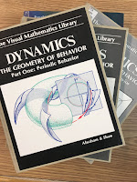
Dynamics: The Geometry of Behavior, by Ralph Abraham and Christopher Shaw, superimposed on Intermediate Physics for Medicine and Biology.
Dynamics: The Geometry of Behavior,
by Ralph Abraham and Christopher Shaw.
When I was working at the National Institutes of Health in the 1990s, I ran across a wonderful series of books from the Visual Mathematics Library that had a big impact on the way I thought about math. Dynamics: The Geometry of Behavior, by Ralph Abraham and Christopher Shaw, was published in four volumes: 1 Periodic Behavior, 2 Chaotic Behavior, 3 Global Behavior, and 4 Bifurcation Behavior. The fascinating feature of these books was that they contained almost no equations; everything was explained in pictures. At first glance, they look like comic books, but on closer inspection you realize that the math is presented in a very accurate and rigorous way. There are lots of plots of phase planes, and drawings of experimental apparatus that are being modeled by the math. There is hardly a page without pictures, and 90% of many pages are filled with illustrations. I highly recommend these books for anyone interested in developing an intuitive feeling for nonlinear dynamics (which should be everyone).

Their foreword begins
During the Renaissance, algebra was resumed from Near Eastern sources, and geometry from the Greek. Scholars of the time became familiar with classical mathematics. When calculus was born in 1665, the new ideas spread quickly through the intellectual circles of Europe. Our history shows the importance of the diffusion of these mathematical ideas, and their effects upon the subsequent development of the sciences and technology.

Today, there is a cultural resistance to mathematical ideas. Due to the widespread impression that mathematics is difficult to understand, or to a structural flaw in our educational system, or perhaps to other mechanisms, mathematics has become an esoteric subject. Intellectuals of all sorts now carry on their discourse in nearly total ignorance of mathematical ideas. We cannot help thinking that this is a critical situation, as we hold the view that mathematical ideas are essential for the future evolution of our society.

The absence of visual representations in the curriculum may be part of the problem, contributing to mathematical illiteracy, and to the math-avoidance reflex. This series is based on the idea that mathematical concepts may be communicated easily in a format which combines visual, verbal, and symbolic representations in tight coordination. It aims to attack math ignorance with an abundance of visual representations.

In sum, the purpose of this series is to encourage the diffusion of mathematical ideas, by presenting them visually.
Dynamics: The Geometry of Behavior, by Ralph Abraham and Christopher Shaw, with Intermediate Physics for Medicine and Biology.
Dynamics: The Geometry of Behavior,
by Ralph Abraham and Christopher Shaw.
In the 4th edition of Intermediate Physics for Medicine and Biology, Russ Hobbie and I do not suppress mathematical expressions. I suspect many of our readers would claim we have too many, rather than too few, equations. Nevertheless, we try to convey our subject in figures as well as math, visually as well as symbolically. We discuss nonlinear dynamics in Chapter 10, and we have some state space figures that are similar to those found in Abraham and Shaw (although they use 4-color figures—green, red, blue, and black—while we use the less attractive black and white). I believe all the illustrations in Abraham and Shaw are hand-drawn, giving them a charm that often is lacking in this age of computer-generated drawings. Unfortunately, Russ and I never cited Abraham and Shaw. One reason I write this blog is to alert our readers to books and articles that don’t appear in the pages of Intermediate Physics for Medicine and Biology.

Dynamics: The Geometry of Behavior is one of those rare gems that you should become familiar with, both for what it can teach and also for its beauty. To learn more about The Visual Mathematics Library, see Ralph Abraham’s webpage.

Friday, December 7, 2012

Lord Rayleigh, Biological Physicist

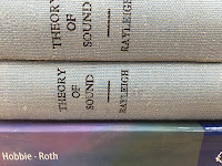
Theory of Sound, by Lord Rayleigh, with Intermediate Physics for Medicine and Biology.
Theory of Sound,
by Lord Rayleigh.
I am a big fan of Victorian physicists. Among my heroes are Faraday, Maxwell, and Kelvin. Another leading Victorian was John William Strutt, also known as Lord Rayleigh (1842–1919). Russ Hobbie and I mention Rayleigh in the 4th edition of Intermediate Physics for Medicine and Biology, in the context of Rayleigh Scattering. In Chapter 15 on the interaction of x-rays with matter, we write
A photon can also scatter elastically from an atom, with none of the electrons leaving their energy levels. This (γ, γ) process is called coherent scattering (sometimes called Rayleigh scattering), and its cross section is σcoh. The entire atom recoils; if one substitutes the atomic mass in Eqs. 15.15 and 15.16, one finds that the atomic recoil kinetic energy is negligible.
In Rayleigh scattering, the oscillating electric field in an electromagnetic wave exerts a force on electrons. These electrons are displaced by this force, and therefore oscillate at the same frequency as the wave. An oscillating charge emits electromagnetic radiation. The net result is scattering of the incident wave. If the electrons are free, this is known as Thomson scattering. If the electrons are bound to an atom, and the frequency of the light is less than the natural frequency of oscillation of the bound electrons, then it is known as Rayleigh scattering. Light scattering is complicated when the wavelength is similar to or smaller than the size of the scatterer, because light scattered from different regions within the particle interfere. However, Rayleigh scattering assumes that the wavelength is large compared to the size of the scatterer, so interference is not important.

Rayleigh scattering not only plays a role in the scattering of x-rays, but also is responsible for the scattering of visible light. The Rayleigh scattering cross section varies as the 4th power of the frequency, or inversely with the 4th power of the wavelength. When we look at the sky, we see the scattered light. Since the short wavelength blue light is scattered much more than the long wavelength red light, the sky appears blue.

Lord Rayleigh made other important contributions to physics. For example, he wrote an influential book on the Theory of Sound, and he won the Nobel Prize in 1904 for his discovery of the element argon. He succeeded Maxwell as the Cavendish Professor of Physics (see this video: https://www.youtube.com/watch?v=tkwLavjqsBI to learn more).


Was Rayleigh a biological physicist? Yes! Rayleigh was one of the first to explain how we localize sound. His Duplex Theory suggests that we can determine the direction a sound came by sensing the arrival time difference at each of our two ears for low frequencies, and sensing the intensity difference between the ears for high frequencies.

Lord Rayleigh was born 170 years ago this fall (November 12, 1842). J. J. Thomson studied under Rayleigh, and Ernest Rutherford studied under Thomson. Previously in this blog, I described how I am descended, academically speaking, from Rutherford. This means Lord Rayleigh is, again academically speaking, my great-great-great-great-great-great grandfather.

Friday, November 30, 2012

A Dangerous Error in the Dilution of 25 Percent Albumin

In Problem 5 in Chapter 5 of the 4th edition of Intermediate Physics for Medicine and Biology, Russ Hobbie and I consider what happens if a drug is administered without carefully considering osmotic effects. The problem refers to a letter to the editor in the New England Journal of Medicine from Donald Steinmuller.
A Dangerous Error in the Dilution of 25 Percent Albumin
To the Editor: Physicians and pharmacists should be alert to a serious error that can occur in the preparation of replacement albumin solutions for plasmapheresis.

Plasmapheresis was performed in an elderly man who had myeloma with renal insufficiency. One plasma volume exchange was ordered, with 5 percent albumin as the replacement solution, with calcium, potassium, and magnesium supplements. Because of the lack of availability of 5 percent albumin, the hospital pharmacy used 25 percent albumin and diluted this solution 1:4 with sterile water to achieve a 5 percent solution. Reference was made to Trissel’s Handbook on Injectable Drugs, the 1994 edition of which states, “A 5% solution may be prepared from the 25% product by adding 1 volume of the 25% albumin to 4 volumes of sterile water or an infusion solution such as dextrose 5% in water or sodium chloride 0.9%.” The pharmacist used sterile water as described by the handbook, resulting in a hypo-osmolar solution that caused severe hemolysis in the patient. The hematocrit dropped 7.3 points, and renal failure developed.

This flagrant error in instructing dilution with water was only partially corrected in the 1996 edition of Trissel's handbook. This edition states, “If sterile water for injection is the diluent, the tonicity of the diluted solution must be considered. Substantial reduction in tonicity creates the potential for hemolysis.” In view of the osmolarity of the 25 percent albumin solution diluted with sterile water (approximately 36 mOsm per liter), one should never use water to dilute 25 percent albumin.

The problem is aggravated by the label on the 25 percent albumin solution. The label states that 100 ml of 25 percent albumin is “osmotically equivalent to 500 ml of plasma.” This statement is not true. It confuses the osmotic and oncotic effects. The oncotic effect of 100 ml of the 25 percent solution is equivalent to 500 ml of plasma, but since the concentration of saline in the 25 percent solution is isotonic with plasma, the osmotic effect of 100 ml of the 25 percent is equivalent to only 100 ml of plasma.

The handbook and product label need to be corrected as soon as possible to prevent this error in the future. Pharmacies and the medical community should be alert to this potentially life-threatening error.
This letter makes the distinction between osmotic pressure and oncotic pressure, that part of the osmotic pressure caused by large colloidal particles such as albumin.

Before publication, the editor sent the letter to experts at the FDA, who responded with a separate letter that immediately followed Steinmuller’s
…Including the case reported by Dr. Steinmuller, the FDA is aware of four cases of hemolysis that have occurred since 1994 during or after plasmapheresis when albumin (human) 25 percent was diluted to a 5 percent solution with the use of sterile water for injection…. The FDA has taken the following steps to advise the medical community of this potentially serious problem. First, the FDA is recommending to manufacturers of albumin (human) 25 percent that the package inserts for their products be revised to include … information on acceptable diluents, such as 0.9 percent sodium chloride or 5 percent dextrose in water…
The FDA letter triggered a third letter from Richard Kravath, of Kings County Hospital Center in Brooklyn
…Although the response by the representatives of the Food and Drug Administration (FDA) is correct with regard to the recommendation that dilution with 5 percent glucose, instead of water alone, would prevent hemolysis, use of this solution would not prevent the possible development of hyponatremia and brain swelling if the solution were used rapidly in large volumes, as in plasma exchange. Owing to its oncotic pressure, the albumin would tend to remain in the plasma compartment, whereas the glucose would rapidly leave the circulation, enter the interstitial fluid and then enter cells, become metabolized, and no longer exert an osmotic effect….

Glucose solutions should not be used to replace plasma or other extracellular fluids. Sodium chloride 0.9 percent (154 mmol per liter) is a reasonable alternative, but a more physiologic solution, one that more closely resembles plasma, would be even better.
The authors of the handbook quoted by Steinmuller also wrote a letter, in which they quoted the corrected 9th edition of their book (Steinmuller had cited the 8th edition) and claimed (somewhat lamely, in my opinion) that “…It is common knowledge that large volumes of very hypotonic solutions should not be administered intravenously….” and “…This situation points to the need for health care practitioners to use up-to-date references...”.

Finally, the FDA experts responded to these last two letters.
…Plasma exchange or plasmapheresis represents a unique circumstance because, in formulating the replacement solution, one must take into account not only the loss of endogenous plasma proteins (principally, but not exclusively, albumin) but also the fact that significant quantities of electrolytes such as sodium and chloride are being removed by the procedure...
This exchange of letters is fascinating, and emphasizes the importance of the osmotic effects that Russ and I discuss in Chapter 5. The original letter by Steinmuller, which highlights an important medical issue, also makes for an instructive homework problem.

Let me conclude by noting an error (now listed in the book errata, downloadable at the book website) in the 4th edition of Intermediate Physics for Medicine and Biology. Russ and I have the title of Steimnuller’s letter incorrect. On page 133 we write “15 percent” when the title of the letter actually says “25 percent”. The homework problem correctly uses 25 %, and needs no change. Below is the corrected citation:
Steinmuller, D. R. (1998) “A Dangerous Error in the Dilution of 25 Percent Albumin,” New England Journal of Medicine, Volume 338, Page 1226.

Friday, November 23, 2012

Marie Curie

Marie Curie (1867–1934) is one of the few scientists who received two Nobel Prizes: for Physics in 1903, and for Chemistry in 1911. Russ Hobbie and I don’t discuss Curie extensively in the 4th edition of Intermediate Physics for Medicine and Biology, but in Chapter 17 on Nuclear Medicine we do introduce the unit of radioactive activity named for her.
The activity A(t) is the number of radioactive transitions (or transformations or disintegrations) per second. The SI unit of activity is the becquerel (Bq):

1 Bq = 1 transition s−1.

The earlier unit of activity, which is still used occasionally, is the curie (Ci):

1 Ci=3.7 × 10¹⁰ Bq,
1 μCi = 3.7 ×10⁴ Bq.
Several excellent articles were published about Marie Curie and her husband/collaborator Pierre Curie for the centennial of her 1898 discovery of radium. Saenger and Adamek’s article in the journal Medical Physics states
Marie Curie’s activities and research left her imprint on nuclear medicine, which continues to this day. Much of her impact is related to the role of women in science, biology, and medicine. She successfully overcame struggles for recognition in the first decades of this century. One of her major achievements was the development of field-radiography for wounded soldiers in World War I. Her continued endeavors to provide radium therapy for cancer was a giant step for humanity. She worked unceasingly in the laboratory to separate and identify radioactive elements of the periodic table. The standardization of these elements resulted in the 1931 report of the International Radium-Standards Commission and the posthumous two-volume Radio-aktivite´.
The abstract of Mould’s article in the British Journal of Radiology begins
This review celebrates the events of 100 years ago to the month of publication of this December 1998 issue of the British Journal of Radiology, when radium was discovered by the Curies. This followed the earlier discovery in November 1895 of X-rays by Röntgen, which has already been reviewed in the British Journal of Radiology [1] and the discovery in March 1896, by Becquerel, of the phenomenon of radioactivity, which introduces this review. This is particularly relevant as Marie Curie was in 1897 a research student in Becquerel’s laboratory. Marie Curie’s life in Poland prior to her 1891 departure for Paris is included in this review as are other aspects of her life and work such as her work in World War I with radiological ambulances (known as “Little Curies”) on the battlefields of France and Belgium, early experiments with radium and the founding of the Institut du Radium in Paris and of the Radium Institute in Warsaw. Wherever possible I have included appropriate quotations in Marie Curie’s own words [2–4] and each section is related in some way to the life and work of Maria or Pierre. This review is completed with details of the re-interment of the bodies of Pierre and Marie on 20 April 1995 in The Panthéon, Paris.
Excellent overviews of Curie’s life and work are provided by the AIP Center for the History of Physics and the Official Website of the Nobel Prize. You can read about the discovery of radium in Maria Curie’s own words here. And for all you dear readers who prefer Saturday morning cartoons to learned articles, watch this; it doesn’t include any complex or controversial stuff like the Langevin affair, but it is enjoyable in its own simple way.

Marie Curie Animated Hero Classics
https://www.youtube.com/watch?v=x-ynt1yY43I&t=205s
Recently, the visual artist/filmmaker/writer Quintan Ana Wikswo was granted access to Marie Curie’s laboratory in Paris for “creating performance films and photographs for ... LUMINOSITY: THE PASSIONS OF MARIE CURIE, a multimedia opera by composer Pamela Madsen.” Wikswo describes her ongoing work and previews some of her photographs in her blog Bumblemoth.
To see her books, her equipment, to stand at her desk, to see her beakers and centrifuges and shelves of chemicals…it’s a kind of searing existential therapy, and anyone visiting Paris should make the effort to spend a few moments at her lab. Why? It’s an antidote, at the very least. I work half-days at her lab, and then explore art museums of Paris in the off hours. The contrast is shocking and disturbing. Inspiring and sorrowful.
At Oakland University, I work in the College of “Arts and Sciences.” Wikswo has found her own niche at the intersection of these two rarely-overlapping endeavors. I look forward to seeing the completed project.