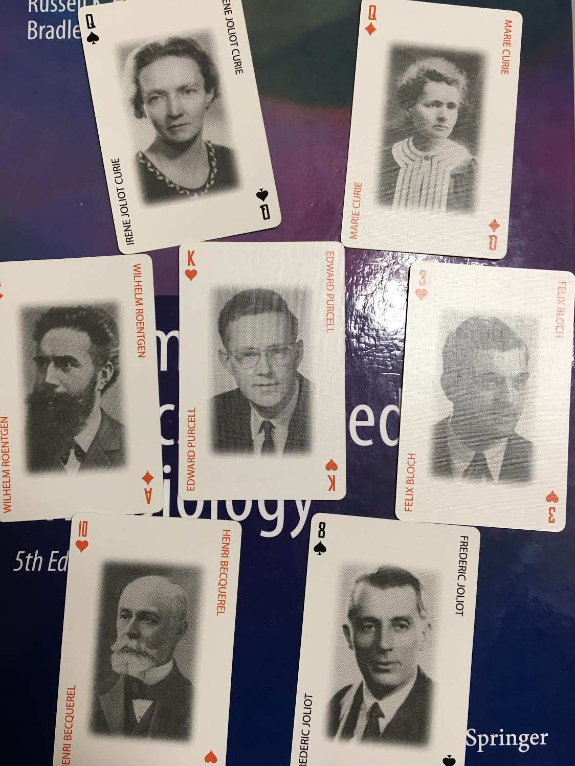
Last Christmas, my daughter Katherine gave me a unique and fascinating present: Physicist Playing Cards. Each card features a picture of an eminent physicist. You can order the cards from the American Institute of Physics at http://store.aip.org. Two decks are available: one of historical physicists, and one of modern physicists. I browsed through the deck of historical physicists and found ten cards that have relevance to biology and medicine. These ten physicists, each a Nobel Prize winner, contributed greatly to the application of physics to the life sciences. Many of these physicists are mentioned in the 4th edition of Intermediate Physics for Medicine and Biology (the appropriate page is given in parentheses).
Queen of Diamonds: Marie Curie, pioneer in the study of radiation (p. 489).
Ace of Diamonds: Wilhelm Roentgen, discoverer of X-rays (p. 440).
Nine of Clubs: William Bragg, analyzed crystal structures using X-ray diffraction (p. 466).
Three of Hearts: Felix Bloch, co-discoverer of nuclear magnetic resonance (p. 519).
Ten of Hearts: Henri Becquerel, discoverer of radioactivity (p. 472).
King of Hearts: Edward Purcell, co-discoverer of nuclear magnetic resonance (p. 519).
Ace of Hearts: Pierre Curie, discoverer of the piezoelectric effect (p. 216).
Eight of Spades: Frederic Joliot, co-creator of the first artificial radioisotope (none).
Nine of Spades: Max von Laue, discoverer of X-ray diffraction (none).
Queen of Spades: Irene Joliot Curie, co-creator of the first artificial radioisotope (none).
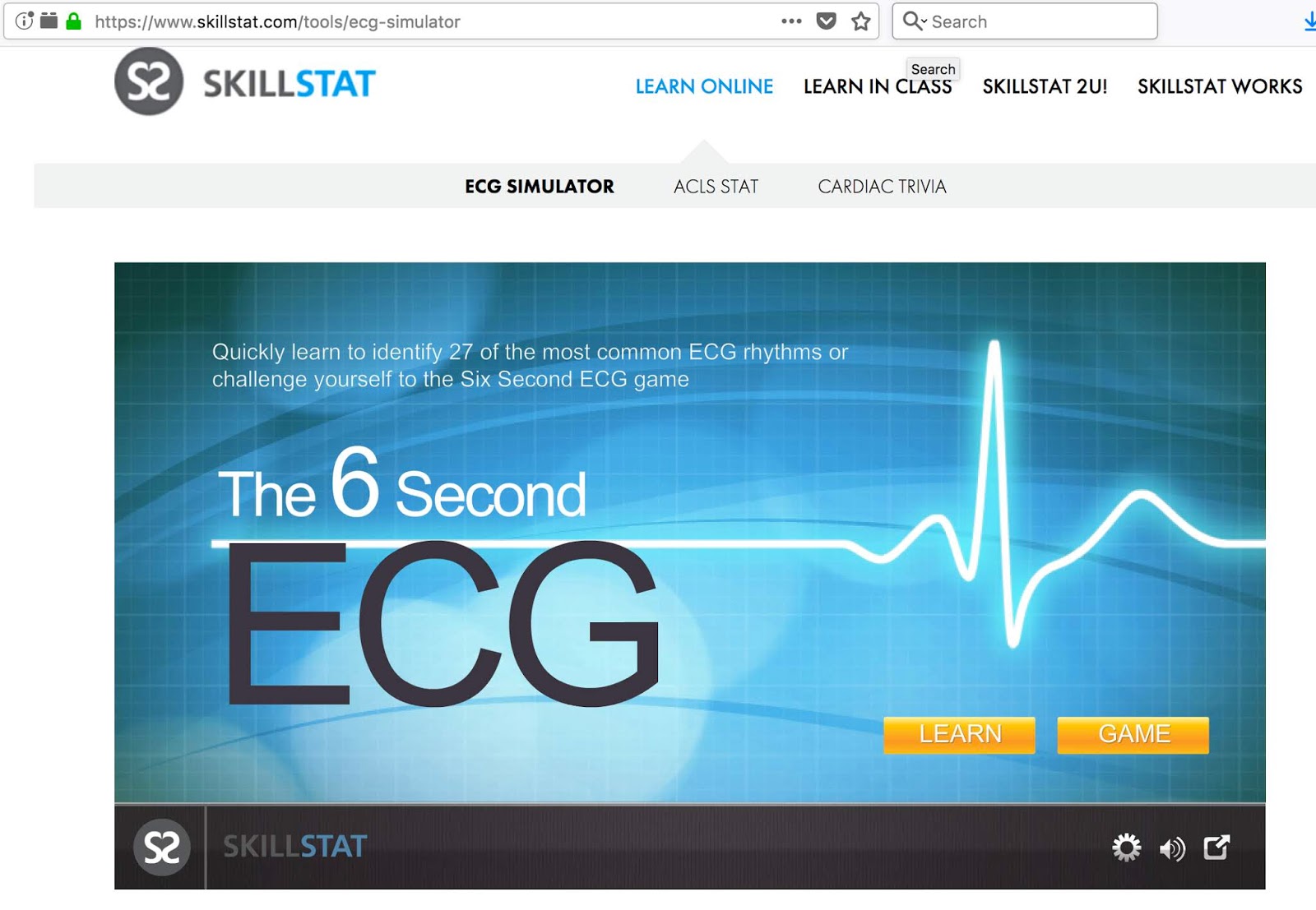
When I teach biological physics, we spend at least one class discussing the electrocardiogram (ECG, sometimes called the EKG for the German term Elektrokardiogramm). The 4th edition of Intermediate Physics for Medicine and Biology covers the ECG in Chapter 7, but students often need additional practice on interpreting the signals from heart arrhythmias. As homework, I require my students to go to the website http://www.skillstat.com/ECG_sim_demo.html and play the ECG simulator game. It tests the students’ knowledge of ECGs in a fun, interactive way. The same website has units on cardiac anatomy, a cardiac dictionary, and cardiac trivia games.
The 1924 Nobel Prize in Physiology or Medicine was awarded to the Dutch physiologist Willem Einthoven (1860–1927) “for his discovery of the mechanism of the electrocardiogram.” The notation of the P, QRS, and T waves (see Fig. 7.17 of our textbook) was developed by Einthoven, as was his interpretation of the ECG using “Einthoven's triangle” (see Fig. 7.19, and page 188). Einthoven’s work is an excellent example of how physics can applied successfully to medicine and biology.
Readers of the 4th edition of Intermediate Physics for Medicine and Biology who are particularly interested in Medical Physics will find the website http://medicalphysicsweb.org interesting. This “community website” is maintained by the Institute of Physics, the United Kingdom’s professional organization for physicists. The IOP created several community websites to “promote innovation, growth and networking,... [and to] provide both a valuable information source and an international forum within which community members can share and exchange their views.”
Medicalphysicsweb provides “a mix of in-depth news, analysis, opinion and primary research papers across the key disciplines of medical physics.” The site contains editorials, job postings, a buyer’s guide, featured journal articles, and research and industry news. Students who are studying from Intermediate Physics for Medicine and Biology will find this website an easy and free way to become familiar with medical physics as a profession. You can become a member (no cost, but there is a registration procedure) and receive a weekly “What's New” newsletter. I highly recommend it.
Often a mathematical physics class will focus on the “big three” partial differential equations of physics: the diffusion equation, the wave equation, and Laplace’s equation. The 4th edition of Intermediate Physics for Medicine and Biology provides a gentle introduction to each of these equations. You won’t find much mathematical theory; Russ Hobbie and I are concerned primarily with exploring their relevance to biology and medicine.
The diffusion equation (Eq. 4.24) is the topic of Chapter 4. Diffusion is not covered in many physics classes, but is crucially important to biology. The diffusion equation is also known by another name: the heat equation. Problem 24 of Chapter 4 explores an extension of this topic: a “reaction-diffusion” equation that governs the nonlinear propagation of calcium waves.
The wave equation (Eq. 13.5) was not discussed much in previous editions of Intermediate Physics for Medicine and Biology, but it is an essential topic in the 4th edition's new Chapter 13 on sound and ultrasound. Here we meet classic wave behavior, such as the relationship between wavelength and frequency, the difference between propagating and standing waves, reflection, and the Doppler effect.
Laplace’s equation (the displayed equation before Eq. 7.44a) is encountered in Chapter 7 on the electrocardiogram. We don’t talk as much about solutions to Laplace’s equation as we do the diffusion and wave equations, but it plays a fundamental role in electrostatics and appears when discussing steady-state solutions to the diffusion equation.
Peter is now the head of the Section on Tissue Biophysics and Biomimetics, which is part of the Eunice Kennedy Shriver National Institute of Child Health and Human Development. The goal of his section is “to understand fundamental physical mechanisms governing tissue-level physiological processes that are essential for life, or necessary to achieve a high quality of life. Examples include understanding the physical basis of nerve excitability and of effective load bearing in cartilage. This entails discovering relationships between physiological function and a tissue's structure, organization, and physical properties. This is done by studying the behavior of biological model systems using novel quantitative approaches (e.g., experimental methods, mathematical models, physical models). Another aim of ours is to transfer these new methodologies to the biomedical research and healthcare communities. An example includes the invention and successful dissemination of diffusion tensor magnetic resonance imaging from the 'bench' to the ‘bedside.’” Diffusion tensor imaging is one of the topics that Russ Hobbie and I added to the 4th edition of Intermediate Physics for Medicine and Biology (see Chapter 18, Section 13). We also wrote a new homework problem that asked the student to show that the trace of the diffusion tensor is independent of fiber direction. We had trouble deciding if this problem belonged in Chapter 4 (on diffusion) or Chapter 18 (on magnetic resonance imaging), and we ended up putting the problem in both chapters (see Problems 4.22 and 18.40). Another homework problem featuring Peter’s work on cartilage appears in Chapter 5 (Problem 5.6).
The Office of NIH History has published an interview with Peter, in which he explains how he developed diffusion tensor imaging. Below is a brief excerpt of this interview, describing the moment Peter conceived the idea of DTI (I make a cameo):
Actually, the first exposure I had to diffusion imaging was a talk that Denis Le Bihan had given. He had recently come to the NIH from France and talked about how diffusion could be used—I think it was in stroke—and I thought it was very interesting, but I didn’t really initially make a connection to it. But in the early 1990s, Denis Le Bihan and, I believe it was Philippe Douek had a poster presentation at one of the NIH research festivals off in a corner in one of the white tents that they had constructed over here in the parking lot East of Building 30. They had done something very novel. They had shown that they could color code different parts of the brain according to what they thought was the orientation of diffusion. That was a poster that resulted in a paper, I think early in the next year, by Denis and Philippe. But I visited that poster and I was there with my friend and colleague, Brad Roth, the guy I was doing the magnetic stimulation with, and I realized that there was something really fundamentally wrong with the approach that Denis and Philippe were using.
In the December 14, 2007 entry to this blog, I discussed how the shutdown of a nuclear reactor at Chalk River, Ontario caused a shortage of an isotope of technetium (Tc-99m) used for medical imaging. Tc-99m is obtained from an isotope of molybdenum, Mo-99, that is produced at a handful of international facilities, including the Canadian reactor. The production and use of Tc-99m in nuclear medicine is discussed in Chapter 17 of the 4th edition of Intermediate Physics for Medicine and Biology.
The most recent (May 2008) issue of Physics Today contains an article by science writer Toni Feder that explores this topic further. Feder writes
The disruption late last year of Mo-99 production in Canada threw the nuclear medicine community into a panic. With a half-life of 66 hours, Mo-99 can’t be stockpiled. The Canadian reactor was down for maintenance, and its startup was delayed because of safety violations. Such was the upset in the nuclear medicine community that the Canadian government stepped in and ordered the reactor to start up despite some remaining safety concerns—and demoted the head of Canada’s nuclear regulatory agency.
The article also discusses plans for the production of Mo-99 in the United States. The US producers will use “low-enriched uranium” (LEU) rather than the “highly enriched uranium” (HEU) used at other facilities, including the Chalk River plant. This would have advantages for nonproliferation, and could reduce the amount of HEU available for production of weapons of mass destruction by terrorist groups. Feder continues
“Our program is to minimize civilian use of HEU,” says Parrish Staples, manager of the National Nuclear Security Administration’s program to convert reactors from HEU to LEU fuel. “The only HEU the US is currently exporting is for production of Mo-99 in foreign production facilities.” The US exports about 25 kg of HEU each year, or about half the total used for making Mo-99, he says. If the US stops exporting HEU, he adds, according to the IAEA [International Atomic Energy Agency] definition “a weapon’s worth of material would be removed [from circulation each year].”
“At some point there will be an incident somewhere in the world that will cause the US to close its borders to radioactive materials for a day, a week, two or three weeks, whatever,” says MURR [The University of Missouri-Columbia Research Reactor] director Ralph Butler. “And when you think that there are tens of thousands of patients per day [in the US] utilizing this diagnostic tool [radioactive isotopes], that’s a huge impact.” The US does not have a Mo-99 source, he adds. “There is a national need, and it’s an opportunity we [at MURR] can meet.”
In January 1989, John Wikswo of Vanderbilt University wrote a review in Physics Today (Volume 42, Pages 75–76) about the 2nd edition of Intermediate Physics for Medicine and Biology. His review began
While there are excellent texts, treatises and reviews of medical and radiological physics and biophysics, none of these provides the breadth and depth required of a guidebook for a physicist or biologist desiring to explore, possibly for the first time, the realm where physics joins medicine and biology. The problem in part is that such a book should develop simultaneously both the physics and the biology without assuming extensive prior knowledge of either, and yet should explore the subject with sophistication and quantitative rigor. In 1977, I was confronting the dilemma of finding no suitable text for the very first physics course I had been assigned to teach, an introductory medical physics course for undergraduate premedical students, when a friend of mine from the Mayo Clinic told me that Russell Hobbie of the University of Minnesota was writing just the book I needed. For two years my students and I learned from typed manuscripts kindly provided by Hobbie, and my colleagues and I have been using the first edition of Intermediate Physics for Medicine and Biology ever since then. This year, we can greet our students with the second edition.
In Chapter 15 of the 4th edition of Intermediate Physics for Medicine and Biology, Russ Hobbie and I discuss the interaction of radiation with matter, a topic that is crucial for understanding the medical use of X-rays. Twenty years ago, Russ wrote a computer program called MacDose that provides a two-dimensional simulation of the photoelectric effect, Compton scattering, and pair production; the primary mechanisms of X-ray interaction. MacDose runs on any Macintosh with OS-9 or earlier, including Classic in OS-X. You can download a copy of MacDose, including a student manual and instructors guide, at our book’s website. To learn more about MacDose, see Hobbie’s article in Computers in Physics (Volume 6, Pages 355–359, 1992).
You can also download a 26 minute Quicktime movie in which Russ demonstrates MacDose and explains various concepts related to the attenuation and absorption of X-rays (you can view the movie on either a PC or a Mac). With help from my daughter Stephanie, I have uploaded this movie onto Youtube. Because of a limit on the duration of Youtube videos, Stephanie had to split the movie into three parts. Search on YouTube for “MacDose” and you should find all three. Then pop some popcorn, pour yourself a drink, find a seat, and watch Hollywood’s leading man Russ Hobbie explain how radiation interacts with matter.
In the 1940s and 50s, Alan Hodgkin and Andrew Huxley discovered the ionic basis for nerve conduction, work that resulted in their sharing the 1963 Nobel Prize in Physiology or Medicine. Chapter 6 in the 4th edition of Intermediate Physics for Medicine and Biology describes the Hodgkin-Huxley model in detail. Yet, no textbook can replace the experience of peering over Hodgkin's shoulder while he performs the voltage clamp experiments on a squid nerve axon that were crucial for their discoveries. Fortunately, a movie was made of these experiments, and clips from it can be found online, at a website for a neurophysiology class at Smith College. I particularly recommend the clip “Dissection and Anatomy” showing the dissection of the giant axon from a squid by J. Z. Young, and “Voltage Clamping” by P. F. Baker and Hodgkin himself.
When I teach Biological Physics at Oakland University, I like to have my students read Hodgkin and Huxley's classic paper “A Quantitative Description of Membrane Current and its Application to Conduction and Excitation in Nerve” (Journal of Physiology, Volume 117, Pages 500–544, 1952). A pdf of this article is available online. However, if you encourage your students to read it, be sure to warn them that the definition of the transmembrane potential is different than is used now, with their definition being the outside minus the inside voltage, and zero being rest. (Nowadays, researchers typically use inside minus outside, with rest corresponding to -65 mV). Writing a program to simulate the Hodgkin and Huxley model is the best way to learn about it (we have a sample of such a program in Fig. 6.38 or our textbook), but those who are not programmers might want to try this applet that allows online simulation of a nerve action potential.
The quotes below are taken from the American Association of Physicists in Medicine Golden Anniversary website.
Many of the greatest inventions in modern medicine were developed by physicists who imported technologies such as X rays, nuclear magnetic resonance, ultrasound, particle accelerators and radioisotope tagging and detection techniques into the medical domain. There they became magnetic resonance imaging (MRI), computerized tomography (CT) scanning, nuclear medicine, positron emission tomography (PET) scanning, and various radiotherapy treatment methods. These contributions have revolutionized medical techniques for imaging the human body and treating disease.
Now, in 2008, the American Association of Physicists in Medicine (AAPM), the premier scientific and professional association of medical physicists, is celebrating its 50th anniversary and is calling attention to the field of medical physics achievements.
In the coming year, the AAPM will be calling attention to the many ways in which medical physics has revolutionized medicine. A few highlights include:
1. USING PARTICLE ACCELERATORS TO DEFEAT CANCER 2. BETTER DETECTION OF BREAST CANCER 3. MATTER/ANTIMATTER COLLISION IMAGING 4. ENSURING THE SAFETY OF PEOPLE WHO GET CT SCANS 5. MEDICAL PHYSICS MOMENTS IN HISTORY
This year, the AAPM journal, Medical Physics, will celebrate the 50th anniversary with a year-long celebration. Every issue published in 2008 will have an article devoted to history and reviews of special topics intended to recognize this anniversary, and will carry the AAPM anniversary logo.
The AAPM is a scientific, educational, and professional nonprofit organization whose mission is to advance the application of physics to the diagnosis and treatment of human disease. The association encourages innovative research and development, helps disseminate scientific and technical information, fosters the education and professional development of medical physicists, and promotes the highest quality medical services for patients. In 2008, AAPM will celebrate its 50th year of serving patients, physicians, and physicists.
I am an emeritus professor of physics at Oakland University, and coauthor of the textbook Intermediate Physics for Medicine and Biology. The purpose of this blog is specifically to support and promote my textbook, and in general to illustrate applications of physics to medicine and biology.